Abstract
Aim
This study aims to present a modified Laboratory Risk Indicator for Necrotizing Fasciitis (m-LRINEC) scoring system and to evaluate its ability in discriminating necrotizing fasciitis (NF) from other severe soft-tissue infections.
Methods
Patients with NF diagnosed by surgical findings in our institution between January 2014 and December 2020 were included as the case group, matched by controls with severe soft-tissue infections other than NF in a ratio of 2:1, based on demographics, calendar time and immunosuppressant status. Patients’ demographics, comorbidities and laboratory test results were extracted from medical records. Logistic regression analyses were used to determine the association with NF after adjustment for confounders, whereby m-LRINEC was developed. Receiver operating characteristics (ROC) curves and the area under the curve (AUC) were used to evaluate its discriminating ability.
Results
There were 177 patients included, 59 in the NF group and 118 in the non-NF group. We added comorbid diabetes and kidney disease to the original LRINEC scoring system, used high-sensitivity C-reactive protein (HCRP) to replace the CRP and redefined the cut-off values for the other four variables, to develop the m-LRINEC system. The cut-off value for m-LRINEC was 17 points, with corresponding sensitivity of 93.2% and specificity of 86.9%, and the AUC was 0.935 (95% CI 0.892 to 0.977; p<0.001).
Conclusion
The m-LRINEC scoring system shows a high sensitivity and specificity in discriminating NF from other severe soft-tissue infections. Patients with an m-LRINEC score of >17 points should have a high index of suspicion for the presence of NF. The validity of the m-LRINEC needs to be confirmed in studies with larger samples and better design.
Keywords: necrotizing fasciitis, factors, biomarkers, scoring system, m-LRINEC
Introduction
Necrotizing fasciitis (NF) is an extremely infrequent soft-tissue infection, with a population-based incidence of 0.24–0.4 per 100,000 person-years.1 This condition involves necrosis of the superficial fascia and subcutaneous tissues, and progresses rapidly, leading to a severe systemic toxicity and even mortality. A high index of suspicion, early diagnosis and aggressive surgical debridement of necrotic tissues, or amputation if necessary, are essential for successful treatment. Nevertheless, according to the literature reports, NF is still associated with a high rate of mortality of 10.9–76%2,3 and an amputation rate of 15.0–30.0%.3–5
Clinically, it is very difficult to distinguish early-course NF from cellulitis or abscesses, despite the continuous advances in techniques and imaging modalities. Magnetic resonance imaging (MRI) has been shown to be useful in early differential diagnosis,6 but routine application of MRI is impractical owing to the limitations of cost and availability. The readily available laboratory biomarkers from routine blood and biochemistry examination, or thereby the risk prediction model, can be an ideal tool for screening or ruling out NF, owing to their low cost, rapid access and availability. In 2004, Wong et al7 developed the Laboratory Risk Indicator for Necrotizing Fasciitis (LRINEC) scoring system, and demonstrated its high sensitivity in both development and validation cohorts (for ≥6 points, 89.9% and 92.9%, respectively) in discriminating NF from other severe soft-tissue infections. However, in the ensuing validity studies, researchers found that the validity of the LRINEC had been overstated, and its sensitivity was 43.2–80.0% for a score of ≥6 and 28.6–68.4% for a score of ≥8 in different settings, countries or regions;1,8–10 some studies even demonstrated it to be non-specific.9,11 However, in other studies, the authors suggested that LRINEC had high sensitivity and specificity. For example, LRINEC of the Oro-Cervical Region (LRINEC-OC) exhibited a sensitivity of 88.5% and a specificity of 93.4% when the cut-off values was set to 6 points12
It is very likely that the great variation in validation results for LRINEC is related to differences in race, region or area, demographics (age, sex, body mass index), morbid medical conditions (diabetes, immunosuppressant status) or study design. For example, substantial evidence was found for significant differences in incidence, clinical characteristics, outcomes and even validation results of LRINEC, between NF patients with and without diabetes.8 In fact, only the use of biomarkers is less likely to lead to the development of a universal screening or diagnostic tool for NF, and other potential factors (eg, diabetes or immunosuppressant status) should also be considered.11 Furthermore, considering these differences from multiple aspects, individualized or customized scoring tools should be developed.
In this study, we made modifications to the original LRINEC based on data from matched cases and controls to develop a new scoring system, namely the modified LRINEC (m-LRINEC). The purpose of this study is to present the m-LRINEC and evaluate its discrimination ability.
Methods
This study was a retrospective nested case–control study and the study protocol was approved by the ethics committee of the Third Hospital of Hebei Medical University prior to commencement (NO.H201701101). It was conducted in accordance with the provisions of the Declaration of Helsinki. Written informed consent was obtained from all participating patients or their relatives in law.
Patients
From January 2014 to December 2020, patients aged 15 years or older who had experienced surgical treatment due to NF were deemed to be eligible for inclusion and assigned to the case group. The definite diagnosis of NF was made based on the operative findings: presence of foul-smelling dishwater-like pus with grayish necrotic tissues, and lack of normal resistance to blunt dissection (finger test). In total, 59 patients with NF were identified and assigned to the case group.
In the same time window, 932 patients with a diagnosis on admission of cellulitis, abscesses or myositis, or other variants of dermohypodermitis other than NF, were treated in our institution, from whom control patients were randomly selected. NF cases were matched in a ratio of 1:2 with the controls using the propensity score method, based on the following variables: age (year), sex, body height (cm), body weight (kg), residential place (urban or rural), admission calendar time and the chronic use of immunosuppressants (yes or no). R software 3.6.3 was used to complete the matching process, using the “MatchIt” package and the greedy matching algorithm with a caliper value of 0.2 standard deviation (SD) of the logit of a propensity score. Finally, 118 control patients were included as the control group.
Data Collection
All data were extracted from the inpatient records, including age, sex, body weight, body height, estimated body mass index (BMI), residential place, admission date, presence of comorbidities, (hypertension, diabetes, cerebrovascular disease, heart disease, pulmonary disease, liver disease, kidney disease, peripheral vascular disease, malignancy), and LRINEC-based biomarkers including high-sensitivity C-reactive protein (HCRP), total white blood cell (WBC) count, hemoglobin level, fasting blood glucose (FBG), sodium concentration and serum creatinine. For the purpose of improving the differential ability and lowering the time-dependent effects of these biomarkers on the development of NF, considering the rapidly progressive course of NF, we selected the biomarker results from within 48 hours before the diagnosis of NF and other soft-tissue infections.
The comorbid diseases or conditions were recorded after patients were admitted. Hypertension and diabetes were diagnosed on the basis of patients’ (or their relatives’) self-reported history combined with the routine admission examinations. A history of cerebrovascular disease (hemorrhagic or ischemic stroke, transient cerebral hemorrhage), heart disease (heart failure, ischemic heart disease, valvular heart disease), pulmonary disease (chronic obstructive pulmonary disease, asthma, pulmonary tuberculosis), liver disease (hepatitis or cirrhosis) and/or kidney disease (renal deficiency caused by multiple conditions) was collected via self-reports by the patients or their relatives.
Statistical Analyses and Development of the Modified LRINEC Scoring System
Continuous variables were presented as mean and SD, and differences were compared by the Student’s t-test or Mann–Whitney U-test, as appropriate. Categorical variables were presented as number and percentage, and compared by the chi-squared test or Fisher’s exact test, as appropriate.
To construct a diagnostic tool, factors should be treated as categorical variables. We conducted receiver operating characteristics (ROC) curve analysis to determine the optimal cut-off value for each variable, corresponding to the maximized Youden index (ie, sensitivity + specificity − 1). Area under the ROC curve (AUC) analysis was used to evaluate the discrimination ability.
Repeated logistic regression analyses were performed to determine the independent effect of each variable on the development of NF, after adjustment for age, sex, BMI, residential place and use of immunosuppressive agents, using the entry method. Odds ratios (ORs) with 95% confidence intervals (95% CIs) indicated the magnitude of the correlations.
For the purpose of constructing the score system, the OR value for each independent variable was rounded up and the total score for each patient was calculated by totaling the score (Table 1). Then, the ROC and the AUC were used to indicate the discrimination ability of m-LRINEC, and the optimal cut-off value and corresponding sensitivity and specificity were determined. A value of p<0.05 was considered to be statistically significant. SPSS 24.0 (IBM, Armonk, NY, USA) was used to perform all statistical analyses.
Table 1.
AUC and Optimal Cut-off Value for Each Variable
| Variable | AUC | 95% CI Lower Limit | 95% CI Upper Limit | p Value | Cut-Off Value | Sensitivity | Specificity |
|---|---|---|---|---|---|---|---|
| HCRP | 0.888 | 0.828 | 0.947 | <0.001 | 55 mg/L | 0.860 | 0.872 |
| WBC | 0.645 | 0.548 | 0.742 | 0.002 | 11.7×10⁹/L | 0.439 | 0.908 |
| FBG | 0.702 | 0.612 | 0.793 | <0.001 | 6.1 mmol/L | 0.667 | 0.697 |
| Creatinine | 0.682 | 0.586 | 0.778 | <0.001 | 82.0 µmol/L | 0.439 | 0.963 |
| Hemoglobin | 0.716 | 0.624 | 0.808 | <0.001 | 110 g/L | 0.672 | 0.754 |
| Sodium | 0.709 | 0.622 | 0.797 | <0.001 | 135 mmol/L | 0.862 | 0.509 |
Abbreviations: AUC, area under the receiver operating characteristics curve; CI, confidence interval; HCRP, high-sensitivity C-reactive protein; WBC, white blood cell; FBG, fasting blood glucose.
Results
In our cohort, consisting of 59 patients with NF and 118 patients with other severe soft-tissue infections, there were predominantly males (123, 69.5%), with an average age of 52.0 years; the BMI was 24.6 kg/m2 and 57.6% of patients were from rural areas. NF cases were most frequently seen in the lower extremities (76.2%, 45/59), sometimes in the trunk (23.8%, 14/59) and never in the upper extremities. Regarding the use of the propensity score matching method, the NF group and the non-NF group exhibited almost exactly the same demographic characteristics. About 3.4% of patients reported chronic use of immunosuppressive agents.
We found a significantly higher prevalence of diabetes and kidney disease in the NF group than in the non-NF group (28.8% vs 14.4% for diabetes, p=0.022; 11.9% vs 1.7% for kidney disease, p=0.004). The NF group tended to have a higher prevalence of malignancy (3.4% vs 0.8%), although the difference was non-significant (p=0.217). We used HCRP to replace CRP, traditionally used in previous studies, because HCRP was more frequently used in our institution. We found that all six variables, both continuous and categorical variables, were significantly different between the NF and non-NF groups (all p=0.001 or p<0.001) (Table 2).
Table 2.
Comparison of Variables Between NF and Non-NF Groups Using Univariate Analyses
| Variable | NF Group (n, % or Mean ± SD) | Non-NF Group (n, % or Mean ± SD) | p Value |
|---|---|---|---|
| Age | 52.1 ± 18.7 | 51.9 ± 19.7 | 0.895 |
| Sex (male) | 41 (69.5) | 82 (69.5) | 1.00 |
| BMI (kg/m2) | 24.6 ± 13.5 | 24.6 ± 13.4 | 0.983 |
| Residential place | 1.00 | ||
| Rural | 34 (57.6) | 68 (57.6) | |
| Urban | 25 (42.4) | 50 (42.4) | |
| Long-term use of immunosuppressive agents | 2 (3.4) | 4 (3.4) | 1.00 |
| Hypertension | 18 (30.5) | 21 (17.8) | 0.054 |
| Diabetes | 17 (28.8) | 17 (14.4) | 0.022 |
| Cerebrovascular disease | 6 (10.2) | 9 (7.6) | 0.567 |
| Heart disease | 9 (15.3) | 18 (15.3) | 1.000 |
| Pulmonary disease | 4 (6.8) | 6 (5.1) | 0.645 |
| Liver disease | 2 (3.4) | 6 (5.1) | 0.609 |
| Kidney disease | 7 (11.9) | 2 (1.7) | 0.004 |
| Peripheral vascular disease | 2 (3.4) | 4 (3.4) | 1.000 |
| Malignancy | 2 (3.4) | 1 (0.8) | 0.217 |
| HCRP | 138.8 ± 84.2 | 77.1 ± 36.5 | <0.001 |
| >55 mg/L | 50 (84.7) | 14 (11.9) | <0.001 |
| WBC | 12 ± 6.9 | 9.4 ± 2.9 | 0.001 |
| >11.7×10⁹/L | 26 (44.1) | 15 (12.7) | <0.001 |
| Hemoglobin | 108.8 ± 25.2 | 127.5 ± 16.3 | 0.001 |
| <110 g/L | 32 (54.2) | 19 (16.1) | <0.001 |
| FBG | 8.0 ± 4.1 | 5.9 ± 1.5 | 0.001 |
| >6.1 mmol/L | 37 (62.7) | 37 (31.4) | <0.001 |
| Sodium | 135 ± 5.7 | 139.4 ± 3.2 | 0.001 |
| <135 mmol/L | 22 (37.3) | 9 (7.6) | <0.001 |
| Creatinine | 103.8 ± 82.2 | 59.4 ± 14.1 | <0.001 |
| >82 µmol/L | 26 (44.1) | 5 (4.2) | <0.001 |
Abbreviations: NF, necrotizing fasciitis; SD, standard deviation; BMI, body mass index; HCRP, high-sensitivity C-reactive protein; WBC, white blood cell; FBG, fasting blood glucose.
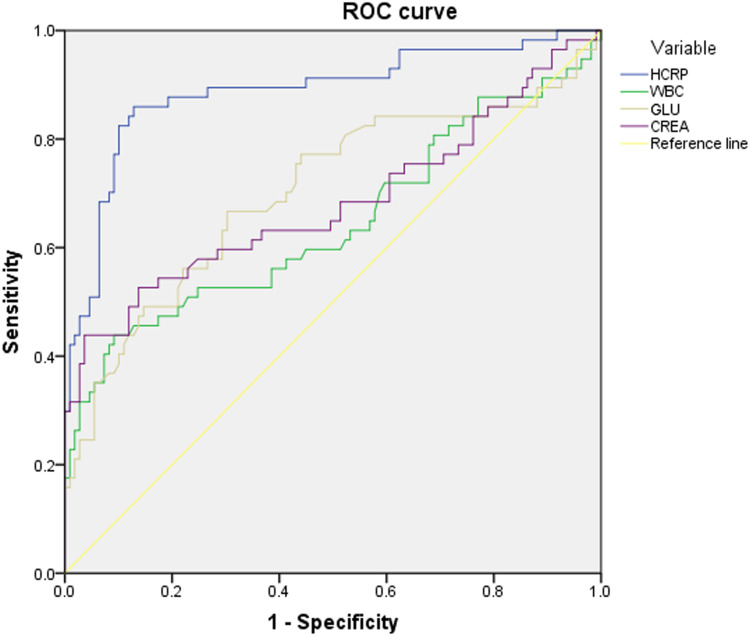
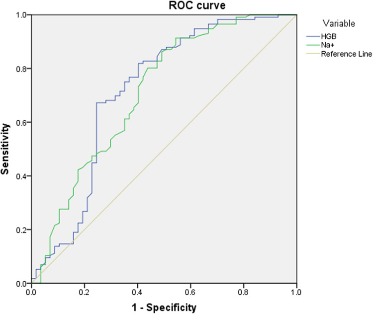
Figures 1 and 2 depict the ROCs for the six variables. All of them exhibited some discrimination ability, with the AUC ranging from 0.645 (95% CI 0.548 to 0.742) for total WBC count to 0.888 (95% CI 0.828 to 0.947) for HCRP level (all p<0.05). The optimal cut-off value for HCRP was 55 mg/L, for WBC count was 11.7×10⁹/L, for FBG was 6.1 mmol/L, for hemoglobin was 110 g/L, and for sodium was 135 mmol/L. Their corresponding sensitivity was at low to high level, from 0.439 for WBC count to 0.862 for sodium, while specificity was at middle to high level, from 0.509 for sodium to 0.963 for serum creatinine (Table 1).
Figure 1.
ROC and AUC for high-sensitivity C-reactive protein (HCRP), white blood cell (WBC), fasting blood glucose (GLU) and creatinine (CREA) levels; from WBC, creatinine, GLU to HCRP, the respective AUC indicated an increasingly larger area, from 0.645 (95% CI 0.548 to 0.742) to 0.888 (95% CI 0.828 to 0.947).
Figure 2.
ROC for hemoglobin level (HGB) and serum sodium (Na+). Their respective AUCs were 0.716 (95% CI 0.624 to 0.808) and 0.709 (95% CI 0.622 to 0.797).
Table 3 describes the association of each variable with NF, based on which the corresponding score was assigned. Presence of HCRP >55 mg/L was assigned the highest score of 12 points, followed by serum creatinine >82 µmol/L (10), comorbid kidney disease (8), sodium (7), WBC count >11.7×10⁹/L (5), gluose >6.1 mmol/L (4) and comorbid diabetes (3).
Table 3.
Assigned Score for Each Variable Based on Their Association Magnitude with Necrotizing Fasciitis
| Variable | Association Magnitude with NF | p | Assigned Score | |
|---|---|---|---|---|
| OR | 95% CI | |||
| HCRP >55 mg/L | 12.27 | 3.029 to 31.789 | <0.001 | 12 |
| WBC >11.7×10⁹/L | 5.14 | 2.56 to 11.42 | <0.001 | 5 |
| FBG >6.1 mmol/L | 3.78 | 1.91 to 7.09 | 0.002 | 4 |
| Creatinine >82 µmol/L | 9.81 | 4.34 to 30.01 | <0.001 | 10 |
| Hemoglobin <110 g/L | 6.18 | 3.31 to 12.5 | 0.001 | 6 |
| Sodium <135 mmol/L | 7.20 | 3.05 to 17.03 | <0.001 | 7 |
| Comorbid diabetes | 2.61 | 1.12 to 5.12 | 0.015 | 3 |
| Comorbid kidney disease | 7.81 | 1.57 to 38.87 | <0.001 | 8 |
Abbreviations: NF, necrotizing fasciitis; OR, odds ratio; CI, confidence interval; HCRP, high-sensitivity C-reactive protein; WBC, white blood cell; FBG, fasting blood glucose.
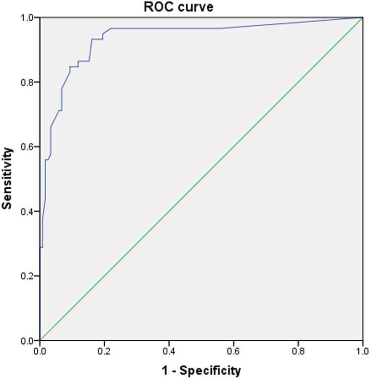
We estimated the total score for each patient based on the assigned score of eight variables, and constructed the combination ROC. The results showed that the m-LRINEC had a sensitivity of 93.2% and a specificity of 86.9%, the cut-off value was 17 points and AUC was 0.935 (95% CI 0.892 to 0.977; p<0.001) (Figure 3).
Figure 3.
ROC for the combination variables (ie, m-LRINEC), which showed a sensitivity of 0.932 and a specificity of 0.869, corresponding to an optimal cut-off value of 17 points and AUC of 93.5%.
Discussion
A high index of suspicion, along with early diagnosis and aggressive surgical treatment, remains the supreme management strategy for NF. The adjunct risk evaluation model based on biomarkers may be useful in the early stages of NF. In this study, on the basis of the original LRINEC proposed by Wong et al,7 we made some modifications to develop the m-LRINEC scoring system. The results showed that the m-LRINEC scoring system exhibited good capacity in discriminating NF from other severe soft-tissue infections, with high sensitivity (93.2%) and specificity (86.9%) when the cut-off value was determined to be 17 points, corresponding to an AUC of 0.935 (95% CI 0.892 to 0.977; p<0.001).
Hippocrates first described NF as long ago as 500 BC, since when multiple terms have been used to describe this disease, such as hospital gangrene, necrotizing erysipelas, streptococcal gangrene and suppurative fasciitis;13 in more recent decades, the term “necrotizing soft tissue infection (NSTI)” was advocated to encompass all forms of the disease process,14 but “necrotizing fasciitis” as a classical name still exists very frequently in practice and research.15,16 Despite improvements in diagnosis and management, this rapidly progressive infection remains associated with high mortality rates of 10.9–76%.2,3 The silver lining is that the mortality is directly proportional to the time to intervention.5,17 Owing to the rapidly progressive nature of NF, it is not practical to repeat MRIs to monitor the disease course. LRINEC, developed by Wong et al7 in 2004 and based on six independently associated biomarkers, has been consistently evaluated for its efficacy in various studies. However, this scoring system has been demonstrated to be neither stable nor reliable, with variable sensitivity from 28.6% to 88.5%.1,8,12,18,19 We believe that these greatly variable results may be attributed to race, ethics, demographics (such as young and elderly patients), the causative bacterial species and, more importantly, the timing of blood sampling for laboratory tests. For example, in Holland’s study,9 the mean age of patients with NF was 40.5 years, which was much younger than in the study by Wong et al,7 and may explain the difference in sensitivity found by Holland.9 Besides, some well-established comorbidities associated with infectious diseases, such as diabetes, kidney disease (renal failure, hypofunction or post-transplant) and long-term use of immunosuppressive agents, are not included in the original LRINEC system, which may also weaken the universality of LRINEC. A direction for future research could be the development of a customized risk assessment scale for application to a certain subgroup of patients.
In the m-LRINEC scoring system, we made several modifications to the LRINEC system. First, we added comorbid diabetes and kidney disease, which were significantly associated with NF, even if the abnormally elevated creatinine and glucose levels were also significant. That seemed to cause a bias due to overestimating the influence of kidney disease or diabetes, but, indeed, these results exactly reflected the greater clinical importance of uncontrolled or poorly controlled diabetes or kidney disease in the development of NF. Therefore, patients with a history of one or both of these conditions, and with abnormally elevated creatinine and/or glucose levels, deserve particular attention regarding the risk of NF. Second, we replaced CRP by HCRP, because the latter is used more often in our practice. In fact, HCRP is more advantageous than CRP only when the concentration is below 10 mg/L, such as in cases of cardiovascular disease or neonatal bacterial infection. Third, we redefined the cut-off values for total WBC count, hemoglobin level, creatinine level and FBG to be 11.7×10⁹/L, 110 g/L, 82 µmol/L and 6.1 mmol/L, respectively. At these cut-off levels, each variable was demonstrated to be capable of discriminating NF from other tissue infections and, furthermore, was identified to be independently associated with NF after adjustment for confounders.
In different clinical or subspecialty settings, researchers have used customized or modified LRINEC (not the present m-LRINEC) models for the auxiliary diagnosis of NF19 or other severe infections,20 or to investigate associations with the prognosis of NF.18 Putnam et al19 developed the pediatric LRINEC (P-LRINEC) score, which was a simplified version of LRINEC only including CRP and sodium, and provided better discrimination ability than LRINEC (AUC 0.70 for LRINEC and 0.84 for P-LRINEC). But the small sample (n=20 each for cases and controls) may affect its validity. Compared to the LRINEC, these modified systems demonstrated better performance, which highlights the importance of individual customization.
The clinical value of the m-LRINEC scoring system was determined by its sensitivity and specificity. As with the description of LRINEC in the study by Wong et al,7 m-LRINEC could also stratify patients into high- and low-risk categories for NF and provide necessary information for the reasonable suspicion of NF. For cases with high suspicion, serial m-LRINEC score monitoring is useful, and hence the empirical use of broad-spectrum antibiotics, which will provide a relatively safe time buffer for definite or surgical treatment to slow down or stop the progression of NF. It is also noted that, in the m-LRINEC system, we dichotomized the biomarker variables, but did not consider some extreme cases, such as the possibility of a total WBC count <4×10⁹/L in leukopenic sepsis21 or even <1.0×10⁹/L in hematological malignancy.22 These cases should alert physicians to the possible presence of life-threatening conditions.
This study has some limitations. First, the sample size is small owing to the rarity of NF. Supposing the larger sample size, other important factors may be identified, such as malignancy, with a prevalence of 3.4% in the NF group versus 0.8% in the non-NF group. Second, the retrospective design had an inherent limitation in data collection, such as patients’ self-reported but unverified comorbidities. Third, this study aimed to develop a modified scoring system based on LRINEC, so we did not include other possibly closely related inflammatory/immune variables, such as reduced serum albumin level due to large consumption in the acute inflammatory response, platelet count, blood coagulation factors or lactate dehydrogenase. Fourth, multiple factors may affect the validity of a scoring system, and hence the generalizability of m-LRINEC may be reduced when used elsewhere.
Conclusions
In summary, we developed the m-LRINEC scoring system using a retrospective nested case–control study. This system demonstrates high sensitivity and specificity in the differential diagnosis of NF and other severe infectious conditions, and may be useful in stratifying patients and providing necessary information for the reasonable suspicion of NF. Studies with larger sample size and better design are required to confirm the validity of m-LRINEC. The development of a customized scoring system for a given subgroup remains a research direction, owing to the multiple factors involved in NF.
Acknowledgments
We are grateful to F Liu and T Zhang of the Department of Orthopedics, and to Y Wang and M Zhan of the Department of Statistics and Applications, for their kind assistance.
Funding Statement
This study was not supported by any funding.
Data Sharing Statement
All the data will be available upon reasonable request to the corresponding author of the present paper.
Ethics Approval and Consent to Participate
The study protocol was approved by the ethics committee of the 3rd Hospital of Hebei Medical University, and informed consent was obtained from all participating patients or their relatives in law.
Consent for Publication
All participating patients or their relatives in law gave their consent for publication of the data.
Disclosure
The authors declare that they have no competing interests.
References
- 1.Al-Hindawi A, McGhee J, Lockey J, Vizcaychipi M. Validation of the laboratory risk indicator for necrotising fasciitis scoring system (LRINEC) in a Northern European population. J Plast Reconstr Aesthet Surg. 2017;70(1):141–143. doi: 10.1016/j.bjps.2016.05.014 [DOI] [PubMed] [Google Scholar]
- 2.McHenry C, Piotrowski J, Petrinic D, Malangoni M. Determinants of mortality for necrotizing soft-tissue infections. Ann Surg. 1995;221(5):558–565. doi: 10.1097/00000658-199505000-00013 [DOI] [PMC free article] [PubMed] [Google Scholar]
- 3.Schwartz S, Kightlinger E, de Virgilio C, de Virgilio M, Kaji A, Neville A. Predictors of mortality and limb loss in necrotizing soft tissue infections. Am Surg. 2013;79(10):1102–1105. doi: 10.1177/000313481307901030 [DOI] [PubMed] [Google Scholar]
- 4.Anaya D, McMahon K, Nathens A, Sullivan S, Foy H, Bulger E. Predictors of mortality and limb loss in necrotizing soft tissue infections. Arch Surg. 2005;140(2):151–7; discussion 158. doi: 10.1001/archsurg.140.2.151 [DOI] [PubMed] [Google Scholar]
- 5.Bilton B, Zibari G, McMillan R, Aultman D, Dunn G, McDonald J. Aggressive surgical management of necrotizing fasciitis serves to decrease mortality: a retrospective study. Am Surg. 1998;64(5):397–401. [PubMed] [Google Scholar]
- 6.Kim MC, Kim S, Cho EB, et al. UtilITY of magnetic resonance imaging for differentiating necrotizing fasciitis from severe cellulitis: a magnetic resonance indicator for necrotizing fasciitis (MRINEC) Algorithm. J Clin Med. 2020;9(9). [DOI] [PMC free article] [PubMed] [Google Scholar]
- 7.Wong CH, Khin LW, Heng KS, Tan KC, Low CO. The LRINEC (Laboratory Risk Indicator for Necrotizing Fasciitis) score: a tool for distinguishing necrotizing fasciitis from other soft tissue infections. Crit Care Med. 2004;32(7):1535–1541. doi: 10.1097/01.CCM.0000129486.35458.7D [DOI] [PubMed] [Google Scholar]
- 8.Tan J, Koh B, Hong C, et al. A comparison of necrotising fasciitis in diabetics and non-diabetics: a review of 127 patients. Bone Joint J. 2016;11:1563–1568. doi: 10.1302/0301-620X.98B11.37526 [DOI] [PubMed] [Google Scholar]
- 9.Holland MJ. Application of the Laboratory Risk Indicator in Necrotising Fasciitis (LRINEC) score to patients in a tropical tertiary referral centre. Anaesth Intensive Care. 2009;37(4):588–592. doi: 10.1177/0310057X0903700416 [DOI] [PubMed] [Google Scholar]
- 10.Abdullah M, McWilliams B, Khan SU. Reliability of the Laboratory Risk Indicator in Necrotising Fasciitis (LRINEC) score. Surgeon. 2019;17(5):309–318. doi: 10.1016/j.surge.2018.08.001 [DOI] [PubMed] [Google Scholar]
- 11.Johnson L, Crisologo P, Sivaganesan S, Caldwell C, Henning J. Evaluation of the Laboratory Risk Indicator for Necrotizing Fasciitis (LRINEC) score for detecting necrotizing soft tissue infections in patients with diabetes and lower extremity infection. Diabetes Res Clin Pract. 2020;171:108520. doi: 10.1016/j.diabres.2020.108520 [DOI] [PubMed] [Google Scholar]
- 12.Ogawa M, Yokoo S, Takayama Y, Kurihara J, Makiguchi T, Shimizu T. Laboratory risk indicator for necrotizing fasciitis of the oro-cervical region (LRINEC-OC): a possible diagnostic tool for emergencies of the oro-cervical region. Emerg Med Int. 2019;2019:1573453. doi: 10.1155/2019/1573453 [DOI] [PMC free article] [PubMed] [Google Scholar]
- 13.Sarani B, Strong M, Pascual J, Schwab CW. Necrotizing fasciitis: current concepts and review of the literature. J Am Coll Surg. 2009;208(2):279–288. doi: 10.1016/j.jamcollsurg.2008.10.032 [DOI] [PubMed] [Google Scholar]
- 14.Goldstein EJC, Anaya DA, Dellinger,EP. Necrotizing soft-tissue infection: diagnosis and management. Clin Infect Dis. 2007;44(5):705–710. doi: 10.1086/511638 [DOI] [PubMed] [Google Scholar]
- 15.Chen LL, Fasolka B, Treacy C. Necrotizing fasciitis: a comprehensive review. Nursing. 2020;50(9):34–40. doi: 10.1097/01.NURSE.0000694752.85118.62 [DOI] [PMC free article] [PubMed] [Google Scholar]
- 16.Leiblein M, Marzi I, Sander AL, Barker JH, Ebert F, Frank J. Necrotizing fasciitis: treatment concepts and clinical results. Eur J Trauma Emerg Surg. 2018;44(2):279–290. doi: 10.1007/s00068-017-0792-8 [DOI] [PubMed] [Google Scholar]
- 17.Voros D, Pissiotis C, Georgantas D, Katsaragakis S, Antoniou S, Papadimitriou J. Role of early and extensive surgery in the treatment of severe necrotizing soft tissue infection. Br J Surg. 1993;80(9):1190–1191. doi: 10.1002/bjs.1800800943 [DOI] [PubMed] [Google Scholar]
- 18.Captain N, Anandi A. A study on validation of modified LRINEC scoring system for the diagnosis of necrotising fasciitis in patients with soft tissue infection. Int Arch Integrated Med. 2020;7(3):35–40. [Google Scholar]
- 19.Putnam LR, Richards MK, Sandvall BK, Hopper RA, Waldhausen JH, Harting MT. Laboratory evaluation for pediatric patients with suspected necrotizing soft tissue infections: a case-control study. J Pediatr Surg. 2016;51(6):1022–1025. doi: 10.1016/j.jpedsurg.2016.02.076 [DOI] [PubMed] [Google Scholar]
- 20.Moon JH. Usefulness of the Modified LRINEC Score in the treatment of patient with deep neck infection. Korean J Otorhinolaryngol-Head Neck Surg. 2015;58(2):115–119. doi: 10.3342/kjorl-hns.2015.58.2.115 [DOI] [Google Scholar]
- 21.Bone RC, Balk RA, Cerra FB, et al. Definitions for sepsis and organ failure and guidelines for the use of innovative therapies in sepsis. The ACCP/SCCM Consensus Conference Committee. American College of Chest Physicians/Society of Critical Care Medicine. Chest. 1992;101(6):1644–1655. doi: 10.1378/chest.101.6.1644 [DOI] [PubMed] [Google Scholar]
- 22.Benoit DD, Vandewoude KH, Decruyenaere JM, Hoste EA, Colardyn FA. Outcome and early prognostic indicators in patients with a hematologic malignancy admitted to the intensive care unit for a life-threatening complication. Crit Care Med. 2003;31(1):104–112. doi: 10.1097/00003246-200301000-00017 [DOI] [PubMed] [Google Scholar]


