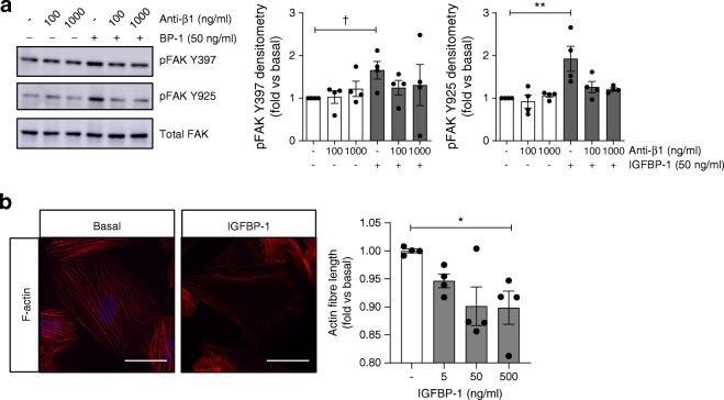
Fig. 6.
IGFBP-1 stimulates FA signalling in human podocytes. (a) Representative western blots and densitometry demonstrating the phosphorylation of FAK Y397 and Y925 in human podocytes stimulated with 50 ng/ml IGFBP-1 for 15 min, with or without pre-treatment with anti-β1-integrin (Anti-β1), P5D2 (100 ng/ml or 1000 ng/ml, 30 min), †p < 0.05, t test vs unstimulated, **p = 0.004, one-way ANOVA with Tukey’s multiple comparison test, n = 4. (b) Representative images (scale bar, 50 μm) and automated quantification of mean F-actin fibre length per cell in human podocytes stimulated with IGFBP-1 at the stated concentrations for 60 min (n = 4, 3 replicates per condition, *p = 0.04, one-way ANOVA with Tukey’s multiple comparison test, 500 ng/ml vs basal). Modest changes in brightness and contrast were uniformly applied to all images for visualisation; unmodified images were used for quantification

