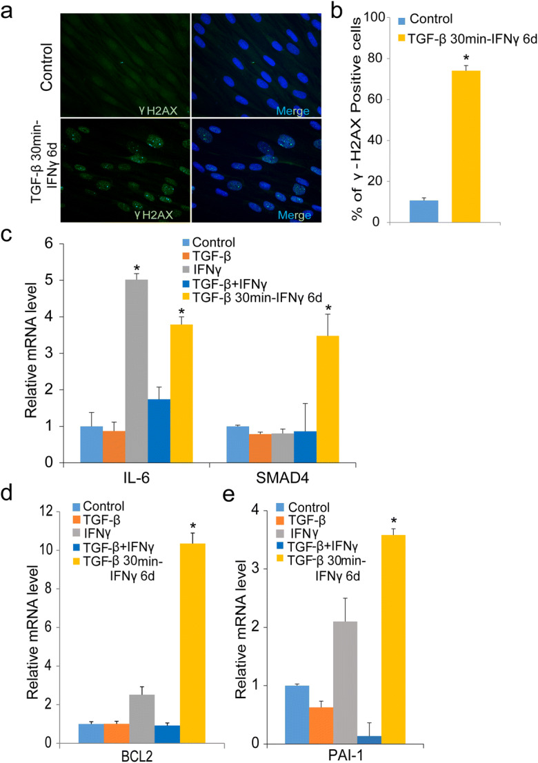
Fig. 6.

DNA damage and increased expression of TGF-β/IFNγ targets in cytokine-treated human fibroblasts. a Immunofluorescence detection of γH2AX foci in untreated or cells treated with TGF-β for 30 min and IFNγ for 6 days; scale bar 10 μm. The γH2AX foci are also shown in merged images of nuclear DAPI staining. b Percentage of γH2AX foci in untreated or cells treated with TGF-β for 30 min and IFNγ for 6 days. c IL-6 and SMAD4 expression determined by real-time PCR in untreated cells or cells treated with TGF-β or IFNγ alone or in combination or with transient TGF-β for 30 min and IFNγ for 6 days. d mRNA expression of the pro-apoptotic gene BCL2 with TGF-β, IFNγ alone, the combination of both, or with transient TGF-β for 30 min and IFNγ for 6 days as measured by real-time PCR. e Real-time PCR analysis of PAI-1 expression in conditions as described *denotes statistical significance (*p < 0.05). Error bars represent SEM
