

Abstract
Objectives:
To clarify the relationship between tooth length and three-dimensional mandibular morphology in a healthy Japanese population.
Materials and Methods:
This study included 181 Japanese adults: 66 men and 115 women. Cone-beam computed tomography (CBCT) images were acquired with a dentofacial cone-beam x-ray CT scanner. Tooth length was measured with open-source OsiriX medical image processing software. Crown height and root length were measured in the maxillary and mandibular central incisors, lateral incisors, canines, first premolars, second premolars, first molars, and second molars. Based on these measurements, principal component (PC) analysis was performed. The following measurements were used to assess three-dimensional mandibular morphology: CD-GO, GO-GN, RCD-LCD, RGO-LGO, RCP-LCP, and the gonial angle. Stepwise multiple regression analysis was performed to examine the associations between three-dimensional mandibular morphology and the patterns of crown and root lengths using the mandibular measurements as explanatory variables and each PC as the dependent variable.
Results:
CD-GO was positively associated with PC1, which represented overall tooth length. RGO-LGO was positively associated with PC2, whereas GO-GN, RCP-LCP, and gonial angle were negatively associated with PC2, which was the axis denoting relatively longer root (+) vs higher crown (−). Being female was associated with PC3, which was the axis denoting relatively longer posterior tooth (+) vs anterior tooth (−).
Conclusions:
The present clinical study effectively used CBCT images and PC analysis to reveal significant correlations between tooth length and mandibular morphology in a modern human population, confirming in part the statement that “large teeth necessitate large jaws.”
Keywords: Tooth, Length, Mandible, Three-dimensional
INTRODUCTION
Although the statement “Large teeth necessitate large jaws, and large jaws, a large body”1 sounds logical, the accuracy of this statement has not been verified to date.2 Dental crowding can be defined as a discrepancy between jaw size and crown width, resulting in overlapping and rotation of the teeth.3 It has been reported4 that crowding is more strongly associated with small jaw size than with large teeth. Both environmental and genetic factors are involved in dental variation.5 Many factors, such as masticatory function, the size of the dentition,6 and persistent sucking habits,7 are involved in mandibular morphology. Mandibular form is a product of both morphogenesis and evolutionary modifications related to mastication.8 Orthodontists need to understand the changes that normally take place in adult craniofacial structures.9 Thus, it is important to understand the relationship between mandibular morphology and tooth size, especially in orthodontics.
Previous studies have demonstrated a relationship between tooth width and mandibular morphology. Anderson et al.10 reported that crown width correlates with alveolar bone and basal bone thickness. Kieser and Groeneveld11 hypothesized that a longer jaw is associated with relatively wider canines and narrower molars. However, little is known about the association between tooth length and mandibular morphology. Smith et al.12 found close associations between crown width, root length, and corpus height in Australian and Maori populations. However, no relationship was found between the cross-sectional shape or proportion of the symphyseal region and variations in canine crown width or height in primates.8 In a micro–computed tomography study of recent modern human dry skulls, Le Cabec et al.13 found no correlation between mandibular anterior tooth root size and jaw size in the symphyseal region. No consistent results have been found relative to the relationship between tooth length and mandibular morphology. In previous studies, mandibular morphology was investigated in two dimensions. Currently, human tooth length and mandibular morphology can be accurately measured with little radiation exposure using dentofacial cone-beam computed tomography (CBCT) images.14,15 In addition, only limited regions of mandibular morphology have been evaluated to date. This problem might also be addressed with the use of dentofacial CBCT images. Furthermore, the small sample size in previous studies remains a problem to be solved.
The aim of this present study was to examine the relationship between tooth length and three-dimensional mandibular morphology in 181 Japanese patients using dentofacial CBCT.
MATERIALS AND METHODS
Patients
This study included patients who underwent CBCT imaging for orthodontic assessment in the Department of Orthodontics at Showa University Dental Hospital. The final cohort included 181 Japanese adults: 66 men (age 18–50 years; mean age: 27.4 years) and 115 women (age 18–57 years; mean: 27.6 years). Patients with congenital disorders such as cleft lip and palate and those with systemic disease were excluded from the study. Those with previous orthodontic treatment, root resorption, or loss of the original crown morphology resulting from caries, trauma, attrition, wear, or dental prosthesis were excluded from measurements.
This study was approved by the ethics committee of Showa University (IRB No. 108) and related committees. All patients provided written informed consent to participate.
CBCT Imaging
CBCT images were acquired with a dentofacial cone-beam x-ray CT scanner (CB MercuRay; Hitachi Medico Technology, Tokyo, Japan, or KaVo 3D eXam; KaVo, Biberach, Germany) in the radiology department. The scanning conditions were 100 kVp, 10 mA, F-mode, 512 slices/scan (slice width: 377 mm), and 9.6-second acquisition time. Measurement of errors between the two CBCT models was verified using the method reported by Katayama et al.,16 which confirmed that the error was very small and nonproblematic.
Tooth Size Measurements
Open-source OsiriX medical image processing software (Pixmeo, Geneva, Switzerland; www.osirix.viewer.com) was used to reconstruct the data, which were exported in DICOM format to a MacBook Pro personal computer (Mac Os X El Capitan 10.11.6, Apple, Cupertino, Calif). For tooth length measurements, multiplanar reconstruction was used to orient the CBCT images, according to a modification of the method of Abeleira et al.17 Each target tooth was positioned according to the method of Abeleira et al.17 The crown height (CH) and root length (RL) were measured in the coronal plane. To measure CH, a line was drawn perpendicular to the line between the buccal and palatal limits of the cementoenamel junction to the incisal edge or to the tip of each cusp in the case of premolars and molars. To measure RL, a line was drawn perpendicular to the line between the buccal and palatal limits of the cementoenamel junction to the apex of the tooth root(s) (Figure 1). In the maxillary and mandibular central incisors (upper [U]1, lower [L]1), lateral incisors (U2, L2), and canines (U3, L3), the distance between the incisal edge and the root apex was measured. In the premolars (U4, U5, L4, L5) the distance between the buccal cusp and buccal root apex was measured. In the upper molars, the distances between the mesiobuccal cusp tip and mesiobuccal root apex (U6M, U7M), between the distobuccal cusp and distobuccal root apex (U6D, U7D), and between the mesiopalatal cusp tip and palatal root apex (U6P, U7P) were measured. In the lower molars, the distances between the mesiobuccal cusp tip (L6M, L7M) and mesial root apex and between the distobuccal cusp tip and distal root apex (L6D, L7D) were measured. The average of left and right measurements for each tooth was used as the crown and root lengths. If only one side was measurable, those measurements were used. If the teeth on both sides were unmeasurable, the value was considered missing. One researcher (YH) evaluated tooth length. To investigate intraoperator error, 25 cases were randomly selected and remeasured under identical conditions in separate sessions with a 2-week interval. Measurement error was estimated with Dahlberg's formula (S2 = ∑d2/2n).18,19
Figure 1.
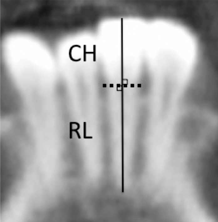
Tooth size measurements on CBCT images. CH indicates crown height; RL, root length.
Mandibular Measurements
Mandibular measurements were made following the method reported by Nakawaki et al.14 The mandibular bone region was segmented from the image data and analyzed with Analyze 3D reconstruction software (Biomedical Imaging Resource, Mayo Clinic and Foundation, Rochester, Minn). Mandibular length and angle were measured by autotracing the outer circumference of the cortical bone on all slides using Analyze. These autotraces were superimposed to prepare an object map for length measurement. Dental crown data were extracted separately from mandibular data because they may be affected by artifacts, such as prostheses. The mandibular measurement items were as follows: CD-GO, GO-GN, RCD-LCD, RGO-LGO, RCP-LCP, and the gonial angle (Figure 2). One researcher (TN) evaluated mandibular measurements.
Figure 2.

Mandibular measurements on CBCT images: CD-GO (mm), GO-GN (mm), RCD-LCD (mm), RGO-LGO (mm), RCP-LCP (mm), and gonial angle (°).
Statistical Analysis
Principal component analysis (PCA) was performed to improve the exploration and visualization of morphological variations by decreasing the dimensionality of the obtained data. Metric tooth-length data were used to create a correlation coefficient matrix. PCA was performed using the correlation coefficient matrix. Eigenvalues, eigenvectors, and principal component (PC) scores were obtained. The PCA of tooth length included individual measurements of both crown height and root length. Stepwise multiple regression analysis was performed to examine the associations between three-dimensional mandibular morphology and the patterns of crown and root lengths using mandibular measurements as explanatory variables and each PC as the dependent variable. Statistical analyses were performed with Statcel3 software (OMS Publishing, Saitama, Japan), with the significance level at 5%.
RESULTS
The tooth measurement error estimated according to Dahlberg's formula was 3% or less for each item, indicating sufficient reproducibility. PCA of tooth measurements revealed that the top six PCs (cumulative contribution: 69.2%) had an eigenvalue greater than 1. This finding indicates that the first six PCs effectively represent the metric data. The patterns of the top three PCs were especially easily interpreted (Figure 3). PC1 had an eigenvalue of 16.85, which contributed 42.1% of the total variance, and represented overall tooth length. PC2 had an eigenvalue of 5.16 (12.9%); positive PC2 scores denoted relatively longer root and shorter crown. PC3 had an eigenvalue of 2.32 (5.81%) and represented a relatively longer posterior tooth (+) vs anterior tooth (−). As for PC4 and higher PCs, however, it is difficult to give a concise interpretation because these PCs could include statistical artifacts generated after extraction of the lower PCs. For mandibular measurements, the mean values and standard deviations are shown in Table 1. The results of the stepwise multiple regression analysis are shown in Table 2. CD-GO was positively associated with PC1 scores. RGO-LGO was positively associated with PC2 scores, whereas GO-GN, RCP-LCP, and gonial angle were negatively associated with PC2 scores. Being female was associated with higher PC3 scores.
Figure 3.

Principal component (PC) loading for each PC. PC analysis of tooth length measurements.
Table 1.
Means and Standard Deviations (SDs) of the Mandibular Measurements from Cone-Beam Computed Tomography (CBCT)
| Males (n = 66) |
Female (n = 115) |
|||
| Mean ± SD |
Range |
Mean ± SD |
Range |
|
| CD-GO, mm | 164.13 ± 13.89 | 124.66–193.87 | 144.17 ± 11.76 | 182.99–111.65 |
| GO-GN, mm | 224.51 ± 14.57 | 185.27–252.31 | 209.20 ± 12.46 | 241.47–173.28 |
| RCD-LCD, mm | 328.44 ± 15.69 | 296.25–374.43 | 313.70 ± 16.25 | 358.30–272.11 |
| RGO-LGO, mm | 255.73 ± 15.60 | 229.21–300.26 | 233.89 ± 13.51 | 271.15–202.17 |
| RCP-LCP, mm | 259.73 ± 14.21 | 232.67–310.37 | 246.12 ± 11.20 | 273.54–216.26 |
| Gonial angle, ° | 121.59 ± 5.88 | 110.30–137.60 | 125.19 ± 8.83 | 192.72–104.45 |
Table 2.
Association Tests Using Multiple Regression Analysesa
| Objective Variable |
Explanatory Variables Selected |
B |
SE |
β |
P |
Adjusted R2 |
| PC1 | 0.0766 | |||||
| CD-GO, mm | 0.071 | 0.020 | 0.289 | .00057*** | ||
| PC2 | 0.1540 | |||||
| GO-GN, mm | −0.045 | 0.015 | −0.292 | .0033** | ||
| RGO-LGO, mm | 0.026 | 0.012 | 0.208 | .034* | ||
| RCP-LCP, mm | −0.042 | 0.015 | −0.266 | .0048** | ||
| Gonial angle, ° | −0.056 | 0.023 | −0.201 | .015* | ||
| PC3 | 0.0611 | |||||
| Sex | 0.818 | 0.259 | 0.261 | .0020** |
Sex, age, CD-GO, GO-GN, RCD-LCD, RGO-LGO, RCP-LCP, and gonial angle were entered as explanatory variables. Variables selected by the stepwise procedure are shown in the table.
P < .05; ** P < .01; *** P < .001.
DISCUSSION
Orthodontists have not been able to answer the very basic question, “How closely are big teeth correlated with jaw size?,” with clear evidence. Obstacles to clarification of this relationship include the small number of samples and the large degree of morphological variation. CBCT provides information about root length in living individuals, regardless of tooth type. PCA has been used to reduce the dimensionality of data and to simplify identification of morphological variations.20 In the present study, the advantages of CBCT and PCA were used to examine the relationships between mandibular morphology and patterns of crown and root length. Correlations between tooth length and three-dimensional mandibular morphology were found.
Previous studies1,2,6,8–10,12,13,21–23 have investigated the relationship between tooth length or width and body height. Among these, several8,12,13,23 have evaluated the relationship between tooth length and mandibular morphology. Garn et al.23 reported correlations between root lengths of the mandibular first premolar through the mandibular second molar and Ar-Gn on 45° oblique jaw radiographs in 122 American adolescents. Le Cabec et al.13 used micro-CT imaging to investigate the correlation between mandibular tooth root length and the size of the symphyseal region in 22 German dry skulls but found no correlations. Smith et al.12 found small correlations between crown width (mesiodistal diameter), root length, and corpus height on dental occlusal films in Maori (circa 1100–1900 ad) and Aboriginal populations from Roonka (7000 bp to 1800 ad). Plavcan and Daegling8 investigated the relationships between mandibular canine crown height, mandibular breadth and height, and symphyseal height in anthropoid primate dry skulls and found no clear correlations. The mandibular morphology measurement regions in the present study were CD-GO, GO-GN, RCD-LCD, RGO-LGO, RCP-LCP, and the gonial angle, according to three-dimensional measurement. In previous reports, human mandibular morphology was investigated by observing only Ar-Gn,23 the symphyseal region height,8 or corpus height.12 In the present study, associations were observed between tooth size and mandibular morphology, not only in the lower alveolar region but also in the entire mandible. Previous studies on tooth length and mandibular morphology in modern humans have evaluated morphometry of the mandible with 45° oblique jaw radiography23 or dental occlusal films,12 whereas CBCT images were used this study. Unfortunately, research on the relationship between tooth length and mandibular morphology has not progressed in recent years. However, the accuracy of distance measurements made with CBCT has recently been reported.15,24,25 In the present study, CBCT images were used to obtain highly accurate three-dimensional mandibular and tooth-length measurements, which may have led to discovery of the novel associations.
Fukase and Suwa26 hypothesized that growth of the anterior mandibular corpus is affected by tooth size and spacing conditions until the time of tooth eruption. They used a microfocal x-ray CT system to investigate the influence of the size and position of developing teeth (measurement of crypt) in determining anterior corpus height in 63 modern Japanese dry skulls of different dental ages. Although the study did not directly compare the root length and mandible form, mandibular height may be affected by root length and eruption distance (vertical distance between upper outer surface of the dental crypt and surface of alveolar bone); however, this issue needs further investigation.26
The combination of PCA of tooth length and three-dimensional morphometry of the entire human mandible revealed that tooth length was associated with the three-dimensional width and morphology of the human mandible. However, the causal relationship between tooth length and mandibular morphology remains unclear. Therefore, further study is necessary to understand the morphogenesis of the tooth-mandible complex.
CONCLUSIONS
RGO-LGO was positively associated, whereas GO-GN, RCP-LCP, and gonial angle were negatively associated, with relatively longer root (+) vs higher crown (−).
Being female was associated with relatively long posterior tooth (+) vs anterior tooth (−).
The results indicate that there are relationships between tooth length and three-dimensional mandibular morphology, confirming in part the statement that “large teeth necessitate large jaws.”
ACKNOWLEDGMENTS
We are deeply grateful to the patients who participated in the present study. We also thank Rebecca Tollefson, DVM, from Edanz Group (www.edanzediting.com/ac), for editing a draft of this manuscript. This work was supported by KAKENHI grant 17K11947.
REFERENCES
- 1.Garn SM, Lewis AB. Tooth size, body size and “giant” fossil man. Am Anthropol. 1958;60(5):874–880. [Google Scholar]
- 2.Reddy S, Shome B, Patil J, Koppolu P. A clinical correlation between stature and posterior tooth length. Pan Afr Med J. 2017;26:17. doi: 10.11604/pamj.2017.26.17.10436. [DOI] [PMC free article] [PubMed] [Google Scholar]
- 3.Howe RP, McNamara JA, Jr, O'Connor KA. An examination of dental crowding and its relationship to tooth size and arch dimension. Am J Orthod. 1983;83(5):363–373. doi: 10.1016/0002-9416(83)90320-2. [DOI] [PubMed] [Google Scholar]
- 4.Corruccini RS. Australian aboriginal tooth succession, interproximal attrition, and Begg's theory. Am J Orthod Dentofacial Orthop. 1990;97(4):349–357. doi: 10.1016/0889-5406(90)70107-N. [DOI] [PubMed] [Google Scholar]
- 5.Townsend G, Hughes T, Luciano M, Bockmann M, Brook A. Genetic and environmental influences on human dental variation: a critical evaluation of studies involving twins. Arch Oral Biol. 2009;54(suppl 1):S45–S51. doi: 10.1016/j.archoralbio.2008.06.009. [DOI] [PMC free article] [PubMed] [Google Scholar]
- 6.Kupczik K, Olejniczak AJ, Skinner MM, Hublin JJ. Molar crown and root size relationship in anthropoid primates. Front Oral Biol. 2009;13:16–22. doi: 10.1159/000242384. [DOI] [PubMed] [Google Scholar]
- 7.Saccomanno S, Antonini G, D'Alatri L, D'Angelantonio M, Fiorita A, Deli R. Causal relationship between malocclusion and oral muscles dysfunction: a model of approach. Eur J Paediatr Dent. 2012;13(4):321–323. [PubMed] [Google Scholar]
- 8.Plavcan JM, Daegling DJ. Interspecific and intraspecific relationships between tooth size and jaw size in primates. J Hum Evol. 2006;51(2):171–184. doi: 10.1016/j.jhevol.2006.02.005. [DOI] [PubMed] [Google Scholar]
- 9.Al-Zubair NM. The relationship between mandibular arch length and widths in a sample of Yemeni subjects with normal dento-skeletal relationship. J Orthod Sci. 2013;2(4):120–123. doi: 10.4103/2278-0203.123198. [DOI] [PMC free article] [PubMed] [Google Scholar]
- 10.Anderson BL, Thompson GW, Popovich F. Evolutionary dental changes. Am J Phys Anthropol. 1975;43(1):95–102. doi: 10.1002/ajpa.1330430113. [DOI] [PubMed] [Google Scholar]
- 11.Kieser JA, Groeneveld HT. Allometric relations of teeth and jaws in man. Am J Phys Anthropol. 1988;77(1):57–67. doi: 10.1002/ajpa.1330770110. [DOI] [PubMed] [Google Scholar]
- 12.Smith P, Wax Y, Adler F. Population variation in tooth, jaw, and root size: a radiographic study of two populations in a high-attrition environment. Am J Phys Anthropol. 1989;79(2):197–206. doi: 10.1002/ajpa.1330790207. [DOI] [PubMed] [Google Scholar]
- 13.Le Cabec A, Kupczik K, Gunz P, Braga J, Hublin JJ. Long anterior mandibular tooth roots in Neanderthals are not the result of their large jaws. J Hum Evol. 2012;63(5):667–681. doi: 10.1016/j.jhevol.2012.07.003. [DOI] [PubMed] [Google Scholar]
- 14.Nakawaki T, Yamaguchi T, Tomita D, et al. Evaluation of mandibular volume classified by vertical skeletal dimensions with cone-beam computed tomography. Angle Orthod. 2016;86(6):949–954. doi: 10.2319/103015-732.1. [DOI] [PMC free article] [PubMed] [Google Scholar]
- 15.Gribel BF, Gribel MN, Frazäo DC, McNamara JA, Jr, Manzi FR. Accuracy and reliability of craniometric measurements on lateral cephalometry and 3D measurements on CBCT scans. Angle Orthod. 2011;81(1):26–35. doi: 10.2319/032210-166.1. [DOI] [PMC free article] [PubMed] [Google Scholar]
- 16.Katayama K, Yamaguchi T, Sugiura M, Haga S, Maki K. Evaluation of mandibular volume using cone-beam computed tomography and correlation with cephalometric values. Angle Orthod. 2014;84(2):337–342. doi: 10.2319/012913-87.1. [DOI] [PMC free article] [PubMed] [Google Scholar]
- 17.Abeleira MT, Outumuro M, Ramos I, Limeres J, Diniz M, Diz P. Dimensions of central incisors, canines, and first molars in subjects with Down syndrome measured on cone-beam computed tomographs. Am J Orthod Dentofacial Orthop. 2014;146(6):765–775. doi: 10.1016/j.ajodo.2014.08.016. [DOI] [PubMed] [Google Scholar]
- 18.Springate SD. The effect of sample size and bias on the reliability of estimates of error: a comparative study of Dahlberg's formula. Eur J Orthod. 2012;34(2):158–163. doi: 10.1093/ejo/cjr010. [DOI] [PubMed] [Google Scholar]
- 19.Harris EF, Smith RN. Accounting for measurement error: a critical but often overlooked process. Arch Oral Biol. 2009;54(suppl 1):S107–S117. doi: 10.1016/j.archoralbio.2008.04.010. [DOI] [PubMed] [Google Scholar]
- 20.Groth D, Hartmann S, Klie S, Selbig J. Principal components analysis. Methods Mol Biol. 2013;930:527–547. doi: 10.1007/978-1-62703-059-5_22. [DOI] [PubMed] [Google Scholar]
- 21.Prabhu S, Acharya AB, Muddapur MV. Are teeth useful in estimating stature? J Forensic Leg Med. 2013;20(5):460–464. doi: 10.1016/j.jflm.2013.02.004. [DOI] [PubMed] [Google Scholar]
- 22.Hossain MZ, Munawar KM, Rahim ZH, Bakri MM. Can stature be estimated from tooth crown dimensions? A study in a sample of South-East Asians. Arch Oral Biol. 2016;64:85–91. doi: 10.1016/j.archoralbio.2016.01.001. [DOI] [PubMed] [Google Scholar]
- 23.Garn SM, Smith BH, Cole PE. Correlations between root length and face size. J Dent Res. 1980;59(2):141. doi: 10.1177/00220345800590021201. [DOI] [PubMed] [Google Scholar]
- 24.Flores-Mir C, Rosenblatt MR, Major PW, Carey JP, Heo G. Measurement accuracy and reliability of tooth length on conventional and CBCT reconstructed panoramic radiographs. Dental Press J Orthod. 2014;19(5):45–53. doi: 10.1590/2176-9451.19.5.045-053.oar. [DOI] [PMC free article] [PubMed] [Google Scholar]
- 25.Timock AM, Cook V, McDonald T, et al. Accuracy and reliability of buccal bone height and thickness measurements from cone-beam computed tomography imaging. Am J Orthod Dentofacial Orthop. 2011;140(5):734–744. doi: 10.1016/j.ajodo.2011.06.021. [DOI] [PubMed] [Google Scholar]
- 26.Fukase H, Suwa G. Influence of size and placement of developing teeth in determining anterior corpus height in prehistoric Jomon and modern Japanese mandibles. Anthropol Sci. 2010;118(2):75–86. [Google Scholar]