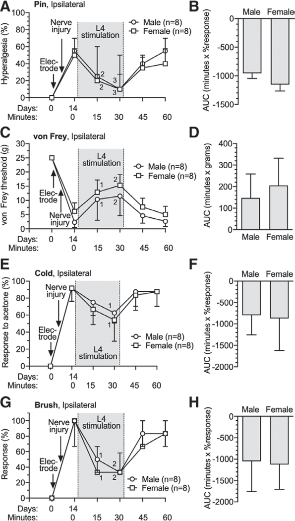
Fig. 3.
Effects of dorsal root ganglion stimulation on male and female rats with tibial nerve injury. Dorsal root ganglion stimulation or spinal cord stimulation electrodes were implanted immediately after the baseline (day 0) behavioral determinations. Time course for effects are shown in the left panels, and area under curve analysis for group comparisons are shown in the right panels, for sensitivity to noxious mechanical stimuli (pin [A, B]), threshold mechanical stimuli (von Frey [C, D]), cold (E, F), and brush (G, H). Results in A, E, and G are median ± interquartile range. Results in B, C, D, F and H are mean ± SD. 1, P < 0.05; 2, P < 0.01; and 3, P < 0.001 compared to data immediately before dorsal root ganglion stimulation by the Dunnett test after two-way repeated measures ANOVA with Greenhouse–Geisser correction; n, number of animals.

