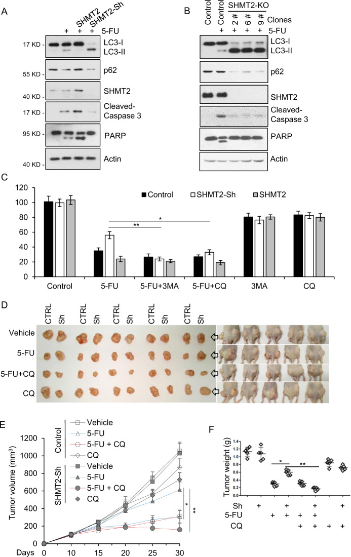
Fig. 4. Inhibition of autophagy induced by low SHMT2 expression sensitizes CRC cells to 5-FU treatment.
A SHMT2 promoted apoptosis and inhibited autophagy in response to 5-FU treatment. Western blot analysis of lysates of HCT116 cells that were transfected with SHMT2 or infected with SHMT2-sh lentivirus and treated with 5-FU (10 μM) for 24 h. The protein levels of SHMT2, p62, LC3, cleaved Caspase 3, PARP, and β-actin (as the internal standard) were assessed with the indicated antibodies. B The protein levels of SHMT2, p62, LC3, cleaved Caspase 3, PARP, and β-actin (as the internal standard) were assessed in SHMT2-KO HCT116 cells. C The indicated cells were treated with 5-FU (2 μM), 3-MA (10 mM) or chloroquine diphosphate salt (CQ, 20 μM) for 4 days and analyzed using the MTT cell viability assay. *P < 0.05, **P < 0.01. D–F The xenograft experiment with Control and SHMT2-sh cells treated with 5-FU or CQ is described in the Methods section. D Xenograft tumors were harvested and photographed. E, F Quantification of the average volumes (E) and weights (F) of the xenograft tumors are shown. Five tumors from individual mice were included in each group; *P < 0.05, **P < 0.01.

