Abstract
In recent years, quantum-dot-like single-photon emitters in atomically thin van der Waals materials have become a promising platform for future on-chip scalable quantum light sources with unique advantages over existing technologies, notably the potential for site-specific engineering. However, the required cryogenic temperatures for the functionality of these sources has been an inhibitor of their full potential. Existing methods to create emitters in 2D materials face fundamental challenges in extending the working temperature while maintaining the emitter’s fabrication yield and purity. In this work, we demonstrate a method of creating site-controlled single-photon emitters in atomically thin WSe2 with high yield utilizing independent and simultaneous strain engineering via nanoscale stressors and defect engineering via electron-beam irradiation. Many of the emitters exhibit biexciton cascaded emission, single-photon purities above 95%, and working temperatures up to 150 K. This methodology, coupled with possible plasmonic or optical micro-cavity integration, furthers the realization of scalable, room-temperature, and high-quality 2D single- and entangled-photon sources.
Subject terms: Two-dimensional materials, Quantum optics, Single photons and quantum effects
Quantum defects in 2D semiconductors are promising quantum light sources, but the required cryogenic temperatures limit their applicability. Here, the authors report a method to create single-photon emitters in monolayer WSe2 operating at temperatures up to 150 K without plasmonic or optical cavities.
Introduction
As the second quantum era emerges in the twenty-first century, what were once considered as tangential quantum effects are instead harnessed in new quantum technology. Single-photon emitters (SPEs) are the heart of many quantum photonic applications1 such as quantum communication, cryptography, metrology, and linear optical quantum computing. Current state-of-the-art semiconducting single-photon sources2—such as InAs self-assembled quantum dots—encounter challenges in scalability and integration with mature photonic technologies due to difficulties in deterministic positioning and growth. In recent years, a new SPE platform3–8 has emerged in two-dimensional (2D) materials, including hexagonal boron nitride and transition metal dichalcogenides (TMDs) such as MoSe2 and WSe2. Monolayer WSe2, in comparison to conventional III–V semiconducting quantum dots, offers several advantages, including a high-photon extraction efficiency due to its atomic thickness, the potential for scalable, site-controlled and deterministic manufacturability9–11, and ease of integration with mature photonic technologies via simple transfer methods12, which has made them an intriguing contender for future technologically relevant quantum light sources.
However, a challenge facing 2D WSe2 SPEs lies in their low working temperature and thermal instability. The ultra-sharp emission lines associated with SPEs that appear ~50–200 meV below the free exciton of WSe2 are observable only at cryogenic temperatures and quench above 30 K due to low confinement potential and quantum yield4,13. Considering that the microscopic origins of these emitters are still not fully understood, their further optimization and engineering become challenging. Recently, it has been hypothesized that SPEs in WSe2 may be attributed to localized intervalley defect-bound excitons that form when the energy of the dark excitonic band of WSe2 is lowered and hybridizes with a valley symmetry-breaking defect state at strained regions14 (Fig. 1). This description clarifies key points regarding the strong brightness and magneto-optic properties of these emitters, which cannot be justified within an only-strain or an only-defect description. Furthermore, it reveals that both strain and defects are pivotal in the creation of SPE. Recent experimental studies15,16 have also confirmed parts of this model, however, the essential link, which is the correlation between defects and quantum emitters, remains elusive. This is partially because defects in 2D TMDs are ubiquitous and difficult to control. The challenge compounds further since most strain engineering methods are also prone to damage or pierce the TMDs in the process9.
Fig. 1. Illustration of a strain and defect-engineered WSe2 single-photon emitter.
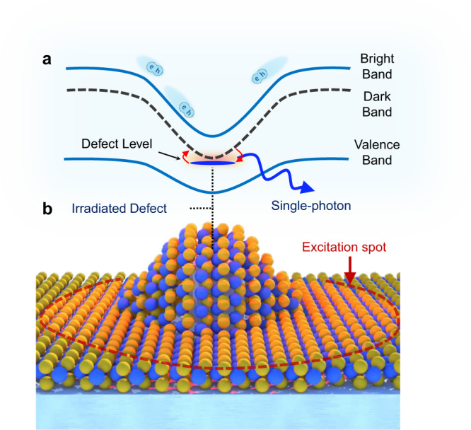
a Denotes the spatial bandgap variations due to strain (solid blue lines represent excitonic bright band and valence band, respectively, dashed gray line represents the dark excitonic band). b Demonstrates an illustration of 1L-WSe2 strained over a SiO2 nanopillar (W atoms in blue and Se in yellow). Neutral excitons, represented as e–h pairs, created within the excitation laser spot (red dashed line) funnel to low-potential strained regions. Note that WSe2 also has a dark-exciton band at energies below the bright exciton, which is optically forbidden due to the transition selection of WSe2. However, at strained regions and in the presence of a defect (top of the irradiated nanopillar, denoted with a blue dash), the dark-exciton can become strain-tuned to a defect-level forming intervalley defect excitons. Given that the defect-level breaks the spin-momentum locking of WSe2, the dark-exciton state can then efficiently recombine through the defect level, giving rise to an exponentially bright single-photon emission line (outgoing blue arrow).
In this study, by using electron-beam (e-beam) irradiation as a controllable method to induce structural defects in WSe2, along with engineering strain fields in encapsulated WSe2 using dielectric nanopillar structures, we decouple the strain and defect engineering processes and prove that there is a direct correlation between defect density and quantum emitters, which provides strong evidence for the intervalley defect exciton model. Moreover, the accurate positioning of defects within the strain field in WSe2 minimizes the non-radiative recombination and achieves higher confinement potential. These, in addition to the possible distinct morphology of our e-beam engineered defects that appear at deeper energies than any other observed SPEs in WSe2, enable engineering SPEs with high yield, high single-photon purity, biexciton-like radiative cascade, and possible working temperatures up to 215 K. This sets the precedent for the highest working temperature of SPEs observed in 2D TMDs without Purcell enhancement (benchmark available in Supplementary Note 1).
Results
Strained WSe2 PL response
A monolayer WSe2 sample encapsulated by h-BN was prepared using dry transfer techniques (see Methods). h-BN flakes, with thickness of less than 5 nm, were chosen to encapsulate the WSe2 not only to isolate the WSe2 from surface states and adsorbates17, but to also act as protective layers that shield the WSe2 from potential damages during the stamping process onto nanopillars. Subsequently, to engineer the necessary strain profile across the 2D flake, the stack was transferred onto a Si/SiO2 substrate array with a predefined nanopillar dielectric array with height and diameter of 200 and 150 nm, respectively, which was previously identified as an optimal aspect ratio for deterministically introducing SPEs without piercing through the materials10. Figure 2a presents the dark-field optical image of the final assembled structure where the brightly illuminated regions correspond to the nanopillar sites. Photoluminescence (PL) spectra of the WSe2 residing on the nanopillars were taken at T = 5 K (Supplementary Fig. 1). Interestingly, no sharp emission peaks that could be associated with single-photon emitters were observed. In contrast, only slightly redshifted neutral exciton (X) and charged exciton (X−) peaks were detected. Given that the strained region is about 2-orders of magnitude smaller than the diffraction-limited spot size, the red-shifted neutral exciton peaks due to strain are dim and cannot be resolved by a far-field diffraction-limited system (see Supplementary Note 2 and Supplementary Figs. 2 and 3), consistent with previous studies18,19. Furthermore, the defect band that usually dominates the PL spectra of 1L-WSe2 is also absent in the PL spectrum both in the strained and unstrained regions for the typical excitation powers and accumulation times used in our experiments. However, a very weak broad-defect band, without any sharp peaks, can be observed when PL intensity at low excitation power is integrated over a long time. Interestingly, the correlation between the defect band and quantum emitters has been hinted in previous studies where the spectral histogram of measured emitters reconstruct the shape of the defect band9,11. Overall, this provides evidence that in WSe2 samples, which are devoid of the defect band and are shielded from the substrate with h-BN encapsulation, single-photon emitters cannot form solely due to the strain in WSe2.
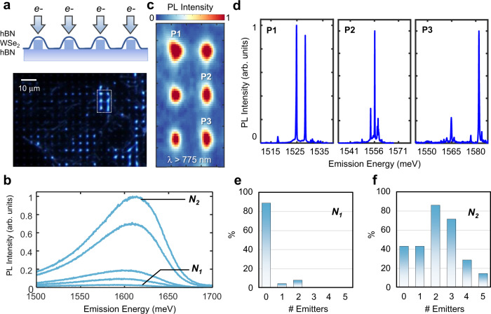
Fig. 2. Site-controlled quantum emitter array via strain and defect engineering.
a Schematic of the fabricated structure and the optical dark-field image of the sample. An stack of h-BN/WSe2/h-BN is transferred on a SiO2 nanopillar array. Bright emission is observed from the top of the pillars due to exciton funneling. Next, top of the pillars are irradiated (e− arrows) to induce structural defects. b PL spectra of unstrained WSe2 defect-bound excitons as a function of irradiation intensity from N1 = 105 electrons/μm to N2 = 106 electrons/μm 10 μW excitation power. c Spectrally integrated PL map of strained and irradiated sites for wavelengths above 775 nm, showing the bright emission associated with single-photon emitters. Some sites host more than one emitter and have reached saturation incident count. The maps resemble the Gaussian spot size of excitation laser due to the localized nature of the emitters. d PL spectrum (1 μW excitation power) captured from different pillar sites showing the sharp emission lines (75 µeV average linewidth) associated with SPEs. Note that many of the sharp emission lines appear in pairs, which indicates the possible formation of exciton-biexciton pairs. e Histogram of number of quantum emitters measured per site by irradiating with e-beam intensity of 105 electrons/μm2. f Histogram of number of quantum emitters measured per site for 106 electrons/μm2.
PL response of defect-induced irradiated WSe2
To further test this hypothesis and examine correlations between defects and quantum emitters, electron-beam irradiation, which has been successfully used to induce structural defects20,21 and phase transformation22 in 2D TMDs, was utilized to induce defects at desired locations. First, an unstrained region away from the nanopillars was irradiated at five different intensities ranging from 8 × 10⁴ electrons/μm2 to 106 electrons/μm2 with an accelerating voltage of 100 keV and an e-beam spot size that is tightly focused (<10 nm) and is held constant to maximize the spatial accuracy. Figure 2b shows the PL spectra of unstrained regions after e-beam irradiation at the energy range corresponding to the WSe2 defect band. At low irradiation intensity (<105 electrons/μm2), the band is very dim and difficult to distinguish. However, as the irradiation intensity increases, the defect band intensifies, which indicates that e-beam irradiation generates optically active defects in WSe2. These observations are consistent with previous studies21 and it should be emphasized that the broad defect band emission retains its qualitative characteristics irrespective of the power used21. It is readily observable that solely with e-beam irradiation, only the defect band can be created, and no sharp quantum emission lines appear in the spectra.
Strain and defect-engineered quantum emitters in WSe2
Next, we irradiated the strained regions at the top of the nanopillars as shown in Fig. 2a with the same nominal intensities. At the lowest intensity, no quantum emitters were observed in our samples. However, at the threshold of 105 electrons/μm2 (denoted as N1), 12% of sites demonstrated at least one quantum emitter (Fig. 2e). As the electron irradiation intensity increases further, both the yield and number of emitters per site increase. At 106 electrons/μm2 (N2), 85% of the emitters demonstrated at least one quantum emitter per site (Fig. 2f). It is worth noting that e-beam irradiation has also been utilized to generate SPEs in h-BN, however, the h-BN emitters are not enabled unless a thermal annealing step is performed. Considering our cryogenic temperature and the fact that emitters also appear at lower energies compared to h-BN emitters at room temperature, the emitters studied here are highly unlikely to originate from h-BN. Lastly, a recent study23 has successfully used helium ion bombardment to create defects exhibiting single-photon emission in 2D MoS2, however, we must emphasize here that WSe2 due to its dark ground-state, is a phenomenological interesting case wherein inducing defects through irradiation alone will not result in SPE creation. Integrated PL maps (Fig. 2b) of six irradiated nanopillars (white dashed box region in Fig. 2a were taken at 5 K for energies below 1.6 eV. These maps provide evidence for the formation of bright emission lines below the WSe2 free exciton. The correlation among e-beam intensity, optical emission intensity of the defect band, and the number of engineered quantum emitters in each site strongly supports the hypothesis that both strain and defects have a fundamental role in single-photon emission in WSe214–16.
Figure 2d shows the PL spectrum at the location of three different irradiated nanopillars. Sharp emission lines associated with SPEs in these materials appear at the energy interval of 1515–1580 meV. Note that this energy interval is approximately 100 meV lower than the usual energy range3–7,9–12 that SPEs appear in WSe2. This suggests that the morphology of the e-beam-induced defects may be distinct from the previously observed native defects and reside deeper in the bandgap.
Observation of biexciton emission cascade
With a closer look at Fig. 2d, it is readily observable that many sharp emission lines appear to form in pairs with an energy spacing of about 3–5 meV. The features of these peaks resemble those of an exciton-biexciton pair in which the inherent fine-structure splitting results in two doublets, but the sign of the splitting is reversed in each pair as a consequence of the emission cascade (Fig. 3a). The polarization-resolved PL spectrum of the pairs further corroborates the radiative cascade (Fig. 3b). The integrated intensities of the pairs show distinct sub-linear and super-linear characteristics as previously observed for exciton-biexciton emissions in WSe224 (Fig. 3c). Similarly, the time-resolved PL (Supplementary Fig. 4) also shows that the decay time (T1) of the exciton (X, T1 = 6.12 ns) feature is almost 1.5 times higher than the biexciton (XX, T1 = 4.01 ns), which is comparable with the previously observed dynamics of biexciton-exciton pairs in WSe224. Note that the physical origin of these exciton-biexciton-like features is still ambiguous; previous studies14 have shown that such features can also be attributed to the hybridization of defect states with localized excitons. Spectral diffusion of both exciton and biexciton was measured in a 5-min period, which showed almost appreciable spectral wandering (Supplementary Fig. 5). Finally, second-order correlation measurements were performed using a Hanbury Brown and Twiss setup25 that confirms both emission lines act as single-photon sources with purities as high as 90% (Fig. 3d).
Fig. 3. Single-emitter photoluminescence and photon antibunching.
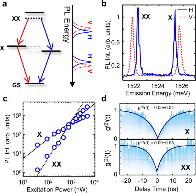
a Schematic of a cascaded biexciton emission proces. GS, X, and XX denotes the ground-state, exciton, and the biexciton states respectively. Biexciton first decays to an exciton either by emitting a vertically (V) or horizontally (H) polarized photon denoted by red or blue arrows respectively. Subsequently, the exciton decays to ground-state by emitting a second vertically or horizontally polarized photon with polarization correlated to the initial emitted photon, completing the emission cascade. The energy splitting between the different linear polarization arises due to the fine-structure splitting. b PL Spectrum of a single-quantum emitter. Two pairs of correlated doublets are observed. c PL intensity vs. Excitation Power: The localized defect exciton shows distinct behavior: Exciton (X) intensity increases sublinearly and begins to saturate at high powers as expected from a two-level system, whereas the biexciton line (XX) intensity increases superlinearly. Dashed lines are linear fit (log (PL Int) = x log (Excitation Power)) to the data with extracted slopes of 0.96 for exciton (X) and 1.587 for biexciton (XX), respectively. d Second-order correlation measurement. Both X and XX exhibit g(2)(0) < 0.1, showing high-purity single-photon emission.
Statistics of the defect and strain engineered sites
Engineering high purity and reproducible single-photon emitters with high yield is a requirement for any scalable photonic technology. By decoupling the strain and defect engineering in our nanopillar irradiation process, we were able to achieve a success rate of over 85% in engineering single-photon emitters per site. Note that, since e-beam irradiation is a repeatable process, subsequent irradiation and dosage optimization processes can be leveraged to achieve near-unity-yield at each site. Figure 4 represents the statistical histograms of the single-photon emitters fabricated using our method. The spectral purity of many of the emitters was measured to be above 95% (with an average purity of 92%, Fig. 4a) with average linewidths of 75 μeV and zero-field splitting of 760 μeV, and the linewidth and purities will likely improve with resonance excitation.
Fig. 4. Statistics of single-emitter properties.
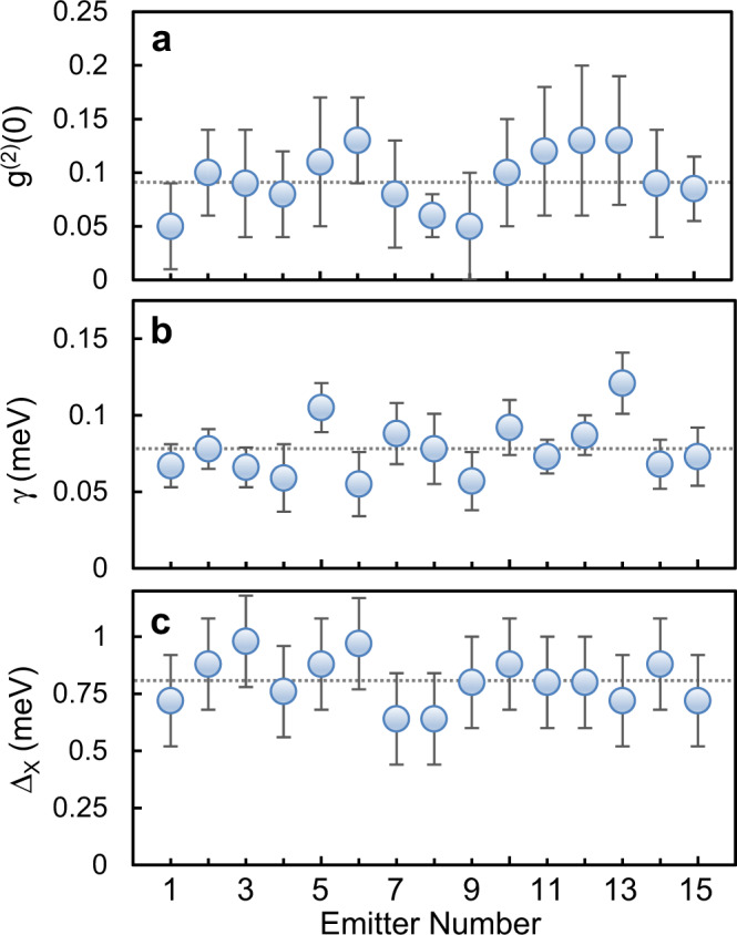
a g(2)(0) distribution. Average g(2)(0) is 0.08, which demonstrates the high purities achieved by this method. b Homogenous linewidth distribution with 75 μeV average. c Zero-field doublet-splitting showing the reproducibility of the method in engineering SPEs with similar characteristics. Horizontal dashed lines denote the average of measurements. Error bars denote the standard deviation from fitting the data.
Furthermore, the energy interval of the observed SPE emission (1515–1580 meV) is smaller than other approaches. This is likely a feature of our decoupled approach—first a broad-defect band is engineered with e-beam irradiation (~1.5–1.7 eV in Fig. 2). The SPEs then appear when a localized stressor red-shifts the neutral exciton energy into resonance with a defect state in the band. Given the defect band range (1.5–1.7 eV) is larger than the spectral range of the emitters (1515–1580 meV), it can be deduced that the variance in the engineered strain profiles sets the limit for the spectral spread of our method. In future experiments, using more precise strain engineering methods may enable engineering identical vdW SPEs.
Temperature evolution of engineered WSe2 emitters
As discussed previously, one of the main disadvantages of native WSe2 single-photon emitters is that their PL intensity and single-photon purity quench at temperatures above 30 K. One of the main contributors to this effect is the defect-bound exciton–phonon interaction, which leads to carrier escape from the confined potential, an effect which is more dominant in shallow quantum emitters. As e-beam-induced defects create deeper states within the bandgap, as evidence by the lower emission energies compared to native defect-based emitters, a higher energy barrier and hence, a higher thermal activation energy is to be expected26. In addition to this, the non-radiative trap-assisted recombination can become the dominant quenching factor at high-trap densities27. In our process the weights of non-radiative recombination pathways are minimized by using exfoliated 2D WSe2 with low-defect density (evident from the absence of the defect band). Conversely, the defects are induced in the sample via a controllable process targeting only the desired strained regions. Hence, our decoupled approach also alleviates this mechanism and enhances the quantum yield of the emitters to further increase the working temperatures. It is worth mentioning that higher confinement potentials can be achieved through more accurate positioning of the defect site within the strain field.
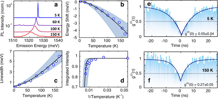
Figure 5a shows the evolution of the single-photon PL emission from 5 to 150 K (Supplementary Note 3 and Supplementary Fig. 6 are dedicated to the quantitative analysis of single-emitter properties). It is readily apparent that the emission line is distinguishable up to 150 K. A ~5 meV redshift is distinguishable up to 150 K (Fig. 5b), which indicates that deep confinement decouples the exciton energy from the temperature dependence of the lattice. Fitting the temperature dependence (Fig. 5b) to a Varshni-form equation (Supplementary Note 3) indicates a temperature coefficient (5.3 × 10⁻⁶ eV K−1), which is smaller than the previously reported values for native defects in CVD grown TMDs13. The evolution of the homogenous linewidth broadening with temperature is indicative of a thermally activated pure dephasing from exciton–phonon scattering and can be seen in Fig. 5c. The Arrhenius plot of the integrated intensity of the SPE vs. temperature shows that the integrated intensity quenches to e−1 at ~215 K (Fig. 5d). The data are fit with an Arrhenius model I(T) = I0/(1 + R exp(−EA/kT)), where R is equal to the ratio of the radiative (Tr) and non-radiative (Tnr) recombination lifetimes and EA is the thermal activation energy that required to dissociate the defect-bound excitons. The fit to the data results in a quantum yield of 5% and an activation energy of 95 meV, suggesting that the higher activation energies observed in this work is the main factor that contributes to high working temperatures of our emitters (see Supplementary Fig. 6 for a comparative analysis). It is worth mentioning that linewidth narrowing and an increase in intensity caused by h-BN encapsulation also contribute to higher working temperatures. However, given that the phonon escape quenching mechanism exponentially affects the PL intensity, whereas linewidth broadening, and initial intensity (I0) have a linear dependence, this enhancement is limited. This is further corroborated by the previous demonstrations of h-BN encapsulated TMD defect-bound excitons where overall a limited enhancement of working temperatures are observed23,28, but the fundamental quenching mechanism is not altered by h-BN encapsulation.
Fig. 5. Statistics of single-photon emission up to 150 K.
a Evolution of the PL spectrum as function of the temperature. b Redshift of the SPE line with temperature increase due to the reduction of the bandgap following Varshni’s empirical relationship. c Homogenous linewidth broadening due to the increase of temperature. d Arhenious plot of the integrated intensity. The PL peak starts to quench around 150 K and reaches e−1 at 215 K. Shaded region in b, c, and d demonstrate the range of recorded parameters for an ensemble of emitters. Open circles demonstrate measured data for a single emitter in the ensemble and the solid black lines are theoretical fit to the data. e Second-order correlation measurement g(2) at T = 5 K and f at 150 K exhibiting g2(0) < 0.3, evidencing that the single-photon nature persists up to our maximum measurement temperature of 150 K and likely persists beyond 200 K. Horizontal dashed lines represents the quantum limit of g(2) at 0.5.
Figure 5e, f show the temperature dependence of the second-order correlation function and demonstrates that the emitters exhibit antibunching up to 150 K with g(2)(0) = 0.27 ± 0.05. Given the high average purity (~75%) of the array of emitters at 150 K (see Supplementary Fig. 7) in combination with the Arrhenius data, it can be expected that the single-photon nature of the SPEs can persist up to 215 K. It can be predicted that our SPE engineering method, in tandem with designs leveraging Purcell enhancement, could enable room temperature single-photon emission.
Discussion
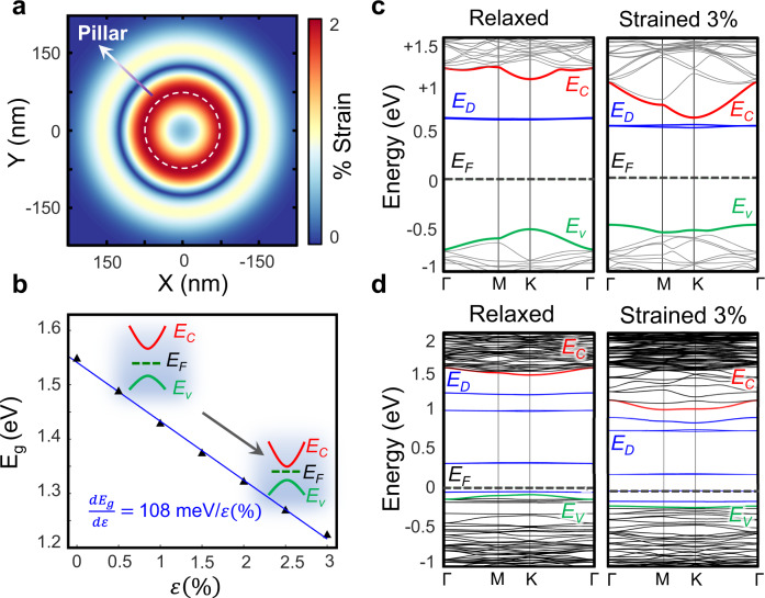
Figure 6 quantitatively summarizes one plausible mechanism behind the single-photon emission as depicted in Fig. 1. The dielectric nanopillar creates a strain profile akin to Fig. 6a in the 2D flake where the strain is at its maximum around the edges of the nanopillar. The applied strain results in the reduction of the bandgap (Fig. 6b), which creates a potential landscape analogous to Fig. 6a. This potential leads to the localization of free exciton states. From e-beam irradiation, defect states appear within the bandgap of the semiconductor in the strained regions. Provided the energy of these defect states at the strained regions is sufficiently close to the localized exciton states, the defects and excitons hybridize and results in the bright single-photon emission peak. While the engineering methods outlined in this work demonstrate the interplay between defects and strain, as predicted by the above-mentioned picture, the physical morphology of the defects responsible for SPE remains elusive. There is a lack of consensus of the defects responsible for single-photon emission, but they have been previously attributed to structures, including selenium vacancies14, tungsten centered vacancies29, oxygen interstitials30, and anti-site defects31. It is also plausible that multiple types of defects may be responsible for single-photon emission as long as they break the valley symmetry, and upon application of strain, introduce defect states with favorable energies close to the conduction band.
Fig. 6. DFT Calculations of strained defect bandstructure.
a Simulated strain profile at the nanopillar location. b Bandgap as a function of applied biaxial tensile strain. The inset band diagram denotes the qualitative movements of conduction band (EC), Valence band (EV), and the Fermi-level (EF) with strain. Note that the conduction band movement is larger with respect to the valence band. c Evolution of the band diagram of selenium vacancy with strain. ED and respective blue bands denote the localized defect levels. Upon the application of strain, Ec and ED come into proximity. d Evolution of the band diagram of WSe6 pore complex vs. strain. After the application of strain, the energy spacing between the conduction band and its closest defect-level decreases. Note that the Fermi-level energy position depends on the size of the supercell.
Given that the defects in this work were created by e-beam irradiation for which the morphology of resulting defects has been extensively studied, the list of possible suspects can be reduced through a systematic, ab initio approach. Studies have shown that e-beam irradiation processes primarily contribute to the generation of chalcogen and double chalcogen vacancies due to a lower knock-off energy20. Furthermore, prolonged exposure of a focused e-beam spot can also lead to the destabilization of the transition metal bond, which causes the transition metal to migrate away from the site, creating more complex vacancy sites such as pore vacancies32,33. In the case of WSe2, the creation of rotational trefoil defect complexes under e-beam irradiation has also been observed34. Therefore, we chose to study five probable defect complexes that might form during the e-beam process, namely selenium single vacancy, selenium double vacancy, tungsten vacancy, pore vacancy, and trefoil vacancies (see Supplementary Note 4 and Supplementary Figs. 8–13 for full details).
Density-functional theory (DFT) calculations were performed using Synopsis QuantumATK35 package by incorporating Perdew–Burke–Ernzerh (PBE) variant of generalized gradient approximation36 (PBE-GGA) exchange-correlation functional (see Methods for details). Note that given the common bandgap underestimation problem in DFT, along with the uncertainties in the exciton binding energies and the magnitude of the confinement potential, our focus is not on a quantitative comparison. Instead, our main criterion is the relative movement of the conduction band and defect levels with respect to each other upon application of strain, which can be assessed using ab initio simulation.
Figure 6c shows the dynamics of selenium single-vacancy levels with applied biaxial tensile strain. It is readily observable that the defect levels appear at favorable energies close to the conduction band and move to the proximity of the conduction band with strain. However, both selenium vacancies and selenium double vacancies are prone to passivation by oxidation37. Given the fact that the SPEs in this work, similar to previous studies, remain functional after many days and temperature cycles, these defects are an unlikely candidate. Conversely, tungsten vacancies do not introduce energies in the vicinity of the conduction band and, therefore, are not energetically favorable (see Supplementary Fig. 11). WSe6 vacancy complex—pore vacancies—on the other hand, form nanopores similar to those observed in MoS232 and WS233 after geometry relaxation (Supplementary Figs. 12 and 13). These vacancies also introduce energies close to the conduction band and move closer upon further application of strain (Fig. 5d). Thus, it is plausible that more complex vacancy structures created by e-beam irradiation, such as nanopores, are responsible for the SPEs observed in the e-beam irradiated sites.
In conclusion, by decoupling the strain and defect engineering process in the design of WSe2 single-photon emitters, we were able to achieve near-unity yield in deterministic positioning of quantum emitters with high purity that can preserve their single-photon nature to well above 150 K. The result indicates that both defects and strain are fundamental to quantum emitters in our WSe2 samples. This method was also successful at deterministic engineering of exciton-biexciton-like features with a radiative cascade that may pave the way for the realization of entangled photon-pair sources. It can be expected that by utilizing the methodology described in this study and integration with plasmonic or microphotonic cavities, our work may set the stage for future scalable elevated-temperature 2D WSe2 quantum light sources.
Methods
Sample preparation
The nanopillar array was fabricated with high-resolution electron-beam lithography following the procedures described in ref. 10. Briefly, a silicon wafer with thermal oxide was cleaved into a 1 cm2 chip followed by a solvent clean. The chip was spin-coated with hydrogen silsesquioxane (HSQ) resist diluted with methyl isobutyl ketone (MIBK). After a 5-min bake at 90 °C, pillar arrays and chip alignment markers were defined via electron-beam lithography and then developed in a 25% tetramethyl ammonium hydroxide (TMAH) solution and rinsed with methanol. A final rapid thermal anneal step at 1000 °C in oxygen converts the HSQ pattern into SiO2. The nanopillars have a nominal diameter of 150 nm and height of 200 nm.
We used a dry viscoelastic transfer technique to create the h-BN-encapsulated WSe2 structure. Intrinsic WSe2 flakes (2D Semiconductors) were exfoliated onto an unpatterned preparation SiO2/silicon substrate for identification and subsequent transfer. Next a thin flake of h-BN is exfoliated onto the transfer stamp, which was used to exfoliate and transfer WSe2 monolayers from thicker flakes on the preparation substrate. Finally, the WSe2/h-BN stack was transferred to an h-BN layer exfoliated on the patterned nanopillar array, which ensures that the WSe2 layer is encapsulated by the h-BN and free of any contaminants or residues during and after the transfer process. Quantum emitters were created using a 100 keV, <10 nm spot size electron-beam to expose the sample at the location of each nanopillar with ~106 electrons/μm2.
Optical spectroscopy
For steady-state micro-photoluminescence (PL) measurements, the sample temperature was held fixed between 5 and 150 K. The emitters were excited non-resonantly with a continuous-wave 633 nm laser focused to a ~1 μm full-width at half-maximum spot size using a long working distance 0.7 numerical aperture microscope objective. The backscattered PL was collected and measured with a spectrometer and TE-cooled charge-coupled device with a spectral resolution of ~30 μeV. For time-resolved PL measurements, a 40 MHz, 635 nm pulsed laser diode was used as the excitation source. The PL was spectrally filtered with ~1 nm bandwidth and detected using a superconducting nanowire single-photon detector and time-tagging electronics with ~40 ps detector temporal resolution. The PL measurements were performed at various average excitation powers ranging from 10 nW to 10 μW. Polarization-resolved spectra were acquired with a half-wave plate and linear polarizer before the spectrometer.
DFT calculations
HGH pseudopotentials with Tier4 basis sets, 3 × 3 × 1 and 3 × 3 × 1 Brillouin zone k-point sampling, 200 Rydberg density mesh cutoffs and a 0.02 eV/Å maximum force constant were used for geometry optimizations and DFT calculations. GGA-HGH Tier4 setup has been widely used in literature to probe the physics of 2D TMDs. WSe2 supercell was geometrically relaxed using the setup discussed above to 0.02 eV/Å maximum force constant. Next, depending on the size of the defect complex, a 5 × 5 or 7 × 7 supercell was constructed, followed by the removal of the appropriate atoms. Next, the geometry was optimized again by constraining the supercell along the in-plane direction to achieve the target strain and allowing for the defective supercell to relax to its final configuration.
Supplementary information
Acknowledgements
We gratefully acknowledge support from the UC Santa Barbara NSF Quantum Foundry funded via the Q-AMASE-i program under award DMR-1906325. G.M. and S.I.A. acknowledge support from the National Science Foundation (ECCS-2032272). K.P. was supported by the ARO (grant W911NF1810366). S.I.A acknowledges support from the California NanoSystems Institute through the Elings Fellowship program. Use was made of computational facilities purchased with funds from the National Science Foundation (CNS-1725797) and administered by the Center for Scientific Computing (CSC). The CSC is supported by the California NanoSystems Institute and the Materials Research Science and Engineering Center (MRSEC; NSF DMR 1720256) at UC Santa Barbara.
Author contributions
G.M. conceived the experiments. G.M., K.P., and S.I.A. analysed the data. K.P. and K.B. performed the DFT analysis. K.P wrote the manuscript. All authors discussed the results and commented on the manuscript at all stages.
Data availability
The data in this manuscript is available upon reasonable request.
Competing interests
The authors declare no competing interests.
Footnotes
Peer review information Nature Communications thanks Toan Tran and the other anonymous reviewers for their contribution to the peer review of this work.
Publisher’s note Springer Nature remains neutral with regard to jurisdictional claims in published maps and institutional affiliations.
Supplementary information
The online version contains supplementary material available at 10.1038/s41467-021-23709-5.
References
- 1.O’Brien JL, Furusawa A, Vuckovic J. Photonic quantum technologies. Nat. Photon. 2009;3:687–695. doi: 10.1038/nphoton.2009.229. [DOI] [Google Scholar]
- 2.Aharonovich I, Englund D, Toth M. Solid-state single-photon emitters. Nat. Photon. 2016;10:631–641. doi: 10.1038/nphoton.2016.186. [DOI] [Google Scholar]
- 3.He Y-M, et al. Single quantum emitters in monolayer semiconductors. Nat. Nanotechnol. 2015;10:497–502. doi: 10.1038/nnano.2015.75. [DOI] [PubMed] [Google Scholar]
- 4.Koperski M, et al. Single photon emitters in exfoliated WSe2 structures. Nat. Nanotechnol. 2015;10:503–506. doi: 10.1038/nnano.2015.67. [DOI] [PubMed] [Google Scholar]
- 5.Srivastava A, et al. Optically active quantum dots in monolayer WSe2. Nat. Nanotechnol. 2015;10:491–496. doi: 10.1038/nnano.2015.60. [DOI] [PubMed] [Google Scholar]
- 6.Tonndorf P, et al. Single-photon emission from localized excitons in an atomically thin semiconductor. Optica. 2015;2:347–352. doi: 10.1364/OPTICA.2.000347. [DOI] [Google Scholar]
- 7.Chakraborty C, Kinnischtzke L, Goodfellow KM, Beams R, Vamivakas AN. Voltage-controlled quantum light from an atomically thin semiconductor. Nat. Nanotechnol. 2015;10:507–511. doi: 10.1038/nnano.2015.79. [DOI] [PubMed] [Google Scholar]
- 8.Tran TT, Bray K, Ford MJ, Toth M, Aharonovich I. Quantum emission from hexagonal boron nitride monolayers. Nat. Nanotechnol. 2016;11:37–41. doi: 10.1038/nnano.2015.242. [DOI] [PubMed] [Google Scholar]
- 9.Branny A, Kumar S, Proux R, Gerardot BD. Deterministic strain-induced arrays of quantum emitters in a two-dimensional semiconductor. Nat. Commun. 2017;8:15053. doi: 10.1038/ncomms15053. [DOI] [PMC free article] [PubMed] [Google Scholar]
- 10.Palacios-Berraquero C, et al. Large-scale quantum-emitter arrays in atomically thin semiconductors. Nat. Commun. 2017;8:15093. doi: 10.1038/ncomms15093. [DOI] [PMC free article] [PubMed] [Google Scholar]
- 11.Rosenberger MR, et al. Quantum calligraphy: writing single-photon emitters in a two-dimensional materials platform. ACS Nano. 2019;13:904–912. doi: 10.1021/acsnano.8b08730. [DOI] [PubMed] [Google Scholar]
- 12.Peyskens F, Chakraborty C, Muneeb M, Van Thourhout D, Englund D. Integration of single photon emitters in 2D layered materials with a silicon nitride photonic chip. Nat. Commun. 2019;10:4435. doi: 10.1038/s41467-019-12421-0. [DOI] [PMC free article] [PubMed] [Google Scholar]
- 13.Luo Y, Liu N, Li X, Hone JC, Strauf S. Single photon emission in WSe2 up 160 K by quantum yield control. 2D Mater. 2019;6:035017. doi: 10.1088/2053-1583/ab15fe. [DOI] [Google Scholar]
- 14.Linhart L, et al. Localized intervalley defect excitons as single-photon emitters in WSe2. Phys. Rev. Lett. 2019;123:146401. doi: 10.1103/PhysRevLett.123.146401. [DOI] [PubMed] [Google Scholar]
- 15.Moon H, et al. Strain-correlated localized exciton energy in atomically thin semiconductors. ACS Photon. 2020;7:1135–1140. doi: 10.1021/acsphotonics.0c00626. [DOI] [Google Scholar]
- 16.Luo Y, Liu N, Kim B, Hone J, Strauf S. Exciton dipole orientation of strain-induced quantum emitters in WSe2. Nano Lett. 2020;20:5119–5126. doi: 10.1021/acs.nanolett.0c01358. [DOI] [PubMed] [Google Scholar]
- 17.Cadiz F, et al. Excitonic linewidth approaching the homogeneous limit in MoS2-based van der Waals heterostructures. Phys. Rev. X. 2017;7:021026. [Google Scholar]
- 18.Darlington TP, et al. Imaging strain-localized excitons in nanoscale bubbles of monolayer WSe2 at room temperature. Nat. Nanotechnol. 2020;15:854–860. doi: 10.1038/s41565-020-0730-5. [DOI] [PubMed] [Google Scholar]
- 19.Tyurnina AV, et al. Strained bubbles in van der Waals heterostructures as local emitters of photoluminescence with adjustable wavelength. ACS Photon. 2019;6:516–524. doi: 10.1021/acsphotonics.8b01497. [DOI] [Google Scholar]
- 20.Komsa HP, Kurasch S, Lehtinen O, Kaiser U, Krasheninnikov AV. From point to extended defects in two-dimensional MoS2: evolution of atomic structure under electron irradiation. Phys. Rev. B. 2013;88:035301. doi: 10.1103/PhysRevB.88.035301. [DOI] [Google Scholar]
- 21.Moody G, et al. Microsecond valley lifetime of defect-bound excitons in monolayer WSe2. Phys. Rev. Lett. 2018;121:057403. doi: 10.1103/PhysRevLett.121.057403. [DOI] [PubMed] [Google Scholar]
- 22.Xie X, et al. Designing artificial 2D crystals with site and size controlled quantum dots. Sci. Rep. 2017;7:9965. doi: 10.1038/s41598-017-08776-3. [DOI] [PMC free article] [PubMed] [Google Scholar]
- 23.Klein J, et al. Site-selectively generated photon emitters in monolayer MoS2 via local helium ion irradiation. Nat. Commun. 2019;10:2755. doi: 10.1038/s41467-019-10632-z. [DOI] [PMC free article] [PubMed] [Google Scholar]
- 24.He YM, et al. Cascaded emission of single photons from the biexciton in monolayered WSe2. Nat. Commun. 2016;7:13409. doi: 10.1038/ncomms13409. [DOI] [PMC free article] [PubMed] [Google Scholar]
- 25.Hanbury Brown R, Twiss RQ. Correlation between photons in two coherent beams of light. Nature. 1956;177:27–29. doi: 10.1038/177027a0. [DOI] [Google Scholar]
- 26.Le Ru,EC, Fack J, Murray R. Temperature and excitation density dependence of the photoluminescence from annealed InAs/GaAs quantum dots. Phys. Rev. B. 2003;67:245318. doi: 10.1103/PhysRevB.67.245318. [DOI] [Google Scholar]
- 27.Chahboun A, et al. Further insight into the temperature quenching of photoluminescence from InAsGaAs self-assembled quantum dots. J. Appl. Phys. 2008;103:083548. doi: 10.1063/1.2913179. [DOI] [Google Scholar]
- 28.Arora A, et al. Dark trions govern the temperature-dependent optical absorption and emission of doped atomically thin semiconductors. Phys. Rev. B. 2020;101:241413. doi: 10.1103/PhysRevB.101.241413. [DOI] [Google Scholar]
- 29.Zhang S, et al. Defect structure of localized excitons in a WSe2 monolayer. Phys. Rev. Lett. 2017;119:046101. doi: 10.1103/PhysRevLett.119.046101. [DOI] [PubMed] [Google Scholar]
- 30.Zheng YJ, et al. Point defects and localized excitons in 2D WSe2. ACS Nano. 2019;13:6050–6059. doi: 10.1021/acsnano.9b02316. [DOI] [PubMed] [Google Scholar]
- 31.Dass CK, et al. Ultra-long lifetimes of single quantum emitters in monolayer WSe2/hBN heterostructures. Adv. Quantum Technol. 2019;2:1900022. doi: 10.1002/qute.201900022. [DOI] [Google Scholar]
- 32.Wang S, et al. Atomic structure and formation mechanism of sub-nanometer pores in 2D monolayer MoS2. Nanoscale. 2017;9:6417–6426. doi: 10.1039/C7NR01127J. [DOI] [PubMed] [Google Scholar]
- 33.Ryu GH, et al. Atomic structure and dynamics of self-limiting sub-nanometer pores in monolayer WS2. ACS Nano. 2018;12:11638–11647. doi: 10.1021/acsnano.8b07051. [DOI] [PubMed] [Google Scholar]
- 34.Lin YC, et al. Three-fold rotational defects in two-dimensional transition metal dichalcogenides. Nat. Commun. 2015;6:6736. doi: 10.1038/ncomms7736. [DOI] [PMC free article] [PubMed] [Google Scholar]
- 35.Smidstrup S, et al. QuantumATK: an integrated platform of electronic and atomic-scale modelling tools. J. Phys. Condens. Matter. 2020;32:015901. doi: 10.1088/1361-648X/ab4007. [DOI] [PubMed] [Google Scholar]
- 36.Perdew JP, Burke K, Ernzerhof M. Generalized gradient approximation made simple. Phys. Rev. Lett. 1996;77:3865–3868. doi: 10.1103/PhysRevLett.77.3865. [DOI] [PubMed] [Google Scholar]
- 37.Lu J, et al. Atomic healing of defects in transition metal dichalcogenides. Nano Lett. 2015;15:3524–3532. doi: 10.1021/acs.nanolett.5b00952. [DOI] [PubMed] [Google Scholar]
Associated Data
This section collects any data citations, data availability statements, or supplementary materials included in this article.
Supplementary Materials
Data Availability Statement
The data in this manuscript is available upon reasonable request.


