

Abstract
Nanomaterials involve an active research and a booming area including different fields (health, environment, electronics, manufacturing, drug delivery). Recently, new concerns are emerging about the risk from increased production and subsequent release into the environment, as they are largely present in consumer products and industrial applications. Our aim was to assess the effects of three different types of cerium oxide nanoparticles (CeO2 NPs) (type 1 defined “as prepared”; type 2 defined “modified”; type 3 defined “commercial”) on zebrafish embryos by Fish Embryo Toxicity test (Z-FET). Immunohistochemical analysis was also performed on treated larvae to evaluate the expression of the following biomarkers: Metallothionein, Heat Shock Protein 70 (HSP70) and 7-Ethoxyresorufin-O-Dietylase (EROD). After 96 h of exposure, there was no lethality, nor were there sub-lethal effects in embryonic development, when compared with the control. No particular positivity was found about Metallothionein and HSP70 expression, while an increased expression of EROD was observed in larvae exposed to the three types of CeO2 NPs compared with the controls. The analyze has confirmed a statistically significant difference (P < 0.001) to EROD biomarker between control group and treated larvae response, finding was higher at 1-mg/l concentration. Further investigations in order to solve conflicting views about potential effects of CeO2 NPs are necessary, also to evaluate its effectiveness in different fields as already reported in literature.
Keywords: cerium oxide, embryo, Danio rerio, nanotoxicity, biomarkers of exposure
Introduction
Nanoparticles (NPs) and nanomaterials (NMs) involve an active and full expansion research area regarding different fields such as health, environment, energy, electronics, industrial and materials production [1–5]. They have gained considerable importance in technological progress due to their physicochemical properties related to their small size, chemical composition (purity, crystallinity), surface structure (large surface area, surface reactivity, surface groups, inorganic or organic coatings), solubility, shape and aggregation, electrical and thermal conductivity, and catalytic activity that improve their performance compared with common materials [6–8].
Although impressive from a physicochemical point of view, the properties of NM raise concerns about adverse effects on biological systems, so that new strategies are emerging to assess risk of NMs due to the increase in their production and their release into the environment, considering they are widely used in consumer products and industrial applications. For these strategies, the main challenge in studying engineered NMs is to predict their behavior comparing it with natural NMs [9].
Several studies have investigated the toxicity of CeO2 NPs, determining to be toxic to daphnids [10], algae [11–13] and bacteria [14] from concentrations in the range of the mg/l.
Jemec et al. [15] have shown sub-lethal effects, respectively, on zebrafish larvae (growth inhibition and malformations) and daphnids (immobility) during exposure with CuO-CeO2 mixed oxides NMs in comparison with pure CeO2. The Authors concluded that none of the four tested NMs caused the mortality of embryos, while the effects on larvae after hatching were found.
Another study focused on the toxicity of two types of CeO2 NPs in aquatic environments and tested on four species, showed that no acute toxicity occurred on any species after short exposure, even at the highest concentrations [16].
Despite the adverse effects, it was also possible to find favorable conditions following the exposure of some NPs. Xia et al. [17], for example, have compared TiO2, ZnO and CeO2 NPs and found toxicity due to exposure to ZnO, which leads to the generation of reactive oxygen species (ROS), inflammation and cell death. On the contrary, CeO2 NPs do not cause inflammation or cytotoxicity but even suppress ROS production, and TiO2 causes neither adverse nor protective effects.
These results demonstrate that metal oxide NPs induce a series of biological responses ranging from cytotoxic to cytoprotective. Because of these conflicting opinions, it is important to pay attention for the environment and all living organisms before using these materials for any application, especially in case of heavy metals, prone to endangering health [18].
In this study, the potential toxic effect of CeO2 NPs was evaluated. Cerium oxide (CeO2), thanks to its peculiar properties, is used in different fields; it is solid at room temperature and white, pale yellow or brownish in color depending on its purity. It is the most widely used cerium compound on the market [19]. In addition, being used as a catalyst in various reactions, it is considered a very useful material for solid oxide fuel cells, thanks to its relatively high conductivity of oxygen ions at intermediate temperatures [20]. Furthermore, the redox properties of CeO2 with an easily formation of Ce4+/Ce3+ species promote a high surface oxygen mobility, which allow to consider this oxide an ideal reactive support for the heterogenous catalysts, usually employed for environmental applications (Volatile Organic Compounds removal) [21] or for the H2 purification in the fuel cell [22]. CeO2 nanoparticulate is often used in automotive catalytic converters because it has the ability to release or absorb oxygen in the exhaust stream of a combustion engine depending on the partial pressure of O2. Through this process, NOx emissions are reduced and carbon monoxide is converted into carbon dioxide: with the addition of CeO2, the fuel burns more cleanly, with a consequent reduction in air pollution [23].
Since CeO2 NPs is insoluble in water and diluted acid, it is commonly used as an abrasive tool: the powder is effective in sanding and polishing other materials. For many years, it has been used for polishing special glass (e.g. telescopic mirrors). It is also used in heat resistant alloy coatings and ceramic coatings [24].
In the biological field, studies have been conducted to develop new potential treatment modalities for disorders induced by oxidative stress. CeO2 NPs have shown, through their antioxidant and regenerative activity, to reduce O2•− and H2O2 levels but also to remove ROS as •OH and RNS as the radical nitric oxide (•NO) and peroxynitrite (O2NO−). This remarkable capacity of CeO2 NPs could see them used as an alternative or co-therapie for diseases and disorders related to oxidative stress and nitrosative stress [25].
The antioxidant properties of CeO2 NPs have feed interest in different fields of medicine, launching a series of studies to evaluate their effectiveness as a treatment in a variety of pathologies, such as neurological [26] and cardiovascular [27]. Colon et al. [28] have evaluated the ability of CeO2 NPs to confer radiation protection on the gastrointestinal epithelium: these studies have shown that CeO2 NPs protect the gastrointestinal epithelium from radiation-induced damage by greasing by free radicals and increasing the production of SOD2 before exposure to radiation.
CeO2 NPs have also shown promising efficacy against endometriosis, so free radicals have been shown to play an important role in pathogenesis. Chaudhury et al. [29] demonstrated that CeO2 NPs mitigates the endometrial lesions induced in the mouse model, decreasing oxidative stress and inhibiting angiogenesis. In addition, it has been observed to protect against adverse effects on endometriosis-related oocytes, which is critical for successful pregnancy.
In ophthalmology, it has been seen that photoreceptor cells are constantly bombarded by photons which, together with the high rate of oxygen metabolism of cells, continuously expose them to high levels of reactive oxygen intermediates (ROIs). Chen et al. [30] showed that these NPs prevent the increase of intracellular concentrations of ROI in the primary cell cultures of the rat retina and, in vivo, prevent vision loss due to light-induced photoreceptor cell degeneration. In anticancer treatment, these NPs have shown toxicity to cancer cells, making them more sensitive to radiation therapy, but also to normal tissues in minimal quantities [31].
Although recent studies have shown that the different dimensions of CeO2 NPs and the relationship between Ce+3 and Ce+4 content could lead to these opposite mechanisms, further research is needed to draw valid conclusions about the adverse or beneficial mechanisms of these NPs [32].
For this reason, it is appropriate to evaluate the environmental fate of CeO2 monitoring its dispersion due to the several applications. Use as a diesel additive combined with a particulate filter can reduce emissions from automobile exhausts by up to 90%, but a part could be emitted causing extremely widespread pollution through exposure to air or it could accumulate in soils [33]. Cerium could also be introduced into landfills and become, if not efficiently removed from waste water flows, a long-term problem for the environment. A better understanding of their toxicity, including interaction with targets and molecular mechanisms, is necessary to mitigate their B-side effects.
The aim of the present work is to provide wider information on CeO2 NP toxicity in aquatic environment. CeO2 NP toxicity was evaluated on zebrafish embryos by Fish Embryo Toxicity test (FET) in which different endpoints are assessed (e.g. inhibition of growth, morphological alterations, mortality). Moreover, an immunohistochemical analysis on zebrafish larvae was performed to evaluate the response to the following biomarkers: Metallothionein (MTs), Heat Shock Protein 70 (HSP70) and 7-ethoxyresorufin-O-diethylase (EROD). A characterization of NPs was also undertaken in order to investigate basic characteristics.
Zebrafish is being increasingly employed as a model system to assess NM toxicity at multiple levels, including mortality, teratogenicity, immunotoxicity, alterations in reproduction and behavior. It is a vertebrate model used for studying development and disease for both pre-clinical studies and toxicological applications due to a range of several characteristics [34–37].
Its small size let to reduce housing requirements as well as the quantity of materials used for testing. A single female of this species is able to spawn around 200 eggs exhibiting a high fecundity rate [34–37]. Zebrafish shares with human genome about 70% similarity [38] underlining a good conservation of major developmental and physiological processes similar to humans. Zebrafish eggs are transparent and develop externally, making them easy to manipulate and suitable for screening some compounds that can disrupt normal development [39].
Materials and Method
Synthesis of CeO2 NPs
Powders of CeO2 NPs were supplied by the CNR-IMM of Catania. In particular, three different types of CeO2 NPs have been tested, indicated as: CeO2 type 1 defined “as-prepared” (mean size 6.8 nm); CeO2 type 2 defined “modified” (mean size 11.3 nm) and CeO2 type 3 defined “commercial” (Sigma-Aldrich N. 202975, size 50 nm).
The as-prepared CeO2 (type 1) was synthesized by chemical precipitation procedure considered one of the most used preparation procedures [4, 40]. Briefly, a cerium nitrate solution was precipitated at pH > 8 and at T = 80°C using a solution of KOH (1 M). After 3 h, the resultant slurry was digested for 24 h, filtered, dried at 100°C for 12 h and finally calcined in air at 450°C for 3 h. The modified CeO2 (type 2) was prepared with the same procedure described above but irradiating the sample after the calcination with a solar lamp for 3 h and in the presence of a hydrogen flow (20 cc/min). This is a simple treatment that allows to increase the surface defects of cerium oxide [4]. All the samples did not shown substantial variations in the morphology being this characterized to a not homogeneous shape of the particles, typical of this preparation method [41, 42].
Characterizations of CeO2 NPs
Morphological analysis by SEM measurements, the structural and textural properties by XRD and N2 adsorption–desorption measurements were performed.
To investigate the surface oxidation state and the surface species present in the NMs, we have done and discussed the XPS characterization. As written above (section Synthesis of CeO2 NPs) considering the preparation method adopted (chemical precipitation with KOH) the morphology of nanoparticles is rough and not-homogeneous, in accordance with the literature data [41, 42], we here report the SEM images of as prepared (Fig. 1A) and modified (Fig. 1B) CeO2, also the commercial sample showed a similar morphology. SEM images was performed by a Jeol JSM-7500F instrument.
Figure 1.
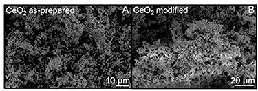
SEM images of as-prepared CeO2 (A) and of modified CeO2 (B).
The X-ray diffraction (XRD) patterns of the samples are reported in Fig. 2. Both the powders showed the XRD signals typical of cerium oxide in the fluorite crystalline phase with the reflections at 2θ values of 28.6 (1 1 1), 33. 1 (2 0 0), 47.4 (2 2 0) and 56.4 (3 1 1) considering the JCPDS Data File. No substantial variation was detected in the as-prepared or modified CeO2 compared with the commercial sample, apart from a slight intensity decrease due to the difference in the average crystallite size. The XRD reveals the high crystallinity of the synthetized samples.
Figure 2.

XRD patterns of the analyzed samples.
The XRD measurements were performed with a PANalytical X’pertPro X-ray diffractometer employing a Cu Kα radiation. The detection of the crystalline phases was made comparing the diffractions with those of standard materials reported in the JCPDS Data File.
The textural properties of the samples are calculated through the N2 physisorption measurements. In particular, Fig. 3A is reported the isotherm curves, whereas in Fig. 3B, the pore size distribution curves. The as-prepared and the modified samples displayed a N2 adsorption–desorption isotherm of type III, with a H3 hysteresis loop (Fig. 3A), indicating the presence of macro-meso slit-shaped pores [43]. The commercial one showed a more remarkable hysteresis indicating the presence of smaller pores [43]. The calculate BET surface area is 40 m2/g for the commercial CeO2, 67 m2/g for the modified CeO2 and 81 m2/g for the as-prepared sample. The decrease of surface area in the modified sample can be reasonably due to the agglomeration of CeO2 particles caused by the treatment used for the synthesis (irradiation under a solar lamp in H2 flow), whereas the lower surface area of the commercial sample is due to the higher mean particle size of this sample (50 nm) compared with the modified (11 nm) and the as-prepared (7 nm) samples. Considering the Barrett, Joyner and Halenda (BJH) pore size distribution curves (Fig. 3B), the mean pore size of the commercial CeO2 is 20 nm, whereas the modified sample showed a mean pore diameter of 58 nm higher compared with the as-prepared CeO2 (36 nm). This pore size increase verified with the modified CeO2 is also strictly related to the peculiar treatment of this latter sample, i.e. the simultaneous utilization of the simulated solar radiation in the H2 stream [4]. The textural properties of the powders were measured by N2 physisorption at −196°C with a Micromeritics Tristar II Plus 3020, outgassing the analyzed materials at 100°C overnight.
Figure 3.

N2 adsorption–desorption isotherms of the CeO2 samples (A); pore size distribution curves (B) of the analyzed samples evaluated with the BJH method.
Figure 4 has reported the XPS spectra of the analyzed samples in the Ce 3d zone.
Figure 4.

XPS spectra of the CeO2 samples in the Ce 3d zone.
In accordance with the literature data [44, 45], the signal at about 917 eV is the typical fingerprint of Ce4+, whereas the component at about 885 eV of Ce3+ is clearly visible only for the modified CeO2 (highlighted by the arrow in the figure), whereas the same signal is absent in the as-prepared CeO2 and in the commercial CeO2. Furthermore, it is possible to note as the ratio between the signals at about 898 and 900 eV (second arrow) is different and shifted in the commercial and as-prepared CeO2. This is another indication of the modification of the ceria surface sites with the higher presence of Ce3+ states in the modified CeO2. The irradiation with solar lamp in H2 stream thus induced the formation of CeO2-x defects on the surface of CeO2.
X-ray photoelectron spectroscopy (XPS) measurements were recorded using a K-Alpha X-ray photoelectron spectrometer utilizing the C 1 peak at 284.9 eV (ascribed to adventitious carbon) as a reference.
Preparation of NP solutions
Powders of CeO2 NPs have been suitably diluted to obtain different concentrations to test. The stock solution of each type of CeO2 NPs was obtained starting from 100-mg/l solution. The dilution water had specific characteristics (temperature 26 ± 1°C; pH range 6.5–8.5). The other lower concentration used in the test was: 10, 1 and 0.1 mg/l. All the solutions have been sonicated (4 cycles of 15 min each one; 40-kHz frequency) and vortexed to ensure a homogeneous dispersion of the NPs that show a natural tendency to aggregate and sediment.
Embryo selection
The Embryo toxicity test was performed according to the OECD guidelines for testing chemicals [46]. Furthermore, protocol procedure was performed according to Pecoraro et al. [47].
The ZFET was conducted on fertilized eggs, coming from the Centre for Experimental Fish Pathology of Sicily (CISS) laboratory, located at the University of Messina. CISS is accredited, since 2006, for use and, since 2010, for production of aquatic organisms for experimental research, and all procedures have been performed following Directive 2010/63/EU; moreover, at CISS laboratory, animal welfare procedures are performed according to Iaria et al. [48, 49]. Zebrafish adults were maintained only in the zebrafish breeding room and reared in a ZebTEC Active Blue Stand Alone system (Tecniplast). In this housing system, the water (disinfected by ultraviolet treatment) derives from reverse osmosis-treated city water. Environmental conditions at the primary enclosure (water tanks) are maintained at 27 ± 1°C, pH 7.2 ± 0.3 and a dissolved oxygen content (DO) of 6.00 ppm for freshwater species. Moreover, animals are exposed to a light/dark cycle (14 light/10 dark) and fed twice daily with Artemia Nauplii (JBL Artemio Pur, BL GmbH & Co. KG, Germany).
Following mating, the eggs were collected in steel grids, placed in the tanks to avoid predation by adults and ensure their collection. Using a stereomicroscope, the fertilized eggs were collected by Pasteur pipettes, while the unfertilized eggs, which did not undergo segmentation or which show irregularities during divisions or chorion lesions, were discarded. The fertilized eggs were transferred to the test solutions for exposure to the test chemical.
Exposure to cerium oxide solutions
According to OECD [46], healthy embryos were placed in 24-well microplates (one embryo per well) in 2-ml solution/well of each type of CeO2 NPs. Control samples (negative control) were incubated only with stock embryo medium and a positive control with 3,4-dichloroaniline (DCA) in water was also done. A controlled room temperature allowed to maintain 26 ± 1°C in wells. Replication was performed for each concentration of CeO2 NPs.
Observation of endpoints
During the exposure period, started within 180 min from fertilization of the eggs and finished at 96 h, four conditions called endpoints were satisfied every 24 h. The endpoints, analyzed by a stereomicroscope, were: embryo coagulation, lack of somite formation, lack of detachment of the tail-bud from the yolk sac and lack of heartbeat. In addition, hatching failure and post-hatching death were also recorded.
Coagulated embryos are recognizable after 24, 48 and 72 h because they have a milky white color and appear dark under the microscope (Fig. 5).
Figure 5.

fertilized eggs; regular development (A) and coagulated embryo after fertilization (B), egg during embryogenesis (C) and coagulated embryo 24 hpf (D).
The formation of somite was examined after 24, 48 and 72 h. After 24 h, about 20 somite should have formed at a temperature of 26 ± 1°C; however, since it is not possible to count the right number, only the presence or absence is recorded, considering that a normally developed embryo shows spontaneous movements (lateral contractions).
The lifting from the yolk, following the lengthening of the posterior part of the body of the embryo, is examined after 24, 48 and 72 h. The presence of the heart beat is recorded after 48 and 72 h.
Failure to hatch also represents an important toxic effect. The hatching rate will not be used to calculate LC50 value; however, if the hatching time is abnormal (for example more than 10% already after 48 h, considering that zebrafish embryos at the temperature of 26°C usually hatch after 72 h), it must be recorded after 96 h and reported.
Immunohistochemical analysis
According to Pecoraro et al. [50], the immunofluorescence protocol was performed on larvae exposed to all concentrations of CeO2 NPs, including controls, to detect positivity to MTs, HSP70 and the EROD biomarkers.
After washing the samples (fixed in paraformaldehyde) in PBS (pH 7.4, 0.1 M), permeabilization is carried out for 15 min in PBS-Triton X-100, which improves the penetration of the antibody, and follows the incubation of the samples with blocking solution to fill nonspecific binding sites of the antibodies for 20 min. The larvae, placed on the slides, are incubated overnight in a humid chamber at 4°C with the primary antibodies: anti-mouse-MTs, anti-mouse-HSP70 (GeneTex, 1:1000) and anti-rabbit-EROD (Abcam, 1:1000). After rinsing in PBS buffer (pH 7.4, 0.1 M) for 10 min, the samples were incubated for 1 h at 4°C in the dark with FITC-conjugated anti-mouse secondary antibody (for MTs and HSP70) and FITC-conjugated anti-rabbit (for EROD); both of them exhibit a green fluorescence. Samples were washed in PBS-Tween20 (2 times for 5 min) at room temperature, dehydrated in increasing alcohol solutions (70°, 80° and 95°) for 1 min each and air dried. Finally, samples were mounted with DAPI and sealed with rubber cement. The observations were made with the NIKON ECLIPSE Ci fluorescence microscope and the images captured with the NIKON DS-Qi2 camera.
Statistical analyses
Image J software [51] has been used to process the images obtained by fluorescence microscope since it calculates the mean value (the sum of the values at all pixels divided by the number of pixels) of a defined area. For each photo, the same area (macro) was analyzed in which the entire larva was considered in order to obtain density histograms. The mean values were compared with GraphPad Software by T-Student test to detect significant differences between the photos of treated larvae at tested concentrations of NPs and the photos of control groups (P < 0.001).
Results
Embryonic development
During the 96 h of test, no significant mortality was observed for both control groups and treated groups. Additionally, control groups showed normal development where the hatching rate and survival rates were above 90%, as described by Kimmel et al. [39] and recommended by the OECD 236 protocol [29]. After 96 h of exposure, there were no significant mortality or sub-lethal effects such as hatching delay, heartbeat alteration and malformation in embryonic development, when compared with control group. The maximum mortality was 2% for the whole test period (Fig. 5). In addition, the control group presented normal development as described by Kimmel et al. [39].
All embryos, exposed to different types and concentration of CeO2 NPs, regularly completed the development. The hatching of the eggs started around 48 h after exposure, took place within the last observation at 72 h. Furthermore, a good reactivity was found, both in the embryos and in the hatched larvae. Cardiovascular activity, indicated by the presence of heartbeat, has also been observed in embryos with a little bit reduced mobility, confirming their viability. Moreover, during observation under the stereomicroscope (Leica M0205C), we have observed the presence of the NPs on the chorion surface of the treated embryos, while there were no NPs on the chorion of control embryos.
Immunohistochemical assay
The results of the immunohistochemical investigation confirm the ability of D. rerio larvae to respond to the presence of metal NPs with the expression of specific detoxification systems; in fact, a positivity has been highlighted compared with the controls of the three biomarkers. We have observed, a higher expression of EROD biomarker in the larvae exposed to the three types of CeO2 NPs, showing a clear difference with the control samples even the samples with lower concentration (Fig. 6).
Figure 6.

EROD antibody-staining of the 96-hpf zebrafish embryos after exposure to different type and concentrations of CeO2 NPs including untreated zebrafish embryos; scale bar: 500 μm.
As regard MTs, no remarkable positivity was found in the expression of this biomarker, so it was weak in the exposed larvae compared with the controls (Fig. 7).
Figure 7.

MT antibody-staining of the 96-hpf zebrafish embryos after exposure to different type and concentrations of CeO2 NPs including untreated zebrafish embryos; scale bar: 500 μm.
Finally, regarding HSP70, they are expressed homogeneously in treated larvae toward different types of NPs and their concentrations, compared with the controls (Fig. 8).
Figure 8.

HSP70 antibody-staining of the 96-hpf zebrafish embryos after exposure to different type and concentrations of CeO2 NPs including untreated zebrafish embryos; scale bar: 500 μm.
The analyze has confirmed a statistically significant difference (P < 0.001) to EROD biomarker between control group and treated larvae response, finding that mean value pixels, given by Image J [51], were higher at 1-mg/l concentration. However, as regard MTs and HSP70 biomarkers, it has not been founded a statistically significant difference.
Discussion
Recent research on CeO2 NPs has reported conflicting results regarding the toxic effects: it has been reported both as an antioxidant and as an inducing agent for the production of ROS through various biological mechanisms [19]. Many researchers have found that CeO2 NPs exploit their ability to eliminate free radicals by acting as antioxidants and mimicking superoxide dismutase, the main catalyst that reduces ROS production and damage to mammalian cells, transforming superoxide into oxygen and hydrogen peroxide. They are also attributed the ability to inhibit cell aging [52], increase the life span of brain cell cultures [53] and prevent retinal disorders leading to blindness [30].
In contrast, other research have shown that CeO2 NPs induce and catalyze ROS generation, reduce the lifespan of nematodes [54], cause liver damage in rats [55] and show a moderate toxicity to various tissues, such as lung tissue [32].
Our results are in agreement with other studies in the literature, for which the exposure of D. rerio larvae to other metal oxide NPs does not show adverse effects on survival and hatching time [57]; the same results were observed by Vranic et al. [58] by exposing D. rerio larvae to SiO2 NPs. The presence of the NPs on the chorion of the treated embryos confirms the role of the chorion as a specific structure that protects the embryo from external influences up to hatching, but at the same time, it is permeable to allow a sufficient supply of gas and transport of nutrients. Chorion barrier function has already been reported for many NPs, compared with metal ions that can cross the membrane, such as SiO2, CuO, ZnO, Co3O4 and NiO NPs [56].
MTs are easily quantifiable exposure biomarkers by immunohistochemical assays and several studies in the literature have shown that D. rerio embryos exposed to AuNPs have exhibited a positive response for anti-MT throughout the body; therefore, metal NPs can induce MTs production [57, 59]. The same NPs were tested by Pan et al. [60] on zebrafish embryos, finding the positive expression of HSP70, also valid biomarkers for exposure from NPs. The activity of etoxyresorufin-O-deethylase (EROD) is considered an important biomarker for aquatic environmental contamination. Cogun et al. [61] assessed the effect of Al2O3 NPs of the EROD activity in a freshwater fish: Oreochromis niloticus. EROD activity in the liver has shown a significant increase in fish exposed to Al2O3 NPs, indicating that EROD activity can be widely used as a biomarker of exposure to heavy metals in fish. Moreover, alterations on enzymatic activities of EROD and other markers of oxidative stress are induced by persistent oxidative stress triggered by pollutants.
Conclusions
As the physiochemical properties of NPs mostly depend on the synthesis procedure, the synthesis method of NPs for biological applications is very important [62] because physical properties (size, surface charge, agglomeration in liquid and coating or residual contamination of the surfactant on the surface) are able to get interactions at the nano-bio interface [63]. Therefore, our investigation has shown that the different types of CeO2 NPs analyzed did not show adverse effects on vitality and hatching time of embryos, but anyway the ability of D. rerio to react at the presence of metallic NPs as highlighted by the expression of biomarkers considered. The toxicological profile of these NPs has not completely been defined, as reported by other previous studies then it seems appropriate to underline the importance that further studies will take, aimed at clarifying the conflicting opinions. This requirement depends not only to assess the potential effects of CeO2, which must be monitored for its several uses, but also by the evaluation of its effectiveness in the different fields as already reported in the literature.
Acknowledgements
R.P. thanks the PON project “AIM” funded by the European Social Found (ESF) CUP: E66C18001300007 for the financial support.
R.F. thanks the PON project “AIM” funded by the European Social Found (ESF) CUP:E66C18001220005 for the financial support.
E.M.S thanks funds University of Catania, Italy, MIUR XXXIV cycle PhD.
Contributor Information
Pecoraro Roberta, Department of Biological, Geological and Environmental Science, University of Catania, via Androne 81, 95124 Catania, Italy.
Scalisi Elena Maria, Department of Biological, Geological and Environmental Science, University of Catania, via Androne 81, 95124 Catania, Italy.
Iaria Carmelo, Department of Chemical, Biological, Pharmacological and Environmental Science, University of Messina, via F. Stagno D’Alcontres 31, Messina 98166, Italy.
Capparucci Fabiano, Department of Chemical, Biological, Pharmacological and Environmental Science, University of Messina, via F. Stagno D’Alcontres 31, Messina 98166, Italy.
Rizza Maria Teresa, Department of Biological, Geological and Environmental Science, University of Catania, via Androne 81, 95124 Catania, Italy.
Ignoto Sara, Department of Biological, Geological and Environmental Science, University of Catania, via Androne 81, 95124 Catania, Italy.
Salvaggio Antonio, Experimental Zooprophylactic Institute of Sicily “A. Mirri”, Palermo 90129, Italy.
Fiorenza Roberto, Department of Chemical Sciences, University of Catania, Viale A. Doria 6, 95125 Catania, Italy.
Impellizzeri Giuliana, CNR-IMM, Via S. Sofia 64, 95123 Catania, Italy.
Brundo Maria Violetta, Department of Biological, Geological and Environmental Science, University of Catania, via Androne 81, 95124 Catania, Italy.
Conflict of Interest Statement
The authors declare no conflict of interest.
CRediT Authorship Contribution Statement
R. Pecoraro: Conceptualization, Investigation, Writing—Review & Editing. Elena M. Scalisi: Conceptualization, Investigation, Writing—Review & Editing. C. Iaria: Resources. F. Capparucci: Resources. Maria T. Rizza: Investigation. Ignoto S: Writing—Review & Editing. A. Salvaggio: Roles/Writing—original draft. R. Fiorenza: Roles/Writing—original draft. G. Impellizzeri: Roles/Writing—original draft. Maria V. Brundo: Conceptualization, Investigation, Supervision, Writing—Review & Editing.
References
- 1. Cantarella M, Di Mauro A, Gulino A et al. Selective photodegradation of paracetamol by molecularly imprinted ZnO nanonuts. Appl Catal B-Environ 2018;238:509–17. [Google Scholar]
- 2. De Crozals G, Bonnet R, Farre C et al. Nanoparticles with multiple properties for biomedical applications: a strategic guide. Nano Today 2016;11:435–63. [Google Scholar]
- 3. Di Mauro A, Zimbone M, Scuderi M et al. Effect of Pt nanoparticles on the photocatalytic activity of ZnO nanofibers. Nanoscale Res Lett 2015;10:484. [DOI] [PMC free article] [PubMed] [Google Scholar]
- 4. Fiorenza R, Balsamo SA, D’Urso L et al. CeO2 for water remediation: comparison of various advanced oxidation processes. Catalysts 2020;10:446. [Google Scholar]
- 5. Huang HL, Huang ZH, Chu YC et al. Application of metallic nanoparticle biochars with ionic liquids for thermal transfer fluids. Chemosphere 2020;250:126219. [DOI] [PubMed] [Google Scholar]
- 6. Jeevanandam J, Barhoum A, Chan YS et al. Review on nanoparticles and nanostructured materials: history, sources, toxicity and regulations. Beilstein J Nanotech 2018;9:1050–74. [DOI] [PMC free article] [PubMed] [Google Scholar]
- 7. Oberdörster G, Oberdörster E, Oberdörster J. Nanotoxicology: an emerging discipline evolving from studies of ultrafine particles. Environ Health Perspect 2005;113:823–83. [DOI] [PMC free article] [PubMed] [Google Scholar]
- 8. Royal Society and Royal Academy of Engineering , 2004. Nanoscience and nanotechnologies: opportunities and uncertainties. RS policy document 19/04. London: The Royal Society. [Google Scholar]
- 9. Wagner S, Gondikas A, Neubauer E et al. Spot the difference: engineered and natural nanoparticles in the environment-release, behavior, and fate. Angew Chem Int Ed Engl 2014;53:12398–419. [DOI] [PubMed] [Google Scholar]
- 10. García A, Espinosa R, Delgado L et al. Acute toxicity of cerium oxide, titanium oxide and iron oxide nanoparticles using standardized tests. Desalination 2011;269:136–41. [Google Scholar]
- 11. Manier N, Garaud M, Delalain P et al. Behaviour of ceria nanoparticles in standardized test media – influence on the results of ecotoxicological tests. J Phys Conf Ser 2011;304:012058. [Google Scholar]
- 12. Manier N, Bado-Nilles A, Delalain P et al. Ecotoxicity of non-aged and aged CeO2 nanomaterials towards freshwater microalgae. Environ Pollut 2013;180:63–70. [DOI] [PubMed] [Google Scholar]
- 13. Rogers NJ, Franklin NM, Apte SC et al. Physico-chemical behaviour and algal toxicity of nanoparticulate CeO2 in freshwater. Environ Chem 2010;7:50. [Google Scholar]
- 14. Thill A, Zeyons O, Spalla O et al. Cytotoxicity of CeO2 nanoparticles for Escherichia coli. Physico-chemical insight of the cytotoxicity mechanism. Environ Sci Technol 2006;40:6151–6. [DOI] [PubMed] [Google Scholar]
- 15. Jemec A, Djinović P, Tišler T et al. Effects of four CeO2 nanocrystalline catalysts on early-life stages of zebrafish Danio rerio and crustacean Daphnia magna. J Hazard Mater 2012;219–220:213–20. [DOI] [PubMed] [Google Scholar]
- 16. Bour A, Mouchet F, Verneuil L et al. Toxicity of CeO2 nanoparticles at different trophic levels – effects on diatoms, chironomids and amphibians. Chemosphere 2015;120:230–6. [DOI] [PubMed] [Google Scholar]
- 17. Xia T, Kovochich M, Liong M et al. Comparison of the mechanism of toxicity of zinc oxide and cerium oxide nanoparticles based on dissolution and oxidative stress properties. ACS Nano 2008;2:2121–34. [DOI] [PMC free article] [PubMed] [Google Scholar]
- 18. Khan I, Saeed K, Khan I. Nanoparticles: properties, applications and toxicities. Arab J Chem 2017;12:908–31. [Google Scholar]
- 19. Dahle JT, Arai Y. Environmental geochemistry of cerium: applications and toxicology of cerium oxide nanoparticles. Int J Environ Res Public Health 2015;12:1253–78. [DOI] [PMC free article] [PubMed] [Google Scholar]
- 20. Kilbourn BT. Cerium and cerium compounds. Kirk-Othmer Encyclopedia of Chemical Technology, 2003, 1–23 [Google Scholar]
- 21. Bellardita M, Fiorenza R, D’Urso L et al. Exploring the photothermo-catalytic performance of Brookite TiO2-CeO2 composites. Catalysts 2020;10:765. [Google Scholar]
- 22. Fiorenza R, Crisafulli C, Scire S. H2 purification through preferential oxidation of CO over ceria supported bimetallic au-based catalysts. Int J Hydrogen Energy 2016;41:19390–8. [Google Scholar]
- 23. Reinhardt K, Winkler H. Cerium mischmetal, cerium alloys, and cerium compounds. Ullmann’s encyclopedia of industrial chemistry, 2000. [Google Scholar]
- 24. Hedrick JB, Sinha SP. Cerium-based polishing compounds: discovery to manufacture. J Alloys Compd 1994;207:377–82. [Google Scholar]
- 25. Nelson BC, Johnson ME, Walker ML et al. Antioxidant cerium oxide nanoparticles in biology and medicine. Antioxidants 2016;5:15. [DOI] [PMC free article] [PubMed] [Google Scholar]
- 26. Estevez AY, Erlichman JS. The potential of cerium oxide nanoparticles (nanoceria) for neurodegenerative disease therapy. Nanomedicine 2014;9:1437–40. [DOI] [PubMed] [Google Scholar]
- 27. Sangomla S, Saifi MA, Khurana A et al. Nanoceria ameliorates doxorubicin induced cardiotoxicity: possible mitigation via reduction of oxidative stress and inflammation. J Trace Elem Med Biol 2018;47:53–62. [DOI] [PubMed] [Google Scholar]
- 28. Colon J, Hsieh N, Ferguson A et al. Cerium oxide nanoparticles protect gastrointestinal epithelium from radiation-induced damage by reduction of reactive oxygen species and upregulation of superoxide dismutase 2. Nanomed-Nanotechnol 2010;6:698–705. [DOI] [PubMed] [Google Scholar]
- 29. Chaudhury K, Babu N, Singh AK et al. Mitigation of endometriosis using regenerative cerium oxide nanoparticles. Nanomedicine: NBM 2013;9:439–48. [DOI] [PubMed] [Google Scholar]
- 30. Chen J, Patil S, Seal S et al. Nanoceria particles prevent ROI-induced blindness. In: Recent advances in retinal degeneration (pp. 53–59). New York, NY: Springer, 2008, 613. [DOI] [PubMed] [Google Scholar]
- 31. Alili L, Sack M, von Montfort C et al. Downregulation of tumor growth and invasion by redox-active nanoparticles. Antioxid Redox Signal 2013;19:765–78. [DOI] [PMC free article] [PubMed] [Google Scholar]
- 32. Park EJ, Choi J, Park YK et al. Oxidative stress induced by cerium oxide nanoparticles in cultured BEAS-2B cells. Toxicology 2008;245:90–100. [DOI] [PubMed] [Google Scholar]
- 33. Park B, Donaldson K, Duffin R et al. Hazard and risk assessment of a nanoparticulate cerium oxide-based diesel fuel additive—a case study. Inhal Toxicol 2008;20:547–66. [DOI] [PubMed] [Google Scholar]
- 34. Dooley K, Zon LI. Zebrafish: a model system for the study of human disease. Curr Opin Genet Dev 2000;10:252–6. [DOI] [PubMed] [Google Scholar]
- 35. Strähle U, Scholz S, Geisler R et al. Zebrafish embryos as an alternative to animal experiments-a commentary on the definition of the onset of protected life stages in animal welfare regulations. Reprod Toxicol 2012;33:128–32. [DOI] [PubMed] [Google Scholar]
- 36. Chakraborty C, Agoramoorthy G. Why zebrafish? Riv Biol 2010;103:25. [PubMed] [Google Scholar]
- 37. Ribas L, Piferrer F. The zebrafish (Danio rerio) as a model organism, with emphasis on applications for finfish aquaculture research. Rev Aquacult 2014;6:209–40. [Google Scholar]
- 38. Howe K, Clark MD, Torroja CF et al. The zebrafish reference genome sequence and its relationship to the human genome. Nature 2013;496:498–503. [DOI] [PMC free article] [PubMed] [Google Scholar]
- 39. Kimmel CB, Ballard WW, Kimmel SR et al. Stages of embryonic development of the zebrafish. Dev Dyn 1995;203:253–310. [DOI] [PubMed] [Google Scholar]
- 40. Fiorenza R, Spitaleri L, Gulino A et al. Ru–Pd bimetallic catalysts supported on CeO2-MnOx oxides as efficient systems for H2 purification through CO preferential oxidation. Catalysts 2018;8:203. [Google Scholar]
- 41. Scirè S, Palmisano L. Cerium and cerium oxide: a brief introduction. In: Cerium Oxide (CeO₂): Synthesis, Properties and Applications (pp. 1–12). Amsterdam, The Netherlands: Elsevier, 2020. [Google Scholar]
- 42. Liu Z, Zhou R, Zheng X. Comparative study of different methods of preparing CuO-CeO2 catalysts for preferential oxidation of CO in excess hydrogen. J Mol Catal A Chem 2007;267:137–42. [Google Scholar]
- 43. Sing KSW, Everet DH, Haul RAW. Reporting physisorption data for gas/solid system with special reference to the determination of surface area and porosity. Pure Appl Chem 1985;57:603–19. [Google Scholar]
- 44. Gamboa-Rosales NK, Ayastuy JL, González-Marcos MP et al. Oxygen-enhanced water gas shift over ceria-supported au-cu bimetallic catalysts prepared by wet impregnation and deposition-precipitation. Int J Hydrogen Energy 2012;37:7005–701. [Google Scholar]
- 45. Fiorenza R, Spitaleri L, Gulino A et al. High-performing au-ag bimetallic catalysts supported on macro-mesoporous CeO2 for preferential oxidation of CO in H2-rich gases. Catalysts 2020;10:49. [Google Scholar]
- 46. OECD . Guideline for the Testing of Chemicals. Fish Embryo Toxicity (FET), N.236, 2013. OECD. [Google Scholar]
- 47. Pecoraro R, Salvaggio A, Marino F et al. Metallic nano-composite toxicity evaluation by zebrafish embryo toxicity test with identification of specific exposure biomarkers. Curr Protoc Toxicol 2017;74:1.14. 1–1.14. 13. [DOI] [PubMed] [Google Scholar]
- 48. Iaria C, Migliore S, Macri D et al. Evidence of Centrocestus formosanus (Nishigori, 1924) in zebrafish (Danio rerio). Zebrafish 2019;16:522–6. [DOI] [PubMed] [Google Scholar]
- 49. Iaria C, Saoca C, Guerrera MC et al. Occurrence of diseases in fish used for experimental research. Lab Anim 2019;5:619–29. [DOI] [PubMed] [Google Scholar]
- 50. Pecoraro R, ’Angelo D D, Filice S et al. Toxicity evaluation of graphene oxide and titania loaded nafion membranes in zebrafish. Front Physiol 2018;8:1039. [DOI] [PMC free article] [PubMed] [Google Scholar]
- 51. Bankhead P. Analyzing fluorescence microscopy images with ImageJ. ImageJ 2014;1:10–1109. [Google Scholar]
- 52. Karakoti A, Singh S, Dowding JM et al. Redox-active radical scavenging nanomaterials. Chem Soc Rev 2010;39:4422–32. [DOI] [PubMed] [Google Scholar]
- 53. Rzigalinski BA, Bailey D, Chow L et al. 2003. Cerium oxide nanoparticles increase the lifespan of cultured brain cells and protect against free radical and mechanical trauma. In: FASEB J (Vol. 17, No. 4, pp. A606-A606). 9650 Rockville Pike, Bethesda, MD 20814-3998 USA: Federation Amer Soc Exp Biol. [Google Scholar]
- 54. Zhang H, He X, Zhang Z et al. Nano-CeO2 exhibits adverse effects at environmental relevant concentrations. Environ Sci Technol 2011;45:3725–30. [DOI] [PubMed] [Google Scholar]
- 55. Nalabotu SK, Kolli MB, Triest WE et al. Intratracheal instillation of cerium oxide nanoparticles induces hepatic toxicity in male Sprague-Dawley rats. Int J Nanomedicine 2011;6:2327–35. [DOI] [PMC free article] [PubMed] [Google Scholar]
- 56. Böhme S, Baccaro M, Schmidt M et al. Metal uptake and distribution in the zebrafish (Danio rerio) embryo: differences between nanoparticles and metal ions. Environ Sci Nano 2017;4:1005–15. [Google Scholar]
- 57. Brundo MV, Pecoraro R, Marino F et al. Toxicity evaluation of new engineered nanomaterials in zebrafish. Front Physiol 2016;7:130. [DOI] [PMC free article] [PubMed] [Google Scholar] [Retracted]
- 58. Vranic S, Shimada Y, Ichihara S et al. Toxicological evaluation of SiO2 nanoparticles by zebrafish embryo toxicity test. Int J Mol Sci 2019;20:882. [DOI] [PMC free article] [PubMed] [Google Scholar]
- 59. Ferrante M, Vassallo M, Mazzola A et al. In vivo exposure of the marine sponge Chondrilla nucula Schmidt, 1862 to cadmium (cd), copper (cu) and lead (Pb) and its potential use for bioremediation purposes. Chemosphere 2018;193:1049–57. [DOI] [PubMed] [Google Scholar]
- 60. Pan Y, Leifert A, Graf M et al. High-sensitivity real-time analysis of nanoparticle toxicity in green fluorescent protein-expressing zebrafish. Small 2013;9:863–9. [DOI] [PubMed] [Google Scholar]
- 61. Cogun HY, Temiz O, Aytekin T et al. 2018. The effect of aluminum nanoparticles on EROD activity in liver of Oreochromis niloticus. Alüminyum Nanopartiküllerin Oreochromis niloticus’ un Karaciğerlerinde EROD Aktivitesi Üzerine Etkisi. In: Congress Chairman.
- 62. Charbgoo F, Ahmad MB, Darroudi M. Cerium oxide nanoparticles: green synthesis and biological applications. Int J Nanomedicine 2017;12:1401. [DOI] [PMC free article] [PubMed] [Google Scholar]
- 63. Dowding JM, Seal S, Self WT. Cerium oxide nanoparticles accelerate the decay of peroxynitrite (ONOO−). Drug Deliv Transl Res 2013;3:375–9. [DOI] [PMC free article] [PubMed] [Google Scholar]