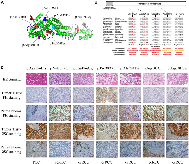
Figure 2.
Analysis of novel FH variants of unknown significance. (A) Structure model representing the FH protein (3e04-China A) and showing the detected mutated amino acid sites differentiated by color (purple, p.Arg101; blue, p.Val139; orange, p.Asn154; brown, p.Pro309; cyan, p.Ala320; and gray, p.His476). (B) FH amino acid conservation across species assessed by multiple sequence alignment and in silico analysis using SIFT, PolyPhen-2, Provean, and mCSM to predict the effects of the 5 missense FH VUSs identified. (C) H & E staining, FH immunohistochemical staining, and protein succination level (2SC) staining of FH VUS–associated tumors and paired normal tissues (×200). Germline FH VUS mutations and tumor histologies are shown. 2SC indicates S-(2-succinyl)cysteine; ccRCC, clear cell renal cell carcinoma; FH, fumarate hydratase; PCC, papillary renal cell carcinoma; VUS, variant of unknown clinical significance.

