Abstract
Background
Canonical nonsense-mediated decay (NMD) is an important splicing-dependent process for mRNA surveillance in mammals. However, processed pseudogenes are not able to trigger NMD due to their lack of introns. It is largely unknown whether they have evolved other surveillance mechanisms.
Results
Here, we find that the RNAs of pseudogenes, especially processed pseudogenes, have dramatically higher m6A levels than their cognate protein-coding genes, associated with de novo m6A peaks and motifs in human cells. Furthermore, pseudogenes have rapidly accumulated m6A motifs during evolution. The m6A sites of pseudogenes are evolutionarily younger than neutral sites and their m6A levels are increasing, supporting the idea that m6A on the RNAs of pseudogenes is under positive selection. We then find that the m6A RNA modification of processed, rather than unprocessed, pseudogenes promotes cytosolic RNA degradation and attenuates interference with the RNAs of their cognate protein-coding genes. We experimentally validate the m6A RNA modification of two processed pseudogenes, DSTNP2 and NAP1L4P1, which promotes the RNA degradation of both pseudogenes and their cognate protein-coding genes DSTN and NAP1L4. In addition, the m6A of DSTNP2 regulation of DSTN is partially dependent on the miRNA miR-362-5p.
Conclusions
Our discovery reveals a novel evolutionary role of m6A RNA modification in cleaning up the unnecessary processed pseudogene transcripts to attenuate their interference with the regulatory network of protein-coding genes.
Supplementary Information
The online version contains supplementary material available at 10.1186/s13059-021-02402-2.
Keywords: Pseudogene, N6-methyladenosine, Natural selection, ceRNA, RNA stability
Background
Nonsense-mediated decay (NMD) is an important process for mRNA surveillance. It degrades mRNAs with premature translation termination codons (PTCs), which are usually generated via nonsense mutations, frameshift mutations, or aberrant splicing [1, 2]. NMD is critical for preventing the formation of truncated proteins, which could be poisonous to cells. Therefore, nonsense mutations that escape NMD often cause dominant-negative effects [1]. In yeast, PTCs are recognized by the presence of downstream sequence element (DSE), which can stimulate NMD [3]. However, the canonical NMD becomes a splicing-dependent process in mammals, which can be triggered as long as the stop codons are more than 50~55 bp upstream of the last exon-exon junction [1]. Although NMD can also be triggered by long 3′UTRs [4], intronless genes are likely insensitive to NMD [5, 6].
As non-functional copies of closely related protein-coding genes, pseudogenes are one of the major class of substrates of NMD due to their accumulated nonsense mutations in the evolution [7, 8]. Although in most cases pseudogenes may still not be very important, increasing evidence indicates that some pseudogenes play important regulatory roles on regulating their protein-coding cognates; dysregulation of pseudogenes are associated with various human diseases including cancer [9]. Due to the high sequence similarities with their protein-coding cognates, pseudogenes often work as competitive endogenous RNAs (ceRNAs), which competitively bind microRNAs (miRNAs) or RNA binding proteins (RBPs) to prevent the degradation (or other processes) of their cognate protein-coding genes, such as PTENP1 [10], BRAFP1 [11], and HMGA1 [12]. Some other pseudogenes can also generate endogenous small interfering RNAs (esiRNA), such as PPM1K [13], or work as trans-acting antisense RNAs, such as nNOSP [14].
There are three classes of pseudogenes according to the unique biogenesis mechanisms: unitary pseudogenes, unprocessed pseudogenes, and processed pseudogenes [15]. Unitary pseudogenes are single-copy genes with spontaneous mutations in the coding regions or regulatory regions, resulting in genes unable to be transcribed or translated into proteins. Unprocessed pseudogenes are originated through gene duplications and subsequent mutations that cause frameshifts or early terminators. Processed pseudogenes, also known as retrotransposed pseudogenes, are generated through retrotransposition of mRNA transcripts, thus do not have introns but may have poly(A) tails. As previously reported, the retrotransposed mRNAs tend to be stable transcripts translated on free cytoplasmic ribosomes [16]. Because canonical NMD is a splicing-dependent process in mammals, processed pseudogenes are not likely the substrates of NMD due to the lack of introns. It is largely unknown whether processed pseudogenes are subject to other RNA surveillance pathways and whether they have acquired novel surveillance mechanisms in the evolutionary history since they diverged from their cognate protein-coding genes.
N6-methyladenosine (m6A) RNA modification is reported in recent years as a novel pathway of degrading RNAs [17, 18]. m6A is a reversible and prevalent internal RNA modification in mRNAs and long noncoding RNAs (lncRNAs). It is installed on “DRACH” motifs of RNAs [19] by m6A methyltransferases complex with METTL3 as the catalytic subunit [17, 18]. Demethylases FTO and ALKBH5 can reverse the modification [17, 18]. In addition, m6A can be specifically regulated through a variety of RNA binding proteins and co-transcriptionally through transcription factors as well as H3K36me3 histone modification [17, 18, 20]. Upon m6A modification on mRNAs, m6A readers such as YTH domain-containing proteins can specifically read the m6A and regulate various post-transcriptional processes of host mRNAs [17, 18], such as promoting the cytosolic degradation [21–23] and nuclear export of mRNAs [24]. In recent years, critical roles of m6A have been reported in a variety of physiological and pathological processes [25–27].
Based on the genome-wide profiling of m6A, the RNAs of pseudogenes are also modified by m6A [28]. However, little is known about the function of the m6A sites on pseudogenes and how they have evolved after separating from their cognate protein-coding genes.
In this study, we found the RNAs of human processed pseudogenes were accumulating novel m6A modifications in company with novel m6A motifs after separating with their cognate protein-coding genes. We found convergent evidence supporting that these recently accumulated m6A motifs had evolved under positive selection. Based on bioinformatic analyses and experimental validation, we have revealed that these m6A sites on the RNAs of processed pseudogenes promoted cytosolic RNA degradation and attenuated their unnecessary interfering with their cognate mRNAs. Our discovery illustrates the evolutionary landscape of an m6A-mediated RNA surveillance mechanism for NMD resistant RNAs.
Results
The RNAs of pseudogenes tend to have higher m6A levels than their cognate mRNAs
In order to study the functions and evolution of m6A on the RNAs of pseudogenes, we first ask whether the RNAs of pseudogenes are methylated differently from their cognate mRNAs. We previously developed m6A-LAIC-seq technology to quantify the m6A levels in transcriptome-wide scale [28]. Here, we took advantage of our previously published m6A-LAIC-seq data of GM12878, which is a human B-lymphoblastoid cell line sequenced deeply in 1000 genome project, as well as H1, which is a human embryonic stem cell line, to study the m6A levels of the RNAs of pseudogenes [28].
Since pseudogenes and their cognate protein-coding genes have similar sequences, cross-mapping can happen frequently when mapping the short-reads of next-generation sequencing to the genome, which will distort the expression patterns of pseudogenes. Here, we improved the mapping procedure to minimize the occurrence of cross-mapping by only allowing perfect matches or mismatches at known SNPs when we aligned the reads to hg19 human genome using HISAT2 [29]. Because the SNPs of GM12878 were mostly included in the known SNP database, the new procedure is more powerful for GM12878 and we mainly focused on this cell line for the downstream analyses. Compared with the conventional HISAT2 mapping procedure allowing 3 mismatches, the new procedure resulted in a reduced number of mapped reads for a large number of genes, reflecting the stricter mapping criteria of the new mapping procedure. In contrast, we observed similar numbers of pseudogenes with more or less mapped reads using the new mapping procedure, suggesting that the stricter new procedure cause re-assignment of mapped reads on pseudogenes and their homologous genomic loci compared with the conventional procedure (Additional file 1: Figure S1a-c). As shown in Additional file 1: Figure S1d and e, we observed dramatic read coverage changes in certain regions of pseudogene RPS7P1 based between the two mapping procedures, due to the re-assignment of reads from its protein-coding cognate RPS7 to pseudogene PRS7P1 in the new mapping procedure; in another case, some reads were re-assigned from pseudogene PPP1R14BP3 to its cognate protein-coding gene PPP1R14B in the new procedure.
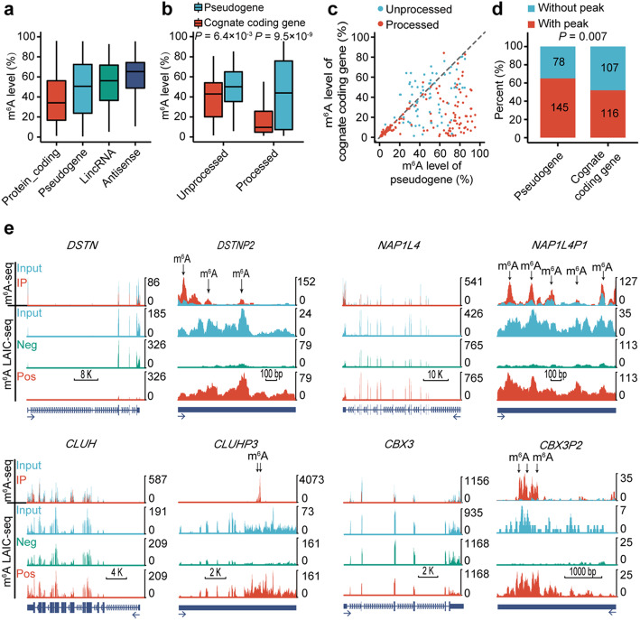
We took advantage of the annotations from GENCODE, Vega, and psiCube [30] databases to compile a list of 12,143 one-to-one pairs of pseudogenes and corresponding protein-coding genes, including 4078 protein-coding genes with different numbers of pseudogenes (Additional file 2: Table S1). Among them, only 223 transcribed pseudogenes with reliable m6A levels were detected in the m6A-LAIC-seq data of GM12878 cell line, reflecting that most pseudogenes are not transcribed [31]. The cognate protein-coding genes of these pseudogenes were enriched in mRNA catabolic process, endoplasmic reticulum localization, and translational initiation, consistent with the previous report about transcribed pseudogene [9] (Additional file 1: Figure S2a, b). We found m6A levels of pseudogenes were higher than protein-coding genes, but lower than lincRNA and antisense RNAs (Fig. 1a).
Fig. 1.
Comparisons of m6A between pseudogenes and their cognate protein-coding genes in GM12878 cell line. a Box plot comparing the m6A levels among different biotypes. b Box plot comparing the m6A levels between pseudogenes and their cognate protein-coding genes for processed pseudogenes and unprocessed pseudogenes respectively. c Scatter plot comparing the m6A levels between pseudogenes and their cognate protein-coding genes for processed pseudogenes and unprocessed pseudogenes respectively. d Stacked bar plot comparing the percent of pseudogenes and their cognate protein-coding genes with or without m6A peaks. The frequencies were displayed in the corresponding boxes. e UCSC genome browser tracks of m6A-seq and m6A-LAIC-seq data showing representative examples of pseudogenes and their cognate protein-coding genes. Read-coverage tracks of input, m6A-negative, and m6A-positive fractions of m6A-LAIC-seq shown along with overlay tracks of m6A-seq (red for RIP and cyan for input; predicted m6A sites in m6A peaks are indicated by arrows). Read coverage (y-axis) of m6A negative and m6A positive are normalized as previously described [28] to reflects the calculated m6A levels (i.e., equal signals in m6A positive (eluate) versus m6A negative (supernatant) indicate m6A levels of 50%), while input and m6A-seq tracks are shown for optimal viewing
We then performed a pairwise comparison between the m6A levels of pseudogenes and their cognate protein-coding genes. We found the m6A levels of pseudogenes were much higher than their cognate protein-coding genes, the differences were much more dramatic in processed pseudogenes than unprocessed pseudogenes (Fig. 1b, c). Consistently, it was more dramatic in intronless pseudogenes than pseudogenes with multiple exons (Additional file 1: Figure S2c, d; Additional file 2: Table S2). Similar results were observed in H1 hESC based on 190 pseudogenes with reliable m6A levels (Additional file 1: Figures S2e, f and S3a-c; Additional file 2: Table S3). These results suggest that the m6A of pseudogenes may get promoted in the evolution after separation from their cognate protein-coding genes.
Because m6A-LAIC-seq quantifies the m6A at gene level, we used the m6A-seq data of GM12878 [32] to test whether the elevation of m6A levels on pseudogenes was due to de novo formation of m6A peaks on pseudogenes or facilitated methylation on ancestral m6A sites. We found 65% of the pseudogenes having m6A peaks; in contrast, 52% of their cognate protein-coding genes had m6A peaks (P = 0.007, two-tailed chi-square test) (Fig. 1d). As shown in Fig. 1e, protein-coding gene DSTN does not have m6A-seq identified m6A peak and the m6A-LAIC-seq data show that the full-length RNAs are mostly in m6A negative fraction. In contrast, m6A-seq identified three m6A peaks on its pseudogene DSTNP2, and the m6A-LAIC-seq data showed that its full-length RNAs were greatly enriched in m6A positive fraction (Fig. 1e). Similar results were also observed for other pairs of protein-coding genes and pseudogenes in GM12878 (Fig. 1e) as well as H1 cell lines (Additional file 1: Figure S3d). The above results suggest that the m6A levels of pseudogenes are increased on novel sites, indicating de novo formation of m6A motifs on pseudogenes. As shown in Additional file 1: Figure S3e, there are pseudogene-specific m6A motifs in the pseudogene-specific peaks of DSTNP2 and NAP1L4P1, suggesting that mutations can generate novel m6A motifs on pseudogenes.
Convergent evidence support the m6A motifs on pseudogenes evolved under positive natural selection
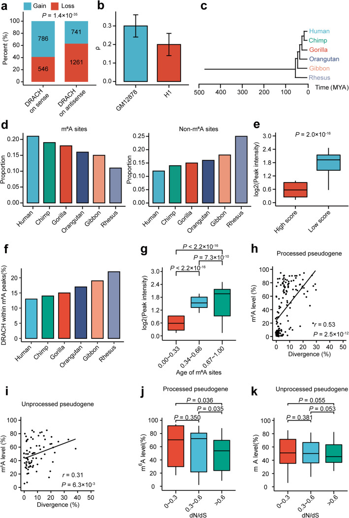
If de novo formation of m6A motifs on pseudogenes is under positive natural selection, we would expect to see a higher probability of obtaining novel m6A motifs than losing m6A motifs on pseudogenes in the evolution. To test this, we used the m6A motifs on the antisense strand as a neutral background, because of equal natural mutation probabilities on both strands. As shown in Fig. 2a, a significantly higher proportion of pseudogene on sense strand than on antisense strand has gained m6A motifs, supporting positive natural selection of the gained m6A motifs on pseudogenes (P = 1.4× 10⁻³⁵, two-tailed chi-square test).
Fig. 2.
Positive natural selection of the m6A on pseudogenes in GM12878 cell line. a Stacked barplot comparing the proportions of pseudogenes gaining/losing m6A motif (DRACH) on sense and antisense strands respectively. b Bar plot showing the estimates of ρ for DRACH m6A motifs for GM12878 and H1 cells respectively. Error bars represent standard errors. c A schematic phylogeny of the species studied for age determination. d The distributions of the ages of m6A sites (left) and control non-m6A DRACH sites (right). The age of each site was based on the most distantly related species in which the site was conserved. e Box plot comparing the peak intensities between m6A sites with high (top 25%, strong constraint) and low (bottom 25%, weak constraint) rejected substitution scores. f Barplot showing the percentages of DRACH motifs within m6A peaks of GM12878 cells for DRACH motif of different ages. g Box plot comparing the peak intensities of m6A sites with different normalized ages (1 represents the oldest, while 0 represents the youngest) of pseudogenes. h, i Scatter plots showing the correlation of the sequence divergences between pseudogenes and their cognate protein-coding genes with the m6A levels of pseudogenes for processed pseudogenes (h) and unprocessed pseudogene (i), respectively. j, k Box plot comparing m6A level of pseudogenes with different ranges of dN/dS ratios of their cognate coding genes for processed (j) and unprocessed (k) pseudogenes, respectively
To further elucidate the evolutionary landscape of the m6A sites on pseudogenes, we took advantage of INSIGHT (Inference of Natural Selection from Interspersed Genomically coHerent elemenTs) to estimate ρ, which represents the proportion of negatively selected m6A sites on pseudogenes. Our estimate of ρ of the m6A sites on pseudogenes was 0.2 ~ 0.3 (Fig. 2b), lower than the previously estimated ρ of 0.33~0.56 for the m6A sites on 3′UTRs of mRNAs [33], suggesting that the m6A on pseudogenes are less conserved. In addition, the estimated ages of the m6A sites, based on a phylogenetic tree of human and representative non-human primates (Fig. 2c), were much younger than the non-m6A “DRACH motifs” on pseudogenes, suggesting that the birth rate of m6A sites is faster than random drift in primates evolution (Fig. 2d). We then calculated the rejected substitution scores, which measure the nucleotide-level constraint [34], to study the relationship between m6A and site conservation. We found that the less conserved m6A sites had significantly higher m6A peak intensities than conserved m6A sites (Fig. 2e). These results are consistent with our finding that there is a selection pressure of gaining new m6A motifs on pseudogenes, further supporting that m6A sites on pseudogenes are positively selected.
We were then interested in whether evolutionarily older pseudogenes tended to have stronger m6A modification. Based on the above calculated ages of these m6A sites, we found that the older the “DRACH” motifs are, the higher proportions of them are within detectable m6A peaks in GM12878 cells (Fig. 2f). In contrast to that only 13% of human-specific “DRACH” motifs were detected in m6A peaks in GM12878 cells, 22% of human and rhesus shared “DRACH” motifs were within m6A peaks detected in the same cell line. Consistently, we found a significant positive correlation between ages and intensities of m6A peaks (Fig. 2g). These results suggest that generating “DRACH” motifs works as the critical first step, followed by long evolution for them to get strong m6A methylation. In addition, we found a significant positive correlation between the sequence divergences and m6A levels for processed pseudogenes (P = 2.5× 10⁻¹², Pearson correlation) and unprocessed pseudogenes (P = 0.006, Pearson correlation) respectively (Fig. 2h, i). Considering that the peak intensities of m6A peaks are overall conserved between human and mouse [35], the above results support that pseudogenes especially processed pseudogenes were originally methylated at a low level but evolved to have high methylation level quickly. Interestingly, highly m6A methylated pseudogenes, especially processed pseudogenes, tend to have cognate protein-coding genes with lower dN/dS ratios (ratio of nonsynonymous substitution rate to synonymous substitution rate), which represent the genes with higher essentiality and stronger functional constraint in the evolution, suggesting that important genes are more likely regulated by the modification of their processed pseudogene transcripts (Fig. 2j, k). On the other hand, we found the highly m6A methylated processed pseudogenes tend to have cognate protein-coding genes with shorter 5′UTRs, longer CDSs and 3′UTRs, lower GC contents, suggesting that these gene features of cognate coding genes may also relate to the m6A evolution of processed pseudogenes (Additional file 1: Figure S4a-d). Similar results were observed in H1 cells (Additional file 1: Figure S5a-l).
m6A facilitates the cytosolic degradation of processed pseudogenes
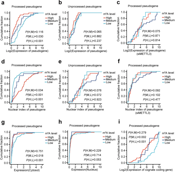
In order to understand the evolutionary pressure for pseudogenes to become highly methylated, we explored the functional consequences of m6A methylation on pseudogenes. m6A has been reported to promote nuclear export [24] and cytosolic degradation [21–23], we first tested whether m6A affected the gene expression of pseudogenes using the input of m6A-LAIC-seq data. We found the highly methylated (m6A level ≥ 0.6) (P < 0.001, two-tailed Wilcoxon test) and moderately methylated (0.6 > m6A level ≥ 0.3) (P = 0.030, two-tailed Wilcoxon test) processed pseudogenes had dramatically lower gene expression than lowly methylated (m6A level < 0.3) processed pseudogenes (Fig. 3a) in GM12878 cells, suggesting that m6A promotes the degradation of processed pseudogenes. Nevertheless, we did not observe a similar result for unprocessed pseudogenes (Fig. 3b), suggesting that the m6A-induced cytosolic degradation might be a specific function to processed pseudogenes. To further confirm whether different expression of processed pseudogenes is due to m6A, we sequenced the input, cytoplasmic, and nuclear RNAs of control and METTL3-knockdown GM12878 cells. Expression differences among the processed pseudogenes with different categories of m6A levels were observed in control cells (Additional file 1: Figure S6a). However, the differences became not significant in METTL3-knockdown cells, indicating the expression differences of processed pseudogenes are dependent on m6A (Fig. 3c).
Fig. 3.
m6A facilitates the cytosolic degradation of processed pseudogenes in the GM12878 cell line. a, b Plot of cumulative fraction of expression for processed (a) and unprocessed (b) pseudogene with high (m6A level ≥ 0.6), medium (0.6 > m6A level ≥ 0.3), and low (m6A level < 0.3) m6A levels, respectively. Two-tailed Wilcoxon P values are indicated at the bottom right corner. c Plot of cumulative fraction of expression for processed pseudogenes with high (m6A level ≥ 0.6), medium (0.6 > m6A level ≥ 0.3), and low (m6A level < 0.3) m6A levels respectively in METTL3 knockdown cells. Two-tailed Wilcoxon P values are indicated at the bottom right corner. d, e Plot of cumulative fraction of nuclear indexes (ratio of expression between the nucleus and cytosol) for processed (d) and unprocessed (e) pseudogenes with high, medium, and low m6A levels, respectively. Two-tailed Wilcoxon P values are indicated at the bottom right corner. f Plot of cumulative fraction of nuclear indexes for processed pseudogenes with high, medium, and low m6A levels respectively in METTL3 knockdown cells. Two-tailed Wilcoxon P values are indicated at the bottom right corner. g, h Plot of cumulative fraction of cytosolic (g) and nuclear (h) gene expression for processed pseudogene with high, medium, and low m6A levels respectively. Two-tailed Wilcoxon P values are indicated at the bottom right corner. i Plot of cumulative fraction of expression of cognate protein-coding genes for processed pseudogene with high, medium, and low m6A levels respectively. Two-tailed Wilcoxon P values are indicated at the top left corner and bottom right corner, respectively
To test whether m6A facilitates the nuclear export of pseudogene RNAs, we took advantage of the CSHL RNA-seq of separated cytosol and nucleus RNAs in GM12878 and H1 cell lines [36]. We found the highly methylated processed pseudogenes had dramatically higher nuclear indexes (ratio of expression between nucleus and cytosol) than moderately methylated (P = 0.034, two-tailed Wilcoxon test) and lowly methylated processed pseudogenes (P < 0.001, two-tailed Wilcoxon test) (Fig. 3d) in GM12878 cell line, suggesting stronger nuclear retention or cytosolic depletion of m6A methylated processed pseudogenes. We did not observe a similar result for unprocessed pseudogenes (Fig. 3e), which is consistent with the above result that the m6A of processed other than unprocessed genes is negatively correlated with gene expression (Fig. 3a, b). Similar results for processed pseudogenes were also observed in control GM12878 cells (Additional file 1: Figure S6b), but the differences were not significant in METTL3-knockdown cells, indicating that the nuclear index differences of processed pseudogenes are due to m6A differences (Fig. 3f). Because m6A has been reported to promote the nuclear export other than nuclear retention of RNAs [21], m6A likely facilitates the cytosolic degradation of processed pseudogenes. We then found the m6A of processed pseudogenes were negatively correlated with the gene expression in cytosol (Fig. 3g), but not significant with the gene expression in nucleus (Fig. 3h), strongly supporting the role of m6A on degrading the processed pseudogenes in cytosol.
Interestingly, we also found that the m6A levels of processed other than unprocessed pseudogenes were negatively correlated with the gene expression of their cognate protein-coding genes (Fig. 3i; Additional file 1: Figure S6c) and positively correlated with the nuclear indexes of their cognate protein-coding genes (Additional file 1: Figure S6d, e), suggesting that the m6A on the RNAs of pseudogenes may affect mRNA expression of their cognate protein-coding genes. Similar results were observed in H1 cells (Additional file 1: Figures S7a-f; S8a-d).
m6A of processed pseudogenes disrupt their crosstalk with their cognate protein-coding genes
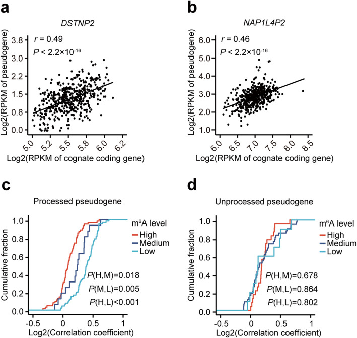
To further address whether m6A of pseudogenes affects the crosstalk between pseudogenes and their cognate protein-coding genes, we analyzed the RNA-seq data of B-lymphoblastoid cell lines (BLCL), the same cell type as GM12878, from 462 European participants of 1000 genome project [37, 38]. We found that 164 out of 223 pairs of pseudogenes and cognate protein-coding genes showed significant correlations (FDR < 0.05) of gene expressions, 74% of them are positive correlations, which is consistent with the positive regulatory roles of ceRNAs (Examples of positive correlations are shown in Fig. 4a, b).
Fig. 4.
m6A of processed pseudogenes disrupt their crosstalk with their cognate protein-coding genes. a, b Scatter plots showing the correlation of the expressions of two representative processed pseudogenes DSTNP2 (a) and NAP1L4P2 (b) with their cognate protein-coding genes among 462 B-lymphoblastoid cell lines. P values of Pearson correlations are indicated at the top left corners. c, d Plot of cumulative fraction of correlation coefficients of gene expressions for processed (c) and unprocessed (d) pseudogenes with their cognate protein-coding genes among 462 B-lymphoblastoid cell lines. Pseudogenes with high, medium, and low m6A levels are shown separately. Two-tailed Wilcoxon P values are indicated at the bottom right corners
To test whether the m6A of pseudogenes affects the crosstalk between pseudogenes and their cognate protein-coding genes, we compared the correlation coefficients of the gene expressions between pseudogenes and their cognate protein-coding genes in three categories of gene pairs with different m6A levels of pseudogenes. We found that the processed pseudogenes with higher m6A levels had significantly lower correlation coefficients (P = 0.018, two-tailed Wilcoxon test) (Fig. 4c), suggesting that m6A of processed pseudogenes disrupt their crosstalk with their cognate protein-coding genes. However, we did not observe a similar result for unprocessed pseudogenes (Fig. 4d).
Experimental validation of the m6A on two processed pseudogenes reduce their crosstalk with their cognate protein-coding genes via promoting the decay of pseudogenes
To experimentally test whether the m6A of pseudogenes affects the expression of their cognate protein-coding genes, we selected two representative processed pseudogenes DSTNP2 and NAP1L4P1 for further validation in GM12878 cell line.
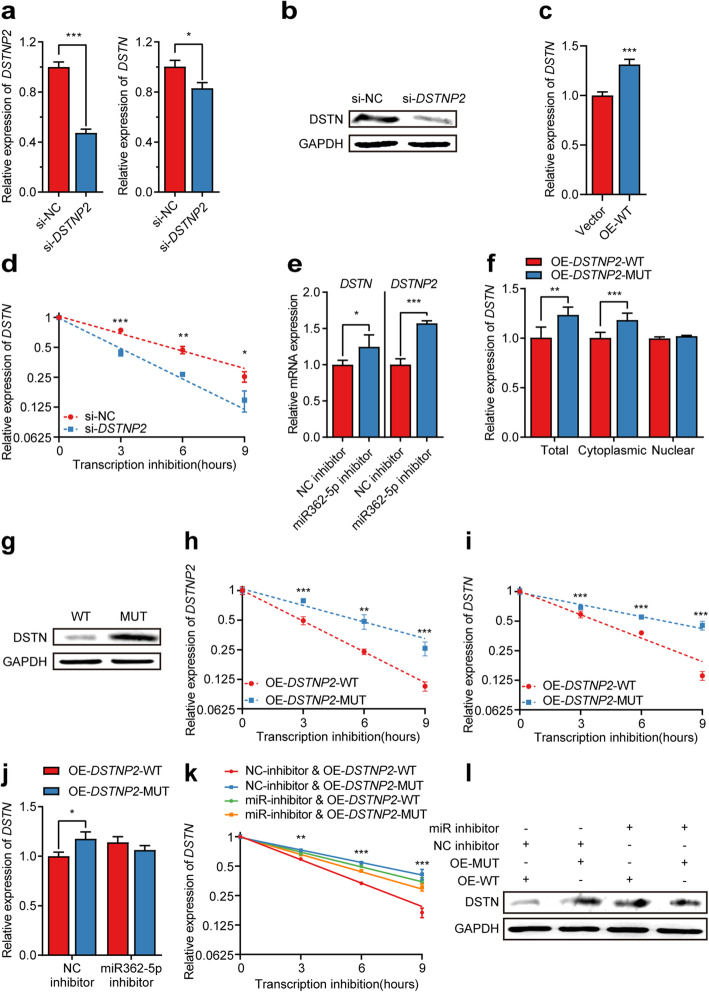
First, we tested whether DSTNP2 can regulate its protein-coding cognate DSTN. We found knockdown of DSTNP2 by siRNA significantly down-regulated mRNA as well as the protein expression of DSTN (Fig. 5a, b; Additional file 1: Figure S10a), while overexpression of DSTNP2 significantly upregulated the expression of DSTN (Fig. 5c), consistent with our observation that their gene expressions were positively correlated in a population of B-lymphoblastoid cell lines (Fig. 4a). In order to test whether DSTNP2 modules DSTN via ceRNA mechanism, we first confirmed that knockdown of DSTNP2 could promote the degradation of DSTN (Fig. 5d). We then predicted the miRNA binding sites and found 6 miRNAs potentially targeting both DSTNP2 and DSTN (Additional file 1: Figure S9a). We selected miR-362-5p for experimental validation due to their higher expression level according to ENCODE miRNA-seq data of GM12878 [39]. We found inhibition of miR-362-5p significantly increased the expression of both DSTNP2 and DSTN, indicating that miR-362-5p targets both DSTNP2 and DSTN (Fig. 5e).
Fig. 5.
Experimental validation of m6A on processed pseudogene DSTNP2 affects the expression of DSTN in GM12878 cell line. a Relative gene expression of DSTNP2 and DSTN after DSTNP2 knockdown using siRNA. * P< 0.05; *** P< 0.001 (two-tailed t test). b Western blot comparing the protein levels of DSTN in control and DSTNP2 knockdown cells. c The relative expression of DSTN upon overexpression empty vector and wild type DSTNP2 respectively. Error bars represent standard errors. *** P< 0.001 (two-tailed t test). d The relative expression of DSTN at different time points after transcription inhibition in control and DSTNP2 knockdown cells respectively. Error bars represent standard errors. * P< 0.05; ** P< 0.01; *** P< 0.001 (two-tailed t test). e The relative expression of DSTN and DSTNP2 after adding control inhibitor versus miR362-5p inhibitor. f Bar plot comparing the relative expression of total, cytoplasmic and nuclear DSTN respectively upon overexpression of wild type versus m6A sites-mutated DSTNP2. g Western blot comparing the protein levels of DSTN upon overexpression of wild type DSTNP2 versus m6A site-mutated DSTNP2. h, i The relative expression of DSTNP2 (h) and DSTN (i) upon overexpression of wild type DSTNP2 versus m6A site-mutated DSTNP2 at different time points after transcription inhibition. Error bars represent standard errors. ** P < 0.01; *** P < 0.001 (two-tailed t test). j The relative expression of DSTN upon overexpression of wild-type DSTNP2 versus m6A site-mutated DSTNP2 after adding control inhibitor and miR362-5p inhibitor respectively. Error bars represent standard errors. * P < 0.05 (two-tailed t test). k The relative expression of DSTN at different time points after transcription inhibition upon overexpression of wild-type DSTNP2 versus m6A site-mutated DSTNP2 after adding control inhibitor and miR362-5p inhibitor respectively. Error bars represent standard errors. ** P< 0.01; *** P< 0.001. l Western blot showing the protein level of DSTN upon overexpression of wild type DSTNP2 versus m6A site-mutated DSTNP2 after adding control inhibitor and miR362-5p inhibitor, respectively
To test whether the m6A of DSTNP2 affects the crosstalk between DSTNP2 and DSTN, we mutated all the 8 m6A sites of DSTNP2 without disrupting the binding sites of miR-362-5p (Additional file 1: Figure S9b), and then overexpressed the wild type and mutant respectively into GM12878 cell line. Compared with wild-type DSTNP2, we found overexpressing DSTNP2 mutant resulted in significantly higher mRNA as well as protein expression of DSTN (Fig. 5f, g; Additional file 1: Figure S10b). We then separated the cytosolic and nuclear RNAs and found the mutations of DSTNP2 m6A sites specifically affected the expression of cytosolic RNAs of DSTN (Fig. 5f). To test whether the m6A of DSTNP2 affects the stabilities of cytoplasmic RNAs, we measured the degradation rates of DSTNP2 and DSTN RNAs. We found that DSTNP2 mutant had significantly higher stability than wild type DSTNP2 (Fig. 5h), and overexpression of DSTNP2 mutant resulted in significantly higher stability of its cognate protein-coding gene DSTN (Fig. 5i; Additional file 1: Figure S10c), suggesting that the m6A of DSTNP2 promotes the degradation of DSTNP2 and indirectly decreases the stability of DSTN. When miR-362-5p was inhibited, overexpression of wild-type and mutant DSTNP2 did not show significantly different effects on the gene expression, RNA stability, and protein expression of DSTN, suggesting that m6A mediated degradation of DSTNP2 reduced the ceRNA effects of DSTNP2 on DSTN (Fig. 5j–l).
Similar results were observed for the other processed pseudogene NAP1L4P1. NAP1L4P1 can also upregulate the expression of NAP1L4 (Additional file 1: Figure S11a, b). Overexpression of NAP1L4P1 m6A-mutant resulted in significantly higher cytoplasmic abundance as well as stability of its cognate protein-coding gene NAP1L4 (Additional file 1: Figure S11c-e), suggesting that the m6A of NAP1L4P1 promotes the degradation of NAP1L4P1 and indirectly decreases the stability of NAP1L4. However, we did not succeed in identifying the miRNA that mediates this process.
Discussion
In this study, we found convergent evidences supporting the adaptive accumulation of m6A sites on human pseudogenes through mutations in the evolution, resulted in higher m6A levels on the RNAs of pseudogenes. Through integrating with public dataset, we realized the m6A on the RNAs of processed other than unprocessed pseudogenes promoted cytosolic RNA degradation and reduced their regulatory effects on their cognate mRNAs mediated by ceRNA mechanism. Our discovery revealed a novel evolutionary role of m6A in cleaning up the unnecessary RNAs of processed pseudogenes to attenuate the interrupting of the regulatory pathway of miRNA on mRNAs. The novel finding in this study also unveiled a mystery of how cells clean transcribed processed pseudogenes through splicing-independent mechanisms.
In general, pseudogenes are nonfunctional, and evolutionarily neural, KA/KS test of pseudogenes indicates low functional constraint of their protein-coding ability [40]. However, carrying a piece of useless DNA is deleterious because it costs energy, there is a tendency of losing pseudogenes in the evolution and the existing pseudogenes tend to be young [41, 42]. On the other hand, transcription of pseudogenes may not be as neutral as previously thought either. It was reported that only half of human transcribed pseudogenes were conserved in rhesus macaque and only 3% of them were conserved in mouse [43], indicating a trend of rapidly losing transcribed pseudogenes in the evolutionary history. Because the sequences of pseudogenes are quite similar as their cognate protein-coding genes, RNA binding proteins and miRNAs may not be able to distinguish them, therefore the transcribed pseudogenes may interrupt the existing regulatory network through mechanisms such as competitive endogenous RNAs [44]. Since most transcribed pseudogenes are evolutionarily young, they are not likely to have obtained important functions, most of these interruptions should be deleterious and degradation of them might be adaptive. For these transcribed pseudogenes, splicing dependent NMD mechanism can recognize the unprocessed pseudogenes and trigger the RNA degradation in the cytoplasm. However, processed pseudogenes cannot degrade through the canonical NMD pathway due to the lack of splicing. In this study, we found that the processed pseudogenes took advantage of the m6A-mediated cytosolic RNA degradation mechanism to get rid of these interrupting RNAs in the evolution. We also found that these methylated m6A motifs were evolutionarily younger than those unmethylated m6A motifs, and there was a specific selective pressure to obtain m6A motif on these pseudogenes, indicating positive selection of m6A on the transcribed pseudogenes.
Though these competitive endogenous RNAs transcribed from pseudogenes are deleterious at the early stage of evolution, it is also possible that some of them become functionally important. For example, the lncRNA Oct4P4 plays an important role in inducing and maintaining the silencing of the ancestral Oct4 gene in differentiating mouse embryonic stem cells (mESCs) [45]. The cellular 5S rRNA pseudogene transcripts, which are unshielded following depletion of their respective binding proteins by the virus, induces RIG-I-mediated antiviral immunity [46]. The TUSC2P (tumor suppressor candidate-2 pseudogenes) promotes TUSC2 function by binding multiple microRNAs, and ectopic expression of TUSC2P and TUSC2 inhibits cell proliferation, survival, migration, invasion, and colony formation, and increases tumor cell death [47]. Pseudogenes regulate mRNAs and lncRNAs via the piRNA pathway in the germline [48]. The Pan-Cancer analysis of pseudogene expression has showed that pseudogenes can be a new paradigm for investigating cancer mechanisms and discovering prognostic biomarkers [49]. In principle, there could be widespread pseudogenes that interfere with the regulatory networks during the long evolutionary history, and thus, important functions of pseudogenes could have the opportunity to get evolved.
Here, we found a novel mechanism of RNA surveillance for processed pseudogenes via m6A RNA methylation. How does this mechanism evolve? In this study, we found a significantly higher probability of obtaining novel m6A motifs on sense strand than antisense strand, strongly suggesting that obtaining m6A sites on pseudogenes tends to be adaptive and accumulating in the evolution. In this way, the elevated m6A on processed pseudogenes are the results of positively selected mutations that produce novel m6A sites. Alternatively, we cannot rule out that there is an unknown specific mechanism, such as RBP-mediated specific regulation of m6A [20, 50], to recognize processed pseudogenes and mark them for cytosolic degradation by m6A RNA modification. However, whether and how the cells mark processed pseudogenes is still unknown and requires further investigation.
The roles of m6A played in evolution especially human evolution are still elusive. Ma et al. reported that newly acquired m6A modifications in humans were under positive selection [51]. However, Liu et al. had contradictory viewpoint that these newly acquired m6A sites were neutral and most m6A sites in protein-coding regions were nonfunctional and nonadaptive [52]. Recently, Zhang et al. reported that a significant fraction of 3′ UTR m6A sites were under negative selection and recently gained 3′ UTR m6A in humans were positively selected [33]. Our study further indicates that m6A on human transcribed pseudogenes are evolutionarily young and evolved under positive selection, indicating that m6A plays more important and widespread roles than expected in human evolution.
Promoting cytosolic RNA degradation is an important molecular role of m6A RNA modification, it plays important roles in diverse physiology process which need to remove existing RNAs. It removes the RNAs of current cell status to facilitate cell fate transition [35]; it cleans the maternal RNAs during the maternal-to-zygotic transition [53]. Removing harmful transcribed pseudogenes is a novel role of m6A mediated RNA decay. In addition, nonsense mutations and frame-shift mutations that occur on intronless protein-coding genes can also produce poisonous truncated proteins, and we found the RNAs of protein-coding genes with single exons and the limited number of exons also showed significantly higher m6A levels than genes with a larger number of exons, suggesting that m6A-mediated RNA degradation may also play roles for RNA surveillance of protein-coding genes with single or a few of exons (Additional file 1: Figure S12a-d).
Conclusions
Our discovery reveals a novel evolutionary role of m6A RNA modification in cleaning up the unnecessary processed pseudogene transcripts to attenuate their interfering with the regulatory network of mRNAs. It provides novel aspect on the importances of m6A in the evolution.
Methods
Data sources
The list of protein-coding genes and corresponding pseudogenes were compiled from GENCODE, Vega, and Pseudogene.org databases [54]. As described, the annotated pseudogenes must have at least one of the disablements, including premature stop codon, frame-shift, truncation at 5′ or 3′ end of CDS, deletion of an internal portion of CDS, and lack of locus-specific transcriptional evidence for process pseudogene [55]. The SNPs of GM12878 were obtained from Genome in a Bottle (GIAB) Consortium [56]. The raw data of nucleus and cytosol RNA-seq of GM12878 and H1 were obtained from ENCODE project [36]. The RNA-seq data of the lymphoblastoid cell lines of 462 individuals from the 1000 Genomes Project were obtained from the Geuvadis project [57]. The m6A-LAIC-seq data of GM12878 and H1 hESC cell lines were obtained from our previous publication [28]. The m6A-seq data of GM12878 cell line [32] and H1 hESC [35] were also obtained from previous publications. The ratio of dN/dS and GC contents were obtained from BioMart database [58]. miRNA binding targets on pseudogenes were obtained from the dreamBase database [59]. miRNA binding targets to protein-coding genes were obtained from the miRWalk database [60]. The microRNA-seq data of GM12878 cells were obtained from ENCODE project [39] (GEO accession: GSE143080).
Processing of sequencing data
The RNA-seq, m6A-LAIC-seq, and m6A-seq reads were aligned to hg19 human genome using Hisat2 [29], known SNPs from the dbSNP database (GM12878 SNPs only for GM12878 data) were provided, and the mismatches at SNP loci were tolerated in the mapping. Only the reads with perfect match except for mismatches at SNPs were allowed. The proper paired and uniquely mapped reads were used for the downstream analyses. To evaluate the accuracy of this alignment procedure, we compared it with the conventional alignment procedure with default parameters of Hisat2.
Gene expression analyses
RPKMs of genes were calculated using StringTie2 [61]. The raw data of nucleus and cytosol RNA-seq of GM12878 and H1 were remapped using the above reprocessing procedure, the RPKMs of nucleus and cytosol were normalized as described in the original paper [36], then the nuclear indexes were calculated as the ratio between normalized RPKMs of nucleus and cytosol.
m6A analyses
We recalculated the m6A levels of all annotated genes including the compiled pseudogenes based on the reprocessed m6A-LAIC-seq data of GM12878 and H1 cells according to the method described in our previously published paper [28]. The m6A peaks were identified based on the reprocessed m6A-seq data of GM12878 [32] and H1 [35] cells according to our previously described method [35]. The single-nucleotide m6A sites were determined by combining the m6A sites predicted by sequence-based m6A site predictors SRAMP [62] and Whistle [63] within m6A peaks regions. The longest transcript of each gene was used in the analyses of gene features.
Evolution analyses
To compare the m6A motifs between pseudogenes and their cognate protein-coding genes, we aligned the pseudogenes and their cognate protein-coding genes using ClustalW2 [64] with default parameters. The DRACH m6A motifs on pseudogenes and their cognate protein-coding genes were counted respectively within the aligned regions covered by m6A peaks of either pseudogenes or their cognate protein-coding genes. The DRACH motifs on the antisense strand were counted in the same way as the background.
The SRAMP [62] and Whistle [63] predicted single-nucleotide m6A sites within m6A peaks of pseudogenes were used to study the natural selection of m6A sites on pseudogenes. We used INSIGHT (Inference of Natural Selection from Interspersed Genomically coHerent elemenTs) [65] to estimate the proportion of m6A sites that are under negative selection. The rejected substitution scores calculated by GERP++ [66] were used to measure the nucleotide-level functional constraint of all m6A sites. The ages of individual m6A sites were determined using a phylostratigraphy approach [67] with pairwise alignments downloaded from the UCSC genome browser, the DRACH motifs outside the m6A peaks on pseudogenes were used as evolutionally neutral control sites.
Cell culture, lentiviral production, and transduction
HEK293T (ATCC® CRL-3216™) cells were cultured in high glucose Dulbecco’s modified Eagle’s medium (Corning), supplemented with 10% FBS (Biological Industries) at 37 °C with 5% CO2. GM12878 cells were cultured in Roswell Park Memorial Institute Medium 1640 (Corning), supplemented with 15% FBS (Biological Industries) at 37 °C with 5% CO2. Both cell lines were obtained from GuangZhou Jennio Biotech Co., Ltd., authenticated and tested for the absence of mycoplasma contamination using Myco-Blue Mycoplasma Detector (Vazyme).
For lentiviral production, 293 T cells were seeded in 6 cm cell culture plates, and 24 hours later, cells were transfected with 6.4 μg of lentiviral backbone, 4.8 μg of psPAX2 (Addgene #12260), and 1.6 μg of pMD2.G (Addgene #12259) using LipoFiter reagent (HANBIO). Lentiviral supernatants were harvested at 48 h and 72 h after transfection and filtered through using a 0.45μm PVDF filter (Millipore) and concentrated using PEG. 5 × 10⁴ GM12878 cells were seeded in a TC-untreated plate and transduced with viral supernatants in the presence of polybrene (8 μg/μL). Twenty-four hours after transduction, cells were selected with puromycin (2 μg/mL).
Construction of plasmid DNA
Wild type and m6A motif mutant DNA sequence of DSTNP2 and NAP1L4P1 genes were synthesized by GENEWIZ company. Lentiviral expression plasmids were generated using ClonExpress II One Step Cloning Kit (Vazyme, C112), by combining PCR-amplified cDNA and EcoR I/BamH I digested pCDH-CMV-MCS-EF1α-CopGFP-T2A-Puro (SBI) backbone.
miRNA inhibitor and siRNA transfection
Cells were seeded in TC-untreated plates and transfected with specific miRNA inhibitors and inhibitor NC or siNC and specific siRNA using LipoFiter reagent (HANBIO). RNA samples and protein samples were harvested at 72 h after transfection for qRT-PCR.
Immunoblotting
Proteins were extracted from cells by incubating with RIPA buffer (Cell Signaling Technology, Cat. 9806) on ice for 10 min and insoluble fraction was removed by centrifugation. Twenty micrograms of extracted protein was separated on 15% SDS-PAGE and transferred to PVDF membrane. Membranes were blocked in 5% BSA in Tris-Buffered Saline with 0.01% Tween 20 (TBS-T) at room temperature for 1 h and incubated overnight with primary antibodies diluted in 1% BSA/TBS-T at 4 °C, followed by incubating with HRP conjugated secondary antibody diluted in TBS-T for 1 h at room temperature, and visualized using Clarity™ Western ECL Substrate (Bio-Rad). The following antibodies were used for immunoblotting: DSTN (1:1000, Abcam, ab192262), GAPDH (1:1000, ab8245).
RNA extraction and real-time quantitative PCR (qPCR)
Total RNA was extracted using the NucleoZol RNA reagent (MACHEREY-NAGEL). And fraction RNA was separated using the Cytoplasmic and Nuclear RNA Purification Kit (NORGEN). One microgram of DNA-free RNA was then reverse-transcribed using HiScript III RT SuperMix for qPCR (+gDNA wiper) (Vazyme, R232). qPCR was carried out using the ChamQ Universal SYBR qPCR Master Mix (Vazyme, Q711) and performed in an LC480 Real-Time PCR System (Roche). Fold-change was calculated using the 2-∆∆CT method. The primers of pseudogenes and their cognate protein-coding genes, which had been appended in Additional file 2: Table S4, were designed at the places with sequence divergences (Additional file 1: Figure S8d).
RNA sequencing
RNA-seq libraries of the input, cytoplasmic, and nuclear RNAs of GM12878 with and without METTL3 knockdown were prepared using the VAHTS® mRNA-seq V2 Library Prep Kit for Illumina from Vazyme and sequenced on Illumina HiSeq 2500 platform to generate 150 bp paired-end reads.
RNA stability assay
The final concentration of 5 μg/mL Actinomycin D (Sigma, A9415) was added to cells to assess RNA stability. After incubation for indicated time points, the cells were collected, and RNA samples were extracted for reverse transcription and qPCR. 18S was used as the reference gene and fold-change was calculated using the 2-∆∆CT method.
Supplementary Information
Additional file 1: Figure S1. The optimization of conventional HISAT2 mapping procedure. Figure S2. Comparisons of m6A between pseudogenes and their cognate protein-coding genes in GM12878 and H1 cell line. Figure S3. Comparisons of m6A between pseudogenes and their cognate protein-coding genes in H1 cell line. Figure S4. Comparisons of pseudogenes m6A among 5′UTRs, CDSs, 3′UTRs and GC contents in their cognate protein-coding genes in GM12878 cell line. Figure S5. Positive natural selection of the m6A on pseudogenes in H1 cell line. Figure S6. m6A facilitates the cytosolic degradation of cognate protein-coding genes of processed pseudogenes in GM12878 cell line. Figure S7. m6A facilitates the cytosolic degradation of processed pseudogenes in H1 cell line. Figure S8. m6A facilitates the cytosolic degradation of cognate protein-coding genes of processed pseudogenes in H1 cell line. Figure S9. The gene regulatory network and primer design principles of processed pseudogenes and cognate protein-coding genes. Figure S10. The uncropped bots of western blots in Fig. 5. Figure S11. Experimental validation of m6A on processed pseudogene NAP1L4P1 affects the expression of NAP1L4 in GM12878 cell line. Figure S12. Correlation between m6A levels and exon numbers of coding genes.
Additional file 2: Table S1. The list of pseudogenes and their cognate protein-coding genes. Table S2. The m6A levels of pseudogenes and their cognate protein-coding genes in GM12878 cell line. Table S3. The m6A levels of pseudogenes and their cognate protein-coding genes in H1 cell line. Table S4. The primers of processed pseudogenes (DSTNP2; NAP1L4P1) and their cognate protein-coding genes (DSTN; NAP1L4). Table S5. The list of collected GEO datasets. Table S6. The list of collected EBI datasets.
Acknowledgements
We are grateful to Prof. Rui Zhang, Prof. Xionglei He, Prof. Yi Xing, and Prof. Jianrong Yang for their constructive discussion and comments.
Peer review information
Andrew Cosgrove was the primary editor of this article and managed its editorial process and peer review in collaboration with the rest of the editorial team.
Review history
The review history is available as Additional file 3.
Authors’ contributions
J.W. conceived and designed the project. L.T. performed bioinformatics analyses. W.C., F.L., and L.W. performed experiments. J.W., L.T., and W.C. wrote the paper. N.C and D.O.W supervised parts of the project. All authors read and approved the final manuscript.
Funding
This study was supported by the National Key R&D Program of China 2018YFA0107200 (JW), the National Natural Science Foundation of China 31771446 (JW), 31970594 (JW), and 31971335 (DOW), Guangzhou Science and Technology Program 201904010181 (JW), AMED 18 dm 0307023 h 0001 (DOW).
Availability of data and materials
The raw sequence data have been deposited in the GEO dataset under the accession number GSE172219 [68]. The accession numbers and links of third-party high-throughput sequencing data obtained from the GEO and EBI database were listed in Additional file 2: Tables S5 and S6, respectively.
Declarations
Ethics approval and consent to participate
Not applicable.
Consent for publication
Not applicable.
Competing interests
The authors declare that they have no competing interests.
Footnotes
Publisher’s Note
Springer Nature remains neutral with regard to jurisdictional claims in published maps and institutional affiliations.
Liqiang Tan and Weisheng Cheng contributed equally to this work.
References
- 1.Brogna S, Wen J. Nonsense-mediated mRNA decay (NMD) mechanisms. Nat Struct Mol Biol. 2009;16(2):107–113. doi: 10.1038/nsmb.1550. [DOI] [PubMed] [Google Scholar]
- 2.Wolin SL, Maquat LE. Cellular RNA surveillance in health and disease. Science. 2019;366(6467):822–827. doi: 10.1126/science.aax2957. [DOI] [PMC free article] [PubMed] [Google Scholar]
- 3.Zhang S, Ruiz-Echevarria MJ, Quan Y, Peltz SW. Identification and characterization of a sequence motif involved in nonsense-mediated mRNA decay. Mol Cell Biol. 1995;15(4):2231–2244. doi: 10.1128/MCB.15.4.2231. [DOI] [PMC free article] [PubMed] [Google Scholar]
- 4.Hogg JR, Goff SP. Upf1 senses 3'UTR length to potentiate mRNA decay. Cell. 2010;143(3):379–389. doi: 10.1016/j.cell.2010.10.005. [DOI] [PMC free article] [PubMed] [Google Scholar]
- 5.Neu-Yilik G, Gehring NH, Thermann R, Frede U, Hentze MW, Kulozik AE. Splicing and 3' end formation in the definition of nonsense-mediated decay-competent human beta-globin mRNPs. EMBO J. 2001;20(3):532–540. doi: 10.1093/emboj/20.3.532. [DOI] [PMC free article] [PubMed] [Google Scholar]
- 6.Maquat LE, Li X. Mammalian heat shock p70 and histone H4 transcripts, which derive from naturally intronless genes, are immune to nonsense-mediated decay. RNA. 2001;7(3):445–456. doi: 10.1017/S1355838201002229. [DOI] [PMC free article] [PubMed] [Google Scholar]
- 7.He F, Li X, Spatrick P, Casillo R, Dong S, Jacobson A. Genome-wide analysis of mRNAs regulated by the nonsense-mediated and 5' to 3' mRNA decay pathways in yeast. Mol Cell. 2003;12(6):1439–1452. doi: 10.1016/S1097-2765(03)00446-5. [DOI] [PubMed] [Google Scholar]
- 8.Mitrovich QM, Anderson P. mRNA surveillance of expressed pseudogenes in C. elegans. Curr Biol. 2005;15(10):963–967. doi: 10.1016/j.cub.2005.04.055. [DOI] [PubMed] [Google Scholar]
- 9.Kalyana-Sundaram S, Kumar-Sinha C, Shankar S, Robinson DR, Wu YM, Cao X, Asangani IA, Kothari V, Prensner JR, Lonigro RJ, Iyer MK, Barrette T, Shanmugam A, Dhanasekaran SM, Palanisamy N, Chinnaiyan AM. Expressed pseudogenes in the transcriptional landscape of human cancers. Cell. 2012;149(7):1622–1634. doi: 10.1016/j.cell.2012.04.041. [DOI] [PMC free article] [PubMed] [Google Scholar]
- 10.Poliseno L, Pandolfi PP. PTEN ceRNA networks in human cancer. Methods. 2015;77-78:41–50. doi: 10.1016/j.ymeth.2015.01.013. [DOI] [PubMed] [Google Scholar]
- 11.Karreth FA, Reschke M, Ruocco A, Ng C, Chapuy B, Leopold V, Sjoberg M, Keane TM, Verma A, Ala U, et al. The BRAF pseudogene functions as a competitive endogenous RNA and induces lymphoma in vivo. Cell. 2015;161(2):319–332. doi: 10.1016/j.cell.2015.02.043. [DOI] [PMC free article] [PubMed] [Google Scholar]
- 12.Chiefari E, Iiritano S, Paonessa F, Le Pera I, Arcidiacono B, Filocamo M, Foti D, Liebhaber SA, Brunetti A. Pseudogene-mediated posttranscriptional silencing of HMGA1 can result in insulin resistance and type 2 diabetes. Nat Commun. 2010;1(1):40. doi: 10.1038/ncomms1040. [DOI] [PubMed] [Google Scholar]
- 13.Chan WL, Yuo CY, Yang WK, Hung SY, Chang YS, Chiu CC, Yeh KT, Huang HD, Chang JG. Transcribed pseudogene psi PPM1K generates endogenous siRNA to suppress oncogenic cell growth in hepatocellular carcinoma. Nucleic Acids Res. 2013;41(6):3734–3747. doi: 10.1093/nar/gkt047. [DOI] [PMC free article] [PubMed] [Google Scholar]
- 14.Korneev SA, Park JH, O'Shea M. Neuronal expression of neural nitric oxide synthase (nNOS) protein is suppressed by an antisense RNA transcribed from an NOS pseudogene. J Neurosci. 1999;19(18):7711–7720. doi: 10.1523/JNEUROSCI.19-18-07711.1999. [DOI] [PMC free article] [PubMed] [Google Scholar]
- 15.Chen X, Wan L, Wang W, Xi WJ, Yang AG, Wang T. Re-recognition of pseudogenes: From molecular to clinical applications. Theranostics. 2020;10(4):1479–1499. doi: 10.7150/thno.40659. [DOI] [PMC free article] [PubMed] [Google Scholar]
- 16.Pavlicek A, Gentles AJ, Paces J, Paces V, Jurka J. Retroposition of processed pseudogenes: the impact of RNA stability and translational control. Trends Genet. 2006;22(2):69–73. doi: 10.1016/j.tig.2005.11.005. [DOI] [PMC free article] [PubMed] [Google Scholar]
- 17.Zaccara S, Ries RJ, Jaffrey SR. Reading, writing and erasing mRNA methylation. Nat Rev Mol Cell Biol. 2019;20(10):608–624. doi: 10.1038/s41580-019-0168-5. [DOI] [PubMed] [Google Scholar]
- 18.Shi H, Wei J, He C. Where, When, and How: Context-Dependent Functions of RNA Methylation Writers, Readers, and Erasers. Mol Cell. 2019;74(4):640–650. doi: 10.1016/j.molcel.2019.04.025. [DOI] [PMC free article] [PubMed] [Google Scholar]
- 19.Linder B, Grozhik AV, Olarerin-George AO, Meydan C, Mason CE, Jaffrey SR. Single-nucleotide-resolution mapping of m6A and m6Am throughout the transcriptome. Nat Methods. 2015;12(8):767–772. doi: 10.1038/nmeth.3453. [DOI] [PMC free article] [PubMed] [Google Scholar]
- 20.An S, Huang W, Huang X, Cun Y, Cheng W, Sun X, Ren Z, Chen Y, Chen W, Wang J. Integrative network analysis identifies cell-specific trans regulators of m6A. Nucleic Acids Res. 2020;48(4):1715–1729. doi: 10.1093/nar/gkz1206. [DOI] [PMC free article] [PubMed] [Google Scholar]
- 21.Du H, Zhao Y, He J, Zhang Y, Xi H, Liu M, Ma J, Wu L. YTHDF2 destabilizes m(6)A-containing RNA through direct recruitment of the CCR4-NOT deadenylase complex. Nat Commun. 2016;7(1):12626. doi: 10.1038/ncomms12626. [DOI] [PMC free article] [PubMed] [Google Scholar]
- 22.Zaccara S, Jaffrey SR. A Unified Model for the Function of YTHDF Proteins in Regulating m(6)A-Modified mRNA. Cell. 2020;181(7):1582–1595. doi: 10.1016/j.cell.2020.05.012. [DOI] [PMC free article] [PubMed] [Google Scholar]
- 23.Wang X, Lu Z, Gomez A, Hon GC, Yue Y, Han D, Fu Y, Parisien M, Dai Q, Jia G, Ren B, Pan T, He C. N6-methyladenosine-dependent regulation of messenger RNA stability. Nature. 2014;505(7481):117–120. doi: 10.1038/nature12730. [DOI] [PMC free article] [PubMed] [Google Scholar]
- 24.Roundtree IA, Luo GZ, Zhang Z, Wang X, Zhou T, Cui Y, et al. YTHDC1 mediates nuclear export of N(6)-methyladenosine methylated mRNAs. Elife. 2017;6. 10.7554/eLife.31311. [DOI] [PMC free article] [PubMed]
- 25.Livneh I, Moshitch-Moshkovitz S, Amariglio N, Rechavi G, Dominissini D. The m(6)A epitranscriptome: transcriptome plasticity in brain development and function. Nat Rev Neurosci. 2020;21(1):36–51. doi: 10.1038/s41583-019-0244-z. [DOI] [PubMed] [Google Scholar]
- 26.Huang H, Weng H. Chen J: m(6)A Modification in Coding and Non-coding RNAs: Roles and Therapeutic Implications in Cancer. Cancer Cell. 2020;37(3):270–288. doi: 10.1016/j.ccell.2020.02.004. [DOI] [PMC free article] [PubMed] [Google Scholar]
- 27.Shulman Z, Stern-Ginossar N. The RNA modification N(6)-methyladenosine as a novel regulator of the immune system. Nat Immunol. 2020;21(5):501–512. doi: 10.1038/s41590-020-0650-4. [DOI] [PubMed] [Google Scholar]
- 28.Molinie B, Wang J, Lim KS, Hillebrand R, Lu ZX, Van Wittenberghe N, Howard BD, Daneshvar K, Mullen AC. Dedon P, et al: m(6)A-LAIC-seq reveals the census and complexity of the m(6)A epitranscriptome. Nat Methods. 2016;13(8):692–698. doi: 10.1038/nmeth.3898. [DOI] [PMC free article] [PubMed] [Google Scholar]
- 29.Kim D, Langmead B, Salzberg SL. HISAT: a fast spliced aligner with low memory requirements. Nat Methods. 2015;12(4):357–360. doi: 10.1038/nmeth.3317. [DOI] [PMC free article] [PubMed] [Google Scholar]
- 30.Sisu C, Pei B, Leng J, Frankish A, Zhang Y, Balasubramanian S, Harte R, Wang D, Rutenberg-Schoenberg M, Clark W, Diekhans M, Rozowsky J, Hubbard T, Harrow J, Gerstein MB. Comparative analysis of pseudogenes across three phyla. Proc Natl Acad Sci U S A. 2014;111(37):13361–13366. doi: 10.1073/pnas.1407293111. [DOI] [PMC free article] [PubMed] [Google Scholar]
- 31.Pink RC, Wicks K, Caley DP, Punch EK, Jacobs L, Carter DR. Pseudogenes: pseudo-functional or key regulators in health and disease? RNA. 2011;17(5):792–798. doi: 10.1261/rna.2658311. [DOI] [PMC free article] [PubMed] [Google Scholar]
- 32.Roost C, Lynch SR, Batista PJ, Qu K, Chang HY, Kool ET. Structure and thermodynamics of N6-methyladenosine in RNA: a spring-loaded base modification. J Am Chem Soc. 2015;137(5):2107–2115. doi: 10.1021/ja513080v. [DOI] [PMC free article] [PubMed] [Google Scholar]
- 33.Zhang H, Shi X, Huang T, Zhao X, Chen W, Gu N, Zhang R. Dynamic landscape and evolution of m6A methylation in human. Nucleic Acids Res. 2020;48(11):6251–6264. doi: 10.1093/nar/gkaa347. [DOI] [PMC free article] [PubMed] [Google Scholar]
- 34.Davydov EV, Goode DL, Sirota M, Cooper GM, Sidow A, Batzoglou S. Identifying a high fraction of the human genome to be under selective constraint using GERP++ PLoS Comput Biol. 2010;6(12):e1001025. doi: 10.1371/journal.pcbi.1001025. [DOI] [PMC free article] [PubMed] [Google Scholar]
- 35.Batista PJ, Molinie B, Wang J, Qu K, Zhang J, Li L, Bouley DM, Lujan E, Haddad B. Daneshvar K, et al: m(6)A RNA modification controls cell fate transition in mammalian embryonic stem cells. Cell Stem Cell. 2014;15(6):707–719. doi: 10.1016/j.stem.2014.09.019. [DOI] [PMC free article] [PubMed] [Google Scholar]
- 36.Djebali S, Davis CA, Merkel A, Dobin A, Lassmann T, Mortazavi A, Tanzer A, Lagarde J, Lin W, Schlesinger F, Xue C, Marinov GK, Khatun J, Williams BA, Zaleski C, Rozowsky J, Röder M, Kokocinski F, Abdelhamid RF, Alioto T, Antoshechkin I, Baer MT, Bar NS, Batut P, Bell K, Bell I, Chakrabortty S, Chen X, Chrast J, Curado J, Derrien T, Drenkow J, Dumais E, Dumais J, Duttagupta R, Falconnet E, Fastuca M, Fejes-Toth K, Ferreira P, Foissac S, Fullwood MJ, Gao H, Gonzalez D, Gordon A, Gunawardena H, Howald C, Jha S, Johnson R, Kapranov P, King B, Kingswood C, Luo OJ, Park E, Persaud K, Preall JB, Ribeca P, Risk B, Robyr D, Sammeth M, Schaffer L, See LH, Shahab A, Skancke J, Suzuki AM, Takahashi H, Tilgner H, Trout D, Walters N, Wang H, Wrobel J, Yu Y, Ruan X, Hayashizaki Y, Harrow J, Gerstein M, Hubbard T, Reymond A, Antonarakis SE, Hannon G, Giddings MC, Ruan Y, Wold B, Carninci P, Guigó R, Gingeras TR. Landscape of transcription in human cells. Nature. 2012;489(7414):101–108. doi: 10.1038/nature11233. [DOI] [PMC free article] [PubMed] [Google Scholar]
- 37.Sudmant PH, Rausch T, Gardner EJ, Handsaker RE, Abyzov A, Huddleston J, Zhang Y, Ye K, Jun G, Fritz MH, et al. An integrated map of structural variation in 2,504 human genomes. Nature. 2015;526:75–81. doi: 10.1038/nature15394. [DOI] [PMC free article] [PubMed] [Google Scholar]
- 38.Genomes Project C, Auton A, Brooks LD, Durbin RM, Garrison EP, Kang HM, Korbel JO, Marchini JL, McCarthy S, McVean GA, Abecasis GR. A global reference for human genetic variation. Nature. 2015;526:68–74. doi: 10.1038/nature15393. [DOI] [PMC free article] [PubMed] [Google Scholar]
- 39.Consortium EP. An integrated encyclopedia of DNA elements in the human genome. Nature. 2012;489(7414):57–74. doi: 10.1038/nature11247. [DOI] [PMC free article] [PubMed] [Google Scholar]
- 40.Torrents D, Suyama M, Zdobnov E, Bork P. A genome-wide survey of human pseudogenes. Genome Res. 2003;13(12):2559–2567. doi: 10.1101/gr.1455503. [DOI] [PMC free article] [PubMed] [Google Scholar]
- 41.Zhang Z, Carriero N, Gerstein M. Comparative analysis of processed pseudogenes in the mouse and human genomes. Trends Genet. 2004;20(2):62–67. doi: 10.1016/j.tig.2003.12.005. [DOI] [PubMed] [Google Scholar]
- 42.Marques AC, Dupanloup I, Vinckenbosch N, Reymond A, Kaessmann H. Emergence of young human genes after a burst of retroposition in primates. PLoS Biol. 2005;3(11):e357. doi: 10.1371/journal.pbio.0030357. [DOI] [PMC free article] [PubMed] [Google Scholar]
- 43.Khachane AN, Harrison PM. Assessing the genomic evidence for conserved transcribed pseudogenes under selection. BMC Genomics. 2009;10(1):435. doi: 10.1186/1471-2164-10-435. [DOI] [PMC free article] [PubMed] [Google Scholar]
- 44.Johnsson P, Ackley A, Vidarsdottir L, Lui WO, Corcoran M, Grander D, Morris KV. A pseudogene long-noncoding-RNA network regulates PTEN transcription and translation in human cells. Nat Struct Mol Biol. 2013;20(4):440–446. doi: 10.1038/nsmb.2516. [DOI] [PMC free article] [PubMed] [Google Scholar]
- 45.Scarola M, Comisso E, Pascolo R, Chiaradia R, Marion RM, Schneider C, Blasco MA, Schoeftner S, Benetti R. Epigenetic silencing of Oct4 by a complex containing SUV39H1 and Oct4 pseudogene lncRNA. Nat Commun. 2015;6(1):7631. doi: 10.1038/ncomms8631. [DOI] [PMC free article] [PubMed] [Google Scholar]
- 46.Chiang JJ, Sparrer KMJ, van Gent M, Lassig C, Huang T, Osterrieder N, Hopfner KP, Gack MU. Viral unmasking of cellular 5S rRNA pseudogene transcripts induces RIG-I-mediated immunity. Nat Immunol. 2018;19(1):53–62. doi: 10.1038/s41590-017-0005-y. [DOI] [PMC free article] [PubMed] [Google Scholar]
- 47.Rutnam ZJ, Du WW, Yang W, Yang X, Yang BB. The pseudogene TUSC2P promotes TUSC2 function by binding multiple microRNAs. Nat Commun. 2014;5(1):2914. doi: 10.1038/ncomms3914. [DOI] [PMC free article] [PubMed] [Google Scholar]
- 48.Watanabe T, Cheng EC, Zhong M, Lin H. Retrotransposons and pseudogenes regulate mRNAs and lncRNAs via the piRNA pathway in the germline. Genome Res. 2015;25(3):368–380. doi: 10.1101/gr.180802.114. [DOI] [PMC free article] [PubMed] [Google Scholar]
- 49.Han L, Yuan Y, Zheng S, Yang Y, Li J, Edgerton ME, Diao L, Xu Y, Verhaak RGW, Liang H. The Pan-Cancer analysis of pseudogene expression reveals biologically and clinically relevant tumour subtypes. Nat Commun. 2014;5(1):3963. doi: 10.1038/ncomms4963. [DOI] [PMC free article] [PubMed] [Google Scholar]
- 50.Wang J. Integrative analyses of transcriptome data reveal the mechanisms of post-transcriptional regulation. Brief Funct Genomics. 2021; (advance online publication). [DOI] [PubMed]
- 51.Ma L, Zhao B, Chen K, Thomas A, Tuteja JH, He X, He C, White KP. Evolution of transcript modification by N(6)-methyladenosine in primates. Genome Res. 2017;27(3):385–392. doi: 10.1101/gr.212563.116. [DOI] [PMC free article] [PubMed] [Google Scholar]
- 52.Liu Z, Zhang J. Most m6A RNA Modifications in Protein-Coding Regions Are Evolutionarily Unconserved and Likely Nonfunctional. Mol Biol Evol. 2018;35(3):666–675. doi: 10.1093/molbev/msx320. [DOI] [PMC free article] [PubMed] [Google Scholar]
- 53.Zhao BS, Wang X, Beadell AV, Lu Z, Shi H, Kuuspalu A, Ho RK. He C: m(6)A-dependent maternal mRNA clearance facilitates zebrafish maternal-to-zygotic transition. Nature. 2017;542(7642):475–478. doi: 10.1038/nature21355. [DOI] [PMC free article] [PubMed] [Google Scholar]
- 54.Karro JE, Yan Y, Zheng D, Zhang Z, Carriero N, Cayting P, Harrrison P, Gerstein M. Pseudogene.org: a comprehensive database and comparison platform for pseudogene annotation. Nucleic Acids Res. 2007;35(suppl_1):D55–D60. doi: 10.1093/nar/gkl851. [DOI] [PMC free article] [PubMed] [Google Scholar]
- 55.Pei B, Sisu C, Frankish A, Howald C, Habegger L, Mu XJ, Harte R, Balasubramanian S, Tanzer A, Diekhans M, Reymond A, Hubbard TJ, Harrow J, Gerstein MB. The GENCODE pseudogene resource. Genome Biol. 2012;13(9):R51. doi: 10.1186/gb-2012-13-9-r51. [DOI] [PMC free article] [PubMed] [Google Scholar]
- 56.Zook JM, Chapman B, Wang J, Mittelman D, Hofmann O, Hide W, Salit M. Integrating human sequence data sets provides a resource of benchmark SNP and indel genotype calls. Nat Biotechnol. 2014;32(3):246–251. doi: 10.1038/nbt.2835. [DOI] [PubMed] [Google Scholar]
- 57.Lappalainen T, Sammeth M, Friedlander MR, t Hoen PA, Monlong J, Rivas MA, Gonzalez-Porta M, Kurbatova N, Griebel T, Ferreira PG, et al. Transcriptome and genome sequencing uncovers functional variation in humans. Nature. 2013;501(7468):506–511. doi: 10.1038/nature12531. [DOI] [PMC free article] [PubMed] [Google Scholar]
- 58.Smedley D, Haider S, Durinck S, Pandini L, Provero P, Allen J, Arnaiz O, Awedh MH, Baldock R, Barbiera G, Bardou P, Beck T, Blake A, Bonierbale M, Brookes AJ, Bucci G, Buetti I, Burge S, Cabau C, Carlson JW, Chelala C, Chrysostomou C, Cittaro D, Collin O, Cordova R, Cutts RJ, Dassi E, Genova AD, Djari A, Esposito A, Estrella H, Eyras E, Fernandez-Banet J, Forbes S, Free RC, Fujisawa T, Gadaleta E, Garcia-Manteiga JM, Goodstein D, Gray K, Guerra-Assunção JA, Haggarty B, Han DJ, Han BW, Harris T, Harshbarger J, Hastings RK, Hayes RD, Hoede C, Hu S, Hu ZL, Hutchins L, Kan Z, Kawaji H, Keliet A, Kerhornou A, Kim S, Kinsella R, Klopp C, Kong L, Lawson D, Lazarevic D, Lee JH, Letellier T, Li CY, Lio P, Liu CJ, Luo J, Maass A, Mariette J, Maurel T, Merella S, Mohamed AM, Moreews F, Nabihoudine I, Ndegwa N, Noirot C, Perez-Llamas C, Primig M, Quattrone A, Quesneville H, Rambaldi D, Reecy J, Riba M, Rosanoff S, Saddiq AA, Salas E, Sallou O, Shepherd R, Simon R, Sperling L, Spooner W, Staines DM, Steinbach D, Stone K, Stupka E, Teague JW, Dayem Ullah AZ, Wang J, Ware D, Wong-Erasmus M, Youens-Clark K, Zadissa A, Zhang SJ, Kasprzyk A. The BioMart community portal: an innovative alternative to large, centralized data repositories. Nucleic Acids Res. 2015;43(W1):W589–W598. doi: 10.1093/nar/gkv350. [DOI] [PMC free article] [PubMed] [Google Scholar]
- 59.Zheng L-L, Zhou K-R, Liu S, Zhang D-Y, Wang Z-L, Chen Z-R, Yang J-H. Qu L-H: dreamBase: DNA modification, RNA regulation and protein binding of expressed pseudogenes in human health and disease. Nucleic Acids Res. 2017;46:D85–D91. doi: 10.1093/nar/gkx972. [DOI] [PMC free article] [PubMed] [Google Scholar]
- 60.Dweep H, Gretz N. miRWalk2.0: a comprehensive atlas of microRNA-target interactions. Nat Methods. 2015;12:697. doi: 10.1038/nmeth.3485. [DOI] [PubMed] [Google Scholar]
- 61.Pertea M, Kim D, Pertea GM, Leek JT, Salzberg SL. Transcript-level expression analysis of RNA-seq experiments with HISAT, StringTie and Ballgown. Nat Protoc. 2016;11(9):1650–1667. doi: 10.1038/nprot.2016.095. [DOI] [PMC free article] [PubMed] [Google Scholar]
- 62.Zhou Y, Zeng P, Li YH, Zhang Z, Cui Q. SRAMP: prediction of mammalian N6-methyladenosine (m6A) sites based on sequence-derived features. Nucleic Acids Res. 2016;44(10):e91. doi: 10.1093/nar/gkw104. [DOI] [PMC free article] [PubMed] [Google Scholar]
- 63.Chen K, Wei Z, Zhang Q, Wu X, Rong R, Lu Z, Su J, de Magalhaes JP, Rigden DJ, Meng J. WHISTLE: a high-accuracy map of the human N6-methyladenosine (m6A) epitranscriptome predicted using a machine learning approach. Nucleic Acids Res. 2019;47(7):e41. doi: 10.1093/nar/gkz074. [DOI] [PMC free article] [PubMed] [Google Scholar]
- 64.Larkin MA, Blackshields G, Brown NP, Chenna R, McGettigan PA, McWilliam H, Valentin F, Wallace IM, Wilm A, Lopez R, et al. Clustal W and Clustal X version 2.0. Bioinformatics. 2007;23(21):2947–2948. doi: 10.1093/bioinformatics/btm404. [DOI] [PubMed] [Google Scholar]
- 65.Gronau I, Arbiza L, Mohammed J, Siepel A. Inference of natural selection from interspersed genomic elements based on polymorphism and divergence. Mol Biol Evol. 2013;30(5):1159–1171. doi: 10.1093/molbev/mst019. [DOI] [PMC free article] [PubMed] [Google Scholar]
- 66.Davydov EV, Goode DL, Sirota M, Cooper GM, Sidow A, Batzoglou S. Identifying a High Fraction of the Human Genome to be under Selective Constraint Using GERP plus. PLoS Comput Biol 4542; 4587; 462; 4829. 2010;6. [DOI] [PMC free article] [PubMed]
- 67.Domazet-Loso T, Brajkovic J, Tautz D. A phylostratigraphy approach to uncover the genomic history of major adaptations in metazoan lineages. Trends Genet. 2007;23(11):533–539. doi: 10.1016/j.tig.2007.08.014. [DOI] [PubMed] [Google Scholar]
- 68.Tan L, Cheng W, Liu F, Wang DO, Wu L, Cao N, Wang J: Positive natural selection of N6-methyladenosine on the RNAs of processed pseudogenes.GSE172219. Gene Expression Omnibus. 2021. https://www.ncbi.nlm.nih.gov/geo/query/acc.cgi?acc=gse172219. [DOI] [PMC free article] [PubMed]
Associated Data
This section collects any data citations, data availability statements, or supplementary materials included in this article.
Supplementary Materials
Additional file 1: Figure S1. The optimization of conventional HISAT2 mapping procedure. Figure S2. Comparisons of m6A between pseudogenes and their cognate protein-coding genes in GM12878 and H1 cell line. Figure S3. Comparisons of m6A between pseudogenes and their cognate protein-coding genes in H1 cell line. Figure S4. Comparisons of pseudogenes m6A among 5′UTRs, CDSs, 3′UTRs and GC contents in their cognate protein-coding genes in GM12878 cell line. Figure S5. Positive natural selection of the m6A on pseudogenes in H1 cell line. Figure S6. m6A facilitates the cytosolic degradation of cognate protein-coding genes of processed pseudogenes in GM12878 cell line. Figure S7. m6A facilitates the cytosolic degradation of processed pseudogenes in H1 cell line. Figure S8. m6A facilitates the cytosolic degradation of cognate protein-coding genes of processed pseudogenes in H1 cell line. Figure S9. The gene regulatory network and primer design principles of processed pseudogenes and cognate protein-coding genes. Figure S10. The uncropped bots of western blots in Fig. 5. Figure S11. Experimental validation of m6A on processed pseudogene NAP1L4P1 affects the expression of NAP1L4 in GM12878 cell line. Figure S12. Correlation between m6A levels and exon numbers of coding genes.
Additional file 2: Table S1. The list of pseudogenes and their cognate protein-coding genes. Table S2. The m6A levels of pseudogenes and their cognate protein-coding genes in GM12878 cell line. Table S3. The m6A levels of pseudogenes and their cognate protein-coding genes in H1 cell line. Table S4. The primers of processed pseudogenes (DSTNP2; NAP1L4P1) and their cognate protein-coding genes (DSTN; NAP1L4). Table S5. The list of collected GEO datasets. Table S6. The list of collected EBI datasets.
Data Availability Statement
The raw sequence data have been deposited in the GEO dataset under the accession number GSE172219 [68]. The accession numbers and links of third-party high-throughput sequencing data obtained from the GEO and EBI database were listed in Additional file 2: Tables S5 and S6, respectively.


