Abstract
We report the case of a previously healthy young woman, who developed a severe form of COVID-19 with massive pneumonia and acute pericarditis in whom constrictive physiology developed rapidly. To our knowledge, this represents the second reported case of SARS-CoV-2 constrictive pericarditis, a rare, but severe cardiac complication.
Keywords: pericardial disease, COVID-19
Background
Considered in the beginning a respiratory illness, with the increase in cases it became evident that SARS-CoV-2 infection affects multiple organs, including the heart. The cardiovascular changes range from asymptomatic increase in myocardial injury markers, contractility disorders, arrhythmias, pericardial disease, vascular insufficiency and sudden cardiac arrest.1 We report the case of a rapidly installed constrictive pericarditis in the setting of COVID-19.
Case presentation
A 27-year-old woman presented to the emergency department for fever, dry cough, chest pain and breathlessness. Symptoms started 3 weeks prior with fever and dry cough while working in Germany, where she was tested negative for COVID-19 and received symptomatic treatment. She had no comorbidities but reported a history of alcohol abuse in remission for 5 years. She denied a personal history of tuberculosis (TB) or any known TB contacts. At presentation, she was tachypnoeic (respiratory rate 30/min), oxygen saturation was 90% and auscultation of the lungs revealed diminished breath sound on the left side. Her pulse was 100/min with a blood pressure of 120/70 mm Hg. Auscultation of the heart was normal.
Investigations
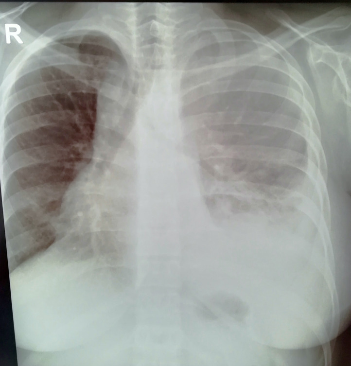
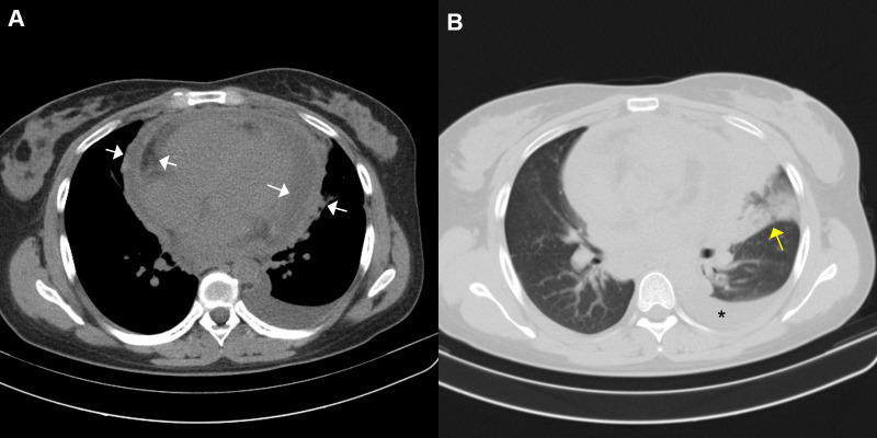
The ECG revealed sinus tachycardia, without other changes. Initial laboratory studies revealed mild leucocytosis with lymphopenia, mild thrombocytosis, and elevated inflammatory markers including: D-dimer 5 µg/mL, fibrinogen 653 mg/dL (200–390 mg/dL), ferritin 950 ng/mL (30–400 ng/mL), lactate dehydrogenase 660 U/L (125–220 U/L), C reactive protein 160 mg/L (0–5 mg/L). The liver and kidney function were within normal values. Chest X-ray showed extensive left pulmonary consolidation with moderate pleural effusion and mild tracheal deviation to the right side (figure 1). A CT scan of the chest showed pneumonia in the left lobe with left pleural effusion, and moderate pericardial effusion (figure 2).
Figure 1.
Chest X-ray showing extensive left pulmonary consolidation with moderate pleural effusion and mild tracheal deviation to right side.
Figure 2.
Chest CT scan showing moderate pericardial effusion (white arrows) (A) and pneumonia in the left lung (yellow arrow) with left pleural effusion (*) (B).
Treatment
Given the high clinical probability of COVID-19, she was admitted to the isolation unit and retested with COVID-19 real-time reverse transcriptase-PCR (RT-PCR) assay, which came positive this time. She was treated with oxygen, corticosteroids, colchicine, low molecular weight heparin and broad-spectrum antibiotic, for 5 days, without improvement.
Outcome and follow-up
Her condition deteriorated progressively with worsening dyspnoea and congestion signs. On day 6, repeated transthoracic echocardiogram revealed pericardial thickening with large fibrinous strands traversing the pericardial space and signs of constrictive pericarditis (video 1). The cardiac chamber dimensions were normal with preserved left ventricular ejection fraction and normal heart valves. Mitral inflow velocities demonstrated significant respiratory variation, increased E/A ratio, and rapid E deceleration time. Dilated non-collapsing inferior vena cava (25 mm diameter), but no features of right atrial or right ventricular collapse were noted. Estimated right atrial pressure was elevated (>20 mm Hg) with abnormal movement of the interventricular septum (septal bounce). Thus, a diagnosis of effusive-constrictive pericarditis was made, and the patient was admitted to the intensive care unit for increasing supplemental oxygen requirements. The doses of corticosteroids were increased, and small doses of diuretics were started.
Video 1.
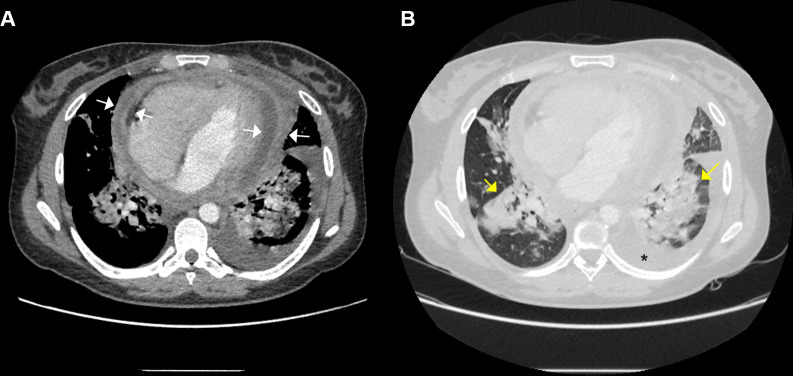
A repeated CT scan suggested worsening of pneumonia with bilateral lung involvement and pericardial thickening (figure 3). Troponin was negative and NT-proBNP was 2200 pg/mL. Diagnostic thoracentesis revealed a transudate, with normal adenosine deaminase levels. Pleural fluid and blood cultures were negative. Testing for HIV antibodies and hepatic viruses were negative.
Figure 3.
Chest CT scan shows pericardial thickening with pericardial effusion (white arrows) (A), and bilateral patchy ground-glass (yellow arrows) and consolidation with moderate pleural effusion on the left side (*) (B).
Diagnostic pericardiocentesis was not done because the pericardial effusion seemed localised near the anterolateral left ventricular wall, without effusion next to the right cavities, so the traditional subxiphoid approach could not be used. Because of the associated risk of serious cardiac injury, we did not consider necessary the use of alternative approaches for pericardiocentesis. Based on the transthoracic echocardiogram and clinical symptomatology, the patient had an indication for pericardiotomy, which was delayed because of her respiratory complications. Also, cardiac catheterisation for further haemodynamic assessment was not possible. On day 7, she was transferred to a Cardiac Surgery Intensive Care Unit in another hospital. On day 9 from admission, she developed severe respiratory insufficiency with persistent hypoxaemia despite increasing supplemental oxygen requirements, and mechanical ventilation was necessary. The evolution was unfavourable, with severe ARDS and hypoxaemia despite optimised ventilation. On day 14, extracorporeal membrane oxygenation was done but unfortunately, the patient died 5 days later.
Discussion
At present, limited data have been published on pericarditis and pericardial effusion in COVID-19,2–8 and only one case of constrictive pericarditis was reported.9
Constrictive pericarditis is characterised by inflammation and fibrotic thickening of the parietal and visceral pericardium, causing a restriction in cardiac filling and leading to hypodiastolic heart failure.10 The most common causes of constrictive pericarditis are cardiac surgery, radiotherapy, tuberculous and purulent pericarditis. Idiopathic and viral pericarditis have a low incidence of constrictive complications.11
Constrictive pericarditis is usually a late complication of chronic pericarditis.10 In rare cases, like the one presented here, it could have a more rapid, subacute evolution. Baker described in 2004 a similar case of a 30-year-old man with acute pericarditis that developed effusive-constrictive physiology in less than 2 weeks. The patient was treated initially with nonsteroidal anti-inflammatory agents and colchicine, but pericardiectomy was needed. The pericardial cavity was filled with fibrinous gelatinous exudates with adhesions to the epicardial surface. Pathological examination noted non-specific findings including fibrosis, chronic inflammation and granulation tissue. The cause of the pericarditis was thought to be idiopathic, presumed viral.11
The concept of effusive-constrictive pericarditis was established in 1971 by Hancock.12 Effusive-constrictive pericarditis is characterised by pericardial effusion, often with cardiac tamponade, and restrictive cardiac filling physiology. Although the gold standard to establish the diagnosis of effusive-constrictive pericarditis has been invasive, involving simultaneous measurement of intrapericardial and intracardiac pressures before and after pericardial drainage, the echocardiography could establish the diagnosis more rapidly.13 14 Echocardiography shows a mixed haemodynamic pattern with elements of constriction and tamponade. The main echocardiographic features are the absence of normalisation in inferior vena cava dilatation and restoration of its respiratory changes, persistently remaining significant respiratory variation of the mitral and tricuspid inflow Doppler pattern, abnormal/paradoxical movement of the interventricular septum (septal bounce) and preserved or increased medial mitral e′ velocity.15
Although the pathophysiology of pericarditis in COVID-19 is unknown, it is hypothesised that it occurs secondary to the systemic inflammatory response and the subsequent cytotoxic and immune-mediated effects related to SARS-CoV-2.16 Proinflammatory cytokines (IL-6, TGF-β) and vascular endothelial growth factor (VEGF) released via the associated angiotensin II pathway could promote inflammation and fibrosis of the pericardium in COVID-19 infection.17 18
In developing countries, and this is the case of Romania, TB remains an important cause of pericarditis and it progresses to constriction more frequently than with viral aetiologies. In TB constrictive pericarditis pathogenic mechanism are similar, with increased ACE levels, resulting in increased enzymatic cleavage of Ac-SDKP and a subsequent dampening in its antifibrotic potential, which potentially contribute to the pathogenesis of constriction. Also, marked elevations of IL-10 and IFN-γ accompanied by low levels of bioactive TGF-β levels in tuberculous pericardial fluid suggest a Th-1 mediated delayed-type hypersensitivity response to the pathogen.19 The lymphocyte Th1 pathway is also involved in COVID-19 infection. It was reported that COVID-19 has higher morbidity and mortality in people with low Th1 immunity.20 In severe cases, studies showed a Th2 response against SARS-CoV-2, rather than a Th1 response.21 New studies are necessary to explain the aggressivity of the virus in some healthy young patients. In our case, the severe evolution could be related to a dysfunction of the innate immune system or by mutations of genes involved in the inflammatory response.
Only one case of SARS-CoV-2 constrictive pericarditis, beside ours, was described in the literature.9 A healthy 29-year-old woman with ARDS developed constrictive pericarditis followed by rapid deterioration of her condition and death. Acute pericarditis could be underdiagnosed in COVID-19 infection. In a study with severe and critical COVID-19 infection, the CT scans described pericardial effusion in 4.8%.22 It is thus important to screen for the presence of pericardial effusion, especially in critically ill patients. Continued observations of the cardiovascular complications of this disease are necessary to fully understand them.
Learning points.
COVID-19 cardiovascular complications are frequent and could be associated with increased mortality and long-term prognostic implications. It is important to raise awareness for other, more rare complications, such as pericardial effusion.
Constrictive pericarditis can occur after any pericardial disease process, but also following acute pericarditis (viral or idiopathic), so it should be suspected in any patient with aggravated symptoms.
This case highlights the potential for patients with COVID-19 to develop life-threatening conditions within a short timeframe.
Patients with moderate or severe forms of COVID-19 should undergo an initial evaluation with an ECG, chest X-ray, and transthoracic echocardiogram. This may facilitate early diagnosis and management of COVID-19 associated pericarditis which may improve outcomes.
Footnotes
Twitter: @LucianPopescu18
Contributors: LP, RD, and AV: acquisition of data; drafting the work; final approval; agreement to be accountable for all aspects of the work. ID: design; final revision; final approval; agreement to be accountable for all aspects of the work.
Funding: The authors have not declared a specific grant for this research from any funding agency in the public, commercial or not-for-profit sectors.
Competing interests: None declared.
Provenance and peer review: Not commissioned; externally peer reviewed.
Ethics statements
Patient consent for publication
Obtained.
References
- 1.Inciardi RM, Lupi L, Zaccone G, et al. Cardiac involvement in a patient with coronavirus disease 2019 (COVID-19). JAMA Cardiol 2020;5:819–24. 10.1001/jamacardio.2020.1096 [DOI] [PMC free article] [PubMed] [Google Scholar]
- 2.Hussain H, Fadel A, Alwaeli H, et al. Coronavirus (COVID-19) fulminant myopericarditis and acute respiratory distress syndrome (ARDS) in a middle-aged male patient. Cureus 2020;12:e8808. 10.7759/cureus.8808 [DOI] [PMC free article] [PubMed] [Google Scholar]
- 3.Hua A, O'Gallagher K, Sado D, et al. Life-Threatening cardiac tamponade complicating myo-pericarditis in COVID-19. Eur Heart J 2020;41:2130. 10.1093/eurheartj/ehaa253 [DOI] [PMC free article] [PubMed] [Google Scholar]
- 4.Dabbagh MF, Aurora L, D'Souza P, et al. Cardiac Tamponade Secondary to COVID-19. JACC Case Rep 2020;2:1326–30. 10.1016/j.jaccas.2020.04.009 [DOI] [PMC free article] [PubMed] [Google Scholar]
- 5.Asif T, Kassab K, Iskander F, et al. Acute pericarditis and cardiac tamponade in a patient with COVID-19: a therapeutic challenge. Eur J Case Rep Intern Med 2020;7:001701. 10.12890/2020_001701 [DOI] [PMC free article] [PubMed] [Google Scholar]
- 6.Marschall A, Concepción Suárez R, Dejuan Bitriá C. Acute pericarditis secondary to COVID-19. pericarditis aguda secundaria a COVID-19. Emergencias 2020;32:221–2. [PubMed] [Google Scholar]
- 7.Kumar R, Kumar J, Daly C, et al. Acute pericarditis as a primary presentation of COVID-19. BMJ Case Rep 2020;13:e237617. 10.1136/bcr-2020-237617 [DOI] [PMC free article] [PubMed] [Google Scholar]
- 8.Walker C, Peyko V, Farrell C, et al. Pericardial effusion and cardiac tamponade requiring pericardial window in an otherwise healthy 30-year-old patient with COVID-19: a case report. J Med Case Rep 2020;14:158. 10.1186/s13256-020-02467-w [DOI] [PMC free article] [PubMed] [Google Scholar]
- 9.SeyedAlinaghi S, Ghadimi M, Gharabaghi MA, et al. Constrictive pericarditis associated with coronavirus disease 2019 (COVID-19): a case report. Infect Disord Drug Targets 2020;20. 10.2174/1871526520666201209145001. [Epub ahead of print: 09 Dec 2020]. [DOI] [PubMed] [Google Scholar]
- 10.Syed FF, Schaff HV, Oh JK. Constrictive pericarditis--a curable diastolic heart failure. Nat Rev Cardiol 2014;11:530–44. 10.1038/nrcardio.2014.100 [DOI] [PubMed] [Google Scholar]
- 11.Baker CM, Orsinelli DA. Subacute effusive-constrictive pericarditis: diagnosis by serial echocardiography. J Am Soc Echocardiogr 2004;17:1204–6. 10.1016/j.echo.2004.06.013 [DOI] [PubMed] [Google Scholar]
- 12.Hancock EW. Subacute effusive-constrictive pericarditis. Circulation 1971;43:183–92. 10.1161/01.CIR.43.2.183 [DOI] [PubMed] [Google Scholar]
- 13.Janus SE, Hoit BD. Effusive-Constrictive pericarditis in the spectrum of pericardial compressive syndromes. Heart 2021;107:450–5. 10.1136/heartjnl-2020-316664 [DOI] [PubMed] [Google Scholar]
- 14.Rasaretnam R, Chanmugam D. Subacute effusive constrictive epicarditis. Br Heart J 1980;44:44–8. 10.1136/hrt.44.1.44 [DOI] [PMC free article] [PubMed] [Google Scholar]
- 15.van der Bijl P, Herbst P, Doubell AF. Redefining effusive-constrictive pericarditis with echocardiography. J Cardiovasc Ultrasound 2016;24:317–23. 10.4250/jcu.2016.24.4.317 [DOI] [PMC free article] [PubMed] [Google Scholar]
- 16.Costela-Ruiz VJ, Illescas-Montes R, Puerta-Puerta JM, et al. SARS-CoV-2 infection: the role of cytokines in COVID-19 disease. Cytokine Growth Factor Rev 2020;54:62–75. 10.1016/j.cytogfr.2020.06.001 [DOI] [PMC free article] [PubMed] [Google Scholar]
- 17.Cizgici AY, Zencirkiran Agus H, Yildiz M. COVID-19 myopericarditis: It should be kept in mind in today’s conditions. Am J Emerg Med 2020;38:1547.e5 10.1016/j.ajem.2020.04.080 [DOI] [PMC free article] [PubMed] [Google Scholar]
- 18.Hirano T, Murakami M. COVID-19: a new virus, but a familiar receptor and cytokine release syndrome. Immunity 2020;52:731–3. 10.1016/j.immuni.2020.04.003 [DOI] [PMC free article] [PubMed] [Google Scholar]
- 19.Ramasamy V, Mayosi BM, Sturrock ED, et al. Established and novel pathophysiological mechanisms of pericardial injury and constrictive pericarditis. World J Cardiol 2018;10:87–96. 10.4330/wjc.v10.i9.87 [DOI] [PMC free article] [PubMed] [Google Scholar]
- 20.Gupta A. Is Immuno-modulation the key to COVID-19 pandemic? Indian J Orthop 2020;54:394–7. 10.1007/s43465-020-00121-7 [DOI] [PMC free article] [PubMed] [Google Scholar]
- 21.Roncati L, Nasillo V, Lusenti B, et al. Signals of Th2 immune response from COVID-19 patients requiring intensive care. Ann Hematol 2020;99:1419–20. 10.1007/s00277-020-04066-7 [DOI] [PMC free article] [PubMed] [Google Scholar]
- 22.Li K, Wu J, Wu F, et al. The clinical and chest CT features associated with severe and critical COVID-19 pneumonia. Invest Radiol 2020;55:327–31. 10.1097/RLI.0000000000000672 [DOI] [PMC free article] [PubMed] [Google Scholar]


