

A 28-year-old lady with Fitzpatrick skin type IV presented with an asymptomatic pigmentation over the tongue for the past 6 months. Examination revealed multiple discrete tan-brown pin-head papules on the anterolateral aspect of the dorsum of the tongue, representing the fungiform papillae. [Figure 1] There was no other mucocutaneous and systemic involvement. Routine laboratory investigations were unremarkable. Dermoscopy (DermLite DL4; polarized mode with 10x magnification) revealed ovoid hyperpigmented projections with dichotomized vessels originating at its base, giving the appearance of “rose petals” [Figure 2]. This clinico-dermoscopic finding is a characteristic of pigmented fungiform papillae of the tongue (PFPT).
Figure 1.
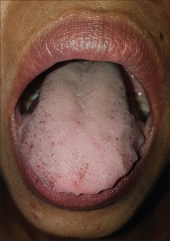
Pigmented fungiform papillae of the tongue
Figure 2.
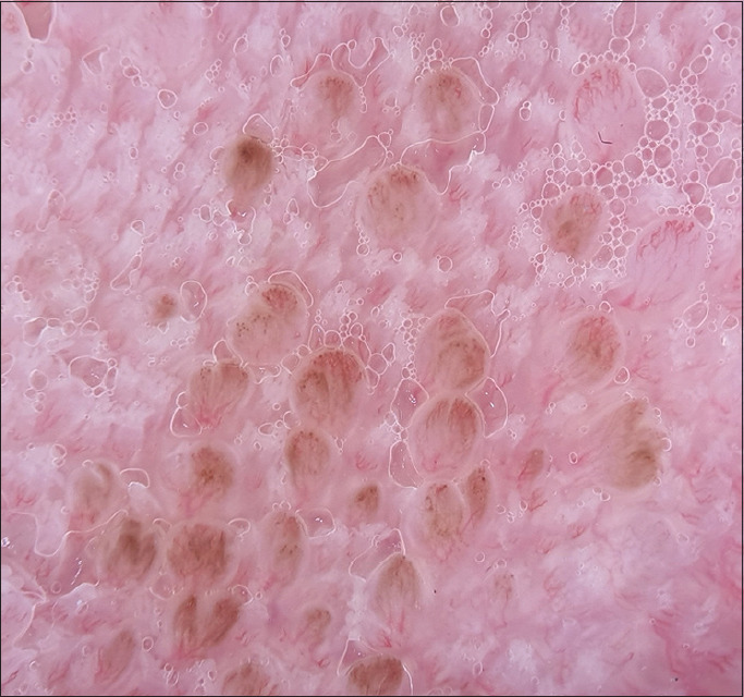
Dermoscopy (DermLite DL4; polarized mode with 10× magnification) showing ovoid hyperpigmented projections with dichotomized vessels originating at its base, giving the appearance of ”rose petals”
Fungiform papillae are discrete projections found on the tongue's dorsal surface involving the tip and anterio-lateral aspect. PFPT is well-circumscribed hyperpigmentation confined to only these papillae.[1] This benign condition is classically noticed in darkly pigmented individuals, manifesting during the second or third decade, although cases involving children have been reported.[2] Although PFPT remains asymptomatic in most patients, associations like linear circumflex ichthyosis, lichen planus, Hori's nevus, melasma, hemochromatosis, scleroderma, pernicious anemia, and iron deficiency anemia have been documented.[3] Dermoscopy typically demonstrates projections with pigmented borders interspersed with dichotomized vessels that originated at their base, giving the aspect a “rose petal” appearance.[4] This correlates with melanophages without any inflammation in the dermis on histopathology; staining positive for melanin with Masson-Fontana but negative for iron with Prussian blue.[5] Such a unique finding will not be visualized in clinical mimickers like oral melanotic macule, melanocytic nevus, Peutz-Jeghers syndrome, Addison disease, and rarely Laughier-Hunziker syndrome.
The characteristic dermoscopic pattern provides an additional clue to help clinicians recognize and differentiate this ethnic, acquired, and benign condition, and avoid unnecessary investigative work-up. To the best of our knowledge, dermoscopic features of PFPT is yet to be described in the Indian population.
Declaration of patient consent
The authors certify that they have obtained all appropriate patient consent forms. In the form the patient(s) has/have given his/her/their consent for his/her/their images and other clinical information to be reported in the journal. The patients understand that their names and initials will not be published and due efforts will be made to conceal their identity, but anonymity cannot be guaranteed.
Financial support and sponsorship
Nil.
Conflicts of interest
There are no conflicts of interest.
References
- 1.Holzwanger JM, Rudolph RI, Heaton CL. Pigmented fungiform papillae of the tongue: A common variant of oral pigmentation. Int J Dermatol. 1974;13:403–8. doi: 10.1111/j.1365-4362.1974.tb05073.x. [DOI] [PubMed] [Google Scholar]
- 2.Sharma S, Sharma S. Pigmentation of the fungiform papillae of the tongue in a child secondary to iron deficiency anaemia: An uncommon occurrence and association. Int J Oral Health Dent. 2016;2:39–42. [Google Scholar]
- 3.Kauzmn A, Pavone M, Blanas N, Bradley G. Pigmented lesions of the oral cavity: Review, differential diagnosis, and case presentations. J Can Dent Assoc. 2004;70:682–3. [PubMed] [Google Scholar]
- 4.Mukamal LV, Ormiga P, Ramos-E-Silva M. Dermoscopy of the pigmented fungiform papillae of the tongue. J Dermatol. 2012;39:397–9. doi: 10.1111/j.1346-8138.2011.01328.x. [DOI] [PubMed] [Google Scholar]
- 5.Werchniak AE, Storm CA, Dinulos JG. Hyperpigmented patches on the tongue of a young girl. Pigmented fungiform papillae of the tongue. Arch Dermatol. 2004;140:1275–80. doi: 10.1001/archderm.140.10.1275-g. [DOI] [PubMed] [Google Scholar]