Abstract
OBJECTIVES
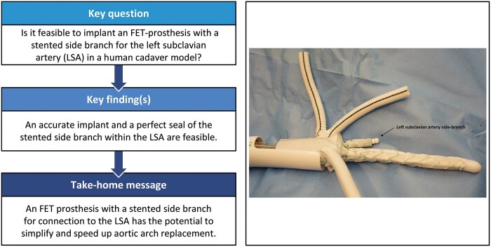
Our goal was to develop a modified frozen elephant trunk (FET) prosthesis with a stented left subclavian artery (LSA) side branch for LSA connection and to perform preclinical testing in a human cadaver model.
METHODS
We measured aortic diameters, distance between and diameters of supra-aortic vessels and the distance from the LSA offspring to the level of the left vertebral artery offspring in 70 patients. Based on these measurements, a novel FET prosthesis was developed (Cryolife/Jotec, Hechingen, Germany) featuring a stented side branch for an intrathoracic LSA connection. The feasibility and ease of implantation were tested in 2 human cadaver models at the Anatomical Institute of the Medical University Graz. A covered stent graft (Advanta V12™ by Atrium Medical Corp., Hudson, NH, USA) was used for an LSA extension.
RESULTS
Accurate deployment of the novel FET prosthesis with anatomical orientation of the stented side branch towards the LSA ostium followed by consecutive stent graft deployment was feasible in both cases. Proximalizing the distal anastomosis level from zone 3 to zone 1 not only diminished the complexity of the procedure but substantially facilitated the completion of the distal anastomosis. A 2.5-cm long extension stent graft was sufficient to seal to the LSA and to maintain left vertebral artery patency in both cases.
CONCLUSIONS
This initial study in human anatomical bodies could demonstrate the feasibility of implanting a newly designed FET prosthesis. This evolution of the FET technique has the potential to substantially ease total aortic arch replacement by proximalization of the distal anastomosis into zone 1 and by shortening spinal and lower body hypothermic circulatory arrest times via a stented side branch to the LSA. This direct connection enables early restoration of systemic perfusion.
Keywords: Anatomy of the aortic arch, Frozen elephant trunk technique, Left subclavian artery side branch stent graft, Total aortic arch replacement
INTRODUCTION
The frozen elephant trunk (FET) technique has emerged as an important treatment strategy for operations of complex aortic arch aneurysms and dissections [1–4]. However, this operative technique represents a complex aortic arch surgical procedure and is associated with long lower body hypothermic circulatory arrest (HCA) times. Consequently, even in highly experienced centres, an operative risk remains, in particular with regard to neurological injury both at the central and at the spinal level [5, 6]. Consequently, technical and conceptual strategies have been developed to reduce these complications to a minimum, such as proximalization of the descending anastomosis from zone 3 to zone 2 [7]. In addition, bilateral and on-top trilateral antegrade selective cerebral perfusion have become standard. One remaining issue is the technique of left subclavian artery (LSA) reimplantation, which is facilitated by using a branched Dacron graft but may even be more completely facilitated by the routine application of a bridging stent graft between the main prosthesis and the native LSA [8].
The goal of this study was to develop a modified FET prosthesis with a stented LSA side branch (FET-LSSB) for LSA connection and to perform preclinical testing in a human cadaver model.
METHODS
Three-stage comprehensive study model
Stage 1: To analyse the morphology of the aortic arch, the size and angulation of the branching vessels and their distances to each other, a customized morphometric computed tomography (CT) analysis model was created and tested on 70 consecutive patients.
Stage 2: In cooperation with Cryolife/Jotec (Hechingen, Germany), a novel FET prosthesis featuring a stented side branch for intrathoracic LSA connection was developed to proximalize the distal anastomosis and to facilitate the surgical procedure.
Stage 3: The FET-LSSB prosthesis was implanted in 2 human cadaver models to test the feasibility of implantation and develop a surgical implantation protocol.
The study was approved by the local ethics committee (EK 061120). The necessity of obtaining informed consent was waived due to the retrospective nature of the analysis.
Stage 1: Morphometric computed tomography analysis of the aortic arch
Between April and September 2018, a total of 70 consecutive patients underwent elective electrocardiographic-gated and contrast-enhanced multislice CT scans of the thoracic aorta. Two patients had to be excluded from the analysis because their images were of insufficient quality. Non-pathological aortic anatomical configurations were found in 56 patients whereas 12 patients had thoracic aortic aneurysms. All scans were performed according to an established institutional protocol with a 2 × 128-slice Somatom Drive Dual Source CT scanner (Siemens Healthcare, Forchheim, Germany). Two experienced readers independently evaluated all data sets for quantitative measurements using the 3mensio Valves™ (Version 7.2, 3mensio Medical Imaging BV, Maastricht, Netherlands). The assessment protocol for the thoracic aorta comprised 3 main steps: (i) determination of the aortic centre line and the diameters of the thoracic aorta; (ii) determination of the take-off point, angulations, diameter and distances of the supra-aortic branches; and (iii) measurement of the length from the LSA to the offspring of the left vertebral artery (LVA) offspring.
With reference points at the aortic valve and the passing of the thoracic aorta through the diaphragm, a ‘stretched vessel view’ was generated allowing exact analysis of the aortic diameters. The take-off points of the supra-aortic branches were defined as the centre of the vessel at the outer aortic curvature, and the distances of the take-off points of the supra-aortic vessels were measured. The take-off angle was defined as the angle between the aortic tangent and the perpendicular line of the respective supra-aortic branch and was assessed in the ‘perpendicular plane’ view. Associated diameters of the supra-aortic branches measured 10 mm after the take-off point to account for tapering at the origin of the vessel. To measure the distance of the LSA to the LVA, a model of the centre line was created with reference points at the aortic origin of the vessel and 10 mm distal to the offspring of the LVA. The distance was then measured in the stretched vessel view. Figure 1 depicts a schematic drawing of the levels of measurements taken for this new FET conceptualization.
Figure 1:
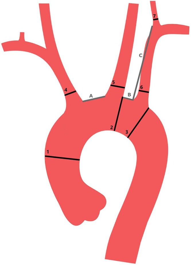
Points at which measurements were taken for conceptualization of a frozen elephant trunk prosthesis with a stented side-branch for the left subclavian artery.
Stage 2: Development of the novel frozen elephant trunk prosthesis with a side branch for the left subclavian artery (frozen elephant trunk prosthesis with a stented side branch for the left subclavian artery)
A new FET prosthesis with a side branch stent graft for the LSA was conceptualized by our group and then built by Cryolife/Jotec. This novel hybrid prosthesis consists of a Dacron graft and a thoracic stent graft connected to the distal end of the Dacron prosthesis. A Dacron collar, which is mounted at the transition between the stent graft component and the Dacron prosthesis, facilitates the accomplishment of the descending anastomosis. A reversed bifurcated graft designed for separate reimplantation of the brachiocephalic artery and the left common carotid artery originates from the Dacron graft. The most important innovation is the endovascular side branch with a length of 2.5 cm and a diameter of 1 cm, which is located just proximal to the Dacron collar on the stent graft. In addition, a perfusion port for distal aortic perfusion is part of this new design (Fig. 2). The deployment sheath is the same as that for the recently released E-vita open NEO prosthesis (Fig. 3).
Figure 2:
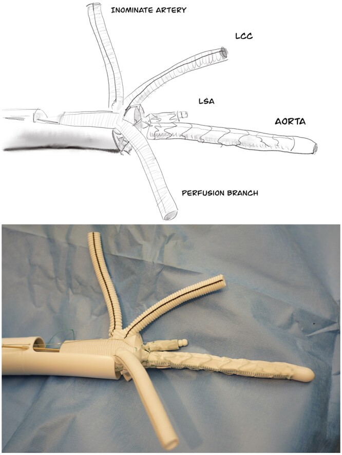
Frozen elephant trunk prosthesis with a stented side-branch for the LSA prosthesis showing the LSA side branch stent. LCC: left common carotid; LSA: left subclavian artery.
Figure 3:
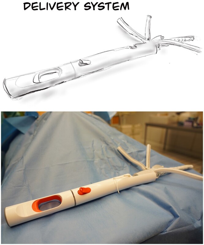
Short introduction and deployment device for the novel prosthesis.
Stage 3: Frozen elephant trunk prosthesis with a stented side branch for the left subclavian artery implant in the human cadaver model
The FET-LSSB prosthesis was implanted in 2 human cadavers at the Anatomical Institute of the Medical University Clinic of Graz. The anatomical bodies were preserved not with formaldehyde, but with Thiel’s solution [9, 10]. This special preservation technique keeps the tissue soft and pliable, which provides excellent conditions for testing the implanted FET-LSSB prosthesis. After a sternotomy, the pericardium was opened, and the brachiocephalic artery was clamped. The ascending aorta was resected up to the brachiocephalic trunk. The left common carotid artery was ligated and transected. A cannula for bilateral antegrade perfusion was inserted. One guide wire was advanced via the left brachial artery into the aortic arch; the second guide wire was inserted in an antegrade fashion into the descending aorta. Thereafter, the FET-LSSB prosthesis was pushed down into the descending aorta, and the LSA side branch was guided into the LSA offspring (Fig. 4). This manoeuvre was performed using a guide wire, which may not be necessary in the clinical setting.
Figure 4:
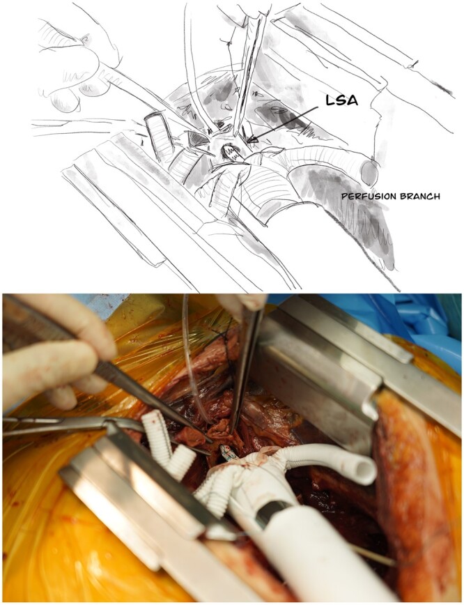
View during implantation showing exact orientation of the side branch towards the LSA ostium. LSA: left subclavian artery.
The hybrid prosthesis was deployed, and the distal anastomosis with the collar of the prosthesis and the native aorta was performed in zone 1 of the aortic arch with a 4–0 polypropylene running suture (Fig. 5). After the distal anastomosis was completed, the Dacron prosthesis was clamped proximal to the side port for distal aortic perfusion, and the left common carotid artery followed by the brachiocephalic artery was anastomosed with a 5–0 polypropylene running suture to the side arms of the arch prosthesis. Thereafter, we performed a proximal anastomosis with the ascending aorta.
Figure 5:
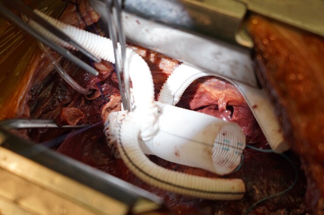
Distal anastomosis is performed in zone 1 of the aortic arch.
Extension stent grafts for the left subclavian artery side branch
It can be assumed that the stented side branch to the LSA will not completely create a seal with the LSA due to the size and length of overlap. Similar to endovascular treatment for thoraco-abdominal aneurysms, additional extension stent grafts may be necessary to prevent endoleak formation. This extension can be performed by sealing the stented side branch with an extension stent graft during HCA. Here, a guide wire is advanced from the left brachial artery into the LSA and into the open aortic arch prosthesis. The tip of the guide wire is crabbed, and a balloon-expandable stent graft (Advanta V12™ by Atrium Medical Corp., Hudson, NH, USA) is inserted over the guide wire in an antegrade fashion and deployed into the stented side branch to achieve complete sealing. Preoperative measurement of the distance between the origin of the LSA and the take-off of the LVA remains decisive.
Another option is sealing the stented side branch with an extension stent graft at the end of the main operation in a hybrid room. A guide wire is advanced via the left brachial artery into the aortic arch under X-ray monitoring. The distance between the take-off of the LVA and the side branch stent is measured using a scaled catheter. After the right size is selected, an extension stent graft is inserted and deployed under angiographic monitoring.
Statistical analyses
Continuous data are expressed either as means and standard deviation (±standard deviation) or median (interquartile range), depending on variable distribution, and were analysed with the ‘Student’s t-test’ or the Wilcoxon rank sum test, respectively. Variable distribution was assessed using the Shapiro–Wilk test and QQ plots. Categorical variables are expressed in absolute numbers and percentages. Depending on the sample size, the χ2 or the Fisher’s exact test was used for comparisons. Results were categorized as statistically significant with an alpha level set at <0.05. All analyses were performed using SPSS, version 25.0 (IBM Corp, Armonk, NY, USA).
RESULTS
Computed tomography mapping of the aortic arch
Patient demographics are shown in Table 1. Whereas no differences between the pathological and non-pathological cohorts were observed with regard to age (pathological cohort: 66.3 ± 10.7 years vs the non-pathological cohort: 69.9 ± 10.8 years; P = 0.0299), sex [female: n = 6 (50%) vs n = 17 (30.4%); P = 0.320] and body mass index (28 ± 6.4 vs 26.33 ± 6.1; P = 0.457), CT angiography measurements of the thoracic aorta revealed a significantly larger mean diameter of the ascending aorta in patients with pathological aortic anatomical variations (43.1 ± 8.1 mm vs 37.9 ± 5.9 mm; P = 0.012). All other diameters, take-off angles and differences between the supra-aortic branches did not differ between the cohorts and are displayed in Table 1. The mean diameter of the LSA was 11.1 ± 3.8 mm in the non-pathological cohort and 12.4 ± 3.7 mm in the pathological cohort (P = 0.789). Furthermore, no differences between the 2 cohorts were found with regard to the angulation of the LSA (26.2° ± 11.5° vs 28.7° ± 12.1°; P = 0.324) or the distance from the take-off of the LSA to the take-off of the LVA (43.3 ± 7.7 vs 40.5 ± 9.3; P = 0.351).
Table 1:
Patient demographics and arch morphometry
| Non-pathological thoracic aorta (n = 56) | Pathological thoracic aorta (n = 12) | P-value | Fig. 1 description | |
|---|---|---|---|---|
| Demographic data | ||||
| Age (years), median ± IQR | 69.9 ± 10.8 | 66.3 ± 10.7 | 0.299 | |
| Height (m), mean ± SD | 1.68 ± 0.08 | 1.72 ± 0.09 | 0.840 | |
| Weight (kg), mean ± SD | 74.0 ± 19.5 | 82.0 ± 15.3 | 0.855 | |
| Female, n (%) | 17 (30) | 6 (50) | 0.320 | |
| BMI (kg/m2), mean ± SD | 26.33 ± 6.1 | 28.0 ± 6.4 | 0.457 | |
| Diameter (mm) | ||||
| Ascending aorta, median ± IQR | 37.9 ± 5.9 | 43.1 ± 8.1 | 0.012 | 1 |
| Aortic arch, mean ± SD | 30.1 ± 3.6 | 31.6 ± 3.5 | 0.211 | 2 |
| Descending aorta, median ± IQR | 28.7 ± 4.0 | 30.8 ± 5.8 | 0.137 | 3 |
| BCT, mean ± SD | 12.2 ± 3.4 | 14.1 ± 4.5 | 0.098 | 4 |
| LCCA, median ± IQR | 7.9 ± 2.8 | 8.1 ± 3.6 | 0.850 | 5 |
| LSA, mean ± SD | 10.8 ± 3.7 | 11.1 ± 3.8 | 0.789 | 6 |
| LVA, mean ± SD | 3.5 ± 1.3 | 4.2 ± 1.4 | 0.136 | |
| Distances (mm) | ||||
| BCT-LCCA, median ± IQR | 2.6 ± 2.4 | 3.1 ± 1.8 | 0.450 | A |
| LCCA-LSA, median ± IQR | 6.2 ± 4.1 | 7.3 ± 5.3 | 0.616 | B |
| LSA origin to LVA origin, mean ± SD | 43.3 ± 7.7 | 40.5 ± 9.3 | 0.351 | C |
| Angulation | ||||
| BCT in degrees, mean ± SD | 43.4 ± 14.0 | 44.2 ± 15.2 | 0.455 | |
| LCCA in degrees, mean ± SD | 25.6 ± 15.1 | 26.1 ± 15.8 | 0.760 | |
| LSA in degrees, mean ± SD | 27.5 ± 16.7 | 28.6 ± 14.2 | 0.378 | |
BCT: brachiocephalic trunk; BMI: body mass index; IQR: interquartile range; LCCA: left common carotid artery; LSA: left subclavian artery; LVA: left vertebral artery; SD: standard deviation.
Frozen elephant trunk prosthesis with a stented side branch for the left subclavian artery implant
Accurate deployment of the prosthesis with placement of the short, stented side branch towards the LSA ostium was feasible in both cases. The FET-LSSB prosthesis was advanced via 2 guide wires: 1 in the descending aorta and 1 in the LSA. Exact deployment of the hybrid prosthesis followed by straightforward implantation of the aortic arch prosthesis was achieved. The distal descending anastomosis was performed in zone 2 in the first case and in zone 1 in the second case. The ‘proximalization’ of the distal anastomosis significantly facilitates and accelerates the accomplishment of the distal descending anastomosis. Thereafter, anastomoses of the left common carotid artery and the innominate artery with the side arms of the hybrid prosthesis were performed with 5–0 polypropylene running sutures. In addition, we managed a perfect seal of the LSA with an additional extension stent graft, which was proven by postoperative angiography (Fig. 6).
Figure 6:
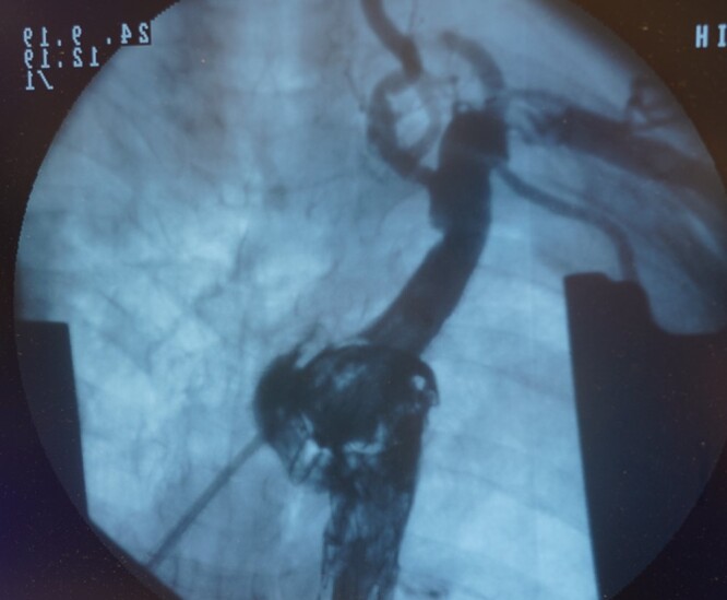
Intraprocedural completion angiography showing no endoleak at the level of the left subclavian artery and regular antegrade perfusion of the left vertebral artery.
DISCUSSION
The use of this newly designed FET prosthesis is feasible, and the deployment of the side branch in the LSA can be carried out with minimal effort. It has the potential to substantially ease total aortic arch replacement by proximalization of the distal anastomosis into zone 1 and by shortening spinal and lower body HCA times via a stented side branch to the LSA. This direct connection with an optional bridging stent graft implant enables early restoration of systemic perfusion.
In the last decade, the FET technique has evolved to an accepted treatment strategy for acute as well as chronic diseases of the aortic arch and the proximal descending aorta [11–14]. However, the complexity of the operation and the significant complications associated with it had a negative impact on the widespread use of this technique [15]. Hemiarch and total arch replacement are traditional treatments of choice for aortic diseases involving the aortic arch. In patients with aortic dissection, residual distal tears and a patent false lumen are associated with late complications in the downstream aorta [16]. As a consequence, hybrid techniques like the FET were developed [17, 18]. The FET concept could demonstrate in patients with aortic dissection an increased rate of false lumen thrombosis and fewer reinterventions in the downstream aorta in the long run [19, 20]. However, FET repair is thought to be more invasive than techniques limited to the proximal aortic arch because longer HCA and cardiopulmonary bypass times are required.
It is important to take into consideration the fact that early postoperative results are substantially influenced by the degree of operative invasiveness. Rylski et al. [21] reported that total arch replacement was a significant risk factor for postoperative in-hospital death in patients with acute type A aortic dissection. Kim et al. [22] reported that a more extensive surgical approach had a higher incidence of pneumonia, postoperative neurological deficit and low cardiac output syndromes compared with the hemiarch replacement approach in acute DeBakey type I aortic dissection. Therefore, it is an important objective to make the FET technique as simple as possible without compromising the advantages of the FET technique itself. Jakob et al. [23] developed with the 3-zone hybrid graft (E-Novia, Cryolife/Jotec, Hechingen, Germany) a novel prosthesis for acute type A aortic dissections with the intention to facilitate the operation, having an easily accessible zone 0 suture line with the benefits of aortic arch and descending aorta true lumen stabilization. Whereas the goal of the E-novia concept is to make the operation for acute type A aortic dissection easier, the FET-LSSB concept focuses on the indications for patients for whom complete aortic arch replacement is necessary, such as those with atherosclerotic aneurysms, penetrating ulcers and chronic dissections.
Traditionally, the distal anastomosis of the FET procedure is performed in aortic arch zone 3 [24]. In patients with an unfavourable chest anatomy, deployment and distal anastomosis of the FET prosthesis distal to the LSA in zone 3 may be cumbersome, particularly in cases of dissection or connective tissue disease. Moreover, an LSA anastomosis can be technically challenging due to the extremely deep intrathoracic localization. For this reason, various groups preferred to perform the LSA anastomosis during HCA [7, 25].
Several modifications have been proposed to facilitate the FET procedure. The complexity of the FET procedure can be decreased by moving the distal anastomosis from zone 3 to zone 2 or 1 [26]. Proximalization of the distal anastomosis facilitates the performance of the anastomosis with the aorta and reduces the incidence of left recurrent laryngeal nerve injury. However, all these modifications require the separate reimplantation of the LSA. This technique can be performed in a single-stage procedure either by direct anastomosis to the LSA or by construction of an extra-anatomic bypass [25–27]. In an elective setting, several centres prefer a two-stage approach with a bypass between the LSA and the left common carotid artery done prior to the FET procedure.
The goal of our concept was to facilitate the total FET procedure by performing a distal anastomosis in aortic arch zone 1 or 2 with simultaneous endovascular treatment of the LSA. These considerations stimulated us to refine the available FET prosthesis with an additional integrated stented side arm for the LSA. This novel FET prosthesis, conceptualized by our group and built by Cryolife/Jotec, enables FET repair in aortic arch zone 1 or 2 without the need for a surgical anastomosis to the LSA. Because cardiovascular surgeons have tried many ways to simplify the FET procedure, we strongly believe that our approach, i.e. integrating a stented side branch in the FET prosthesis, is a technically easy way to overcome LSA reimplantation issues. Pichlmaier et al. [8] recently described a novel stent-bridging technique for the anastomoses to the supra-aortic vessels. According to their method, the anastomoses to the LSA and to the left common carotid artery were performed by placing only 2 to 4 aligning single sutures, followed by a covered stent to bridge the anastomosis.
Our idea might represent a further evolution of this concept by avoiding additional sutures at the level of the LSA. In the experimental setting, we inserted the extension stent graft in an antegrade fashion via the open aortic arch. In the clinical setting, we recommend checking by angiography the sealing of the side branch stent at the end of the operation. If additional sealing is required, an extension stent graft can be implanted via the brachial artery. Based on our anatomical studies of the aortic arch and supra-aortic vessels, we designed the stented side branch for the LSA with a maximum length of 2.5 cm. We can thus ensure a safe distance to the LVA, which usually originates with a distance of 4 cm to the take-off of the LSA. We recommend that such procedures be carried out in a hybrid operating room, a requirement that is valid for any complex aortic procedure. In patients with an isolated origin of the LVA from the aortic arch, the FET-LSSB prosthesis can only be used if a vertebral-carotid artery transposition is carried out.
In the initial phase of the clinical application of this novel FET prosthesis, the indication will be restricted to patients with chronic conditions like penetrating ulcers, atherosclerotic aneurysms and chronic dissections. The FET-LSSB prosthesis will be custom-made, and the diameter and the length of the stented side branch for the LSA will be adapted to the individual patient according to preoperative CT measurements. As we gain more experience in patients with chronic indications, we plan to expand this concept to include acute indications and a standardized length and diameter of the stented side branch.
Limitations and strengths
This is an early feasibility study, and the clinical proof of concept has yet to be completed. However, this approach has the potential to clear up one of the last technical grey zones of this approach and may thereby help to lower the threshold of physicians able to apply the technique in the right clinical scenario by simultaneously reducing the technical demand to a minimum and adding a safety net via the stented side branch.
CONCLUSIONS
The use of this newly designed FET prosthesis is feasible, and the deployment of the side branch in the LSA can be carried out safely with minimal effort. It has the potential to substantially ease total aortic arch replacement by proximalization of the distal anastomosis into zone 1 and by shortening spinal and lower body HCA times via a stented side branch to the LSA. This direct connection enables early reconstitution of systemic perfusion.
ACKNOWLEDGEMENTS
The authors acknowledge the substantial support of Thomas Bogenschütz (Jotec/Cryolife) in the development of a ‘next generation’ hybrid prosthesis for aortic arch replacement.
Conflict of interest: Martin Grabenwöger is a consultant for Jotec/Cryolife. All other authors declared no conflicts of interest.
Author contributions
Martin Grabenwöger: Conceptualization; Investigation; Writing—original draft. Markus Mach: Conceptualization; Formal analysis; Writing—review & editing. Heinrich Mächler: Conceptualization; Methodology; Resources. Zsuzsanna Arnold: Formal analysis; Writing—original draft. Harald Pisarik: Data curation; Investigation. Sandra Folkmann: Data curation; Investigation. Marie-Luise Harrer: Project administration; Validation. Daniela Geisler: Data curation; Visualization. Reinhard Moidl: Validation. Bernhard Winkler: Funding acquisition. Johannes Bonatti: Validation. Martin Czerny: Writing—review & editing. Gabriel Weiss: Visualization; Writing— review & editing.
Reviewer information
European Journal of Cardio-Thoracic Surgery thanks Aung Oo, Sven Peterss and the other, anonymous reviewer(s) for their contribution to the peer review process of this article.
ABBREVIATIONS
- CT
Computed tomography
- FET
Frozen elephant trunk
- FET-LSSB
Frozen elephant trunk prosthesis with a stented side branch for the left subclavian artery
- HCA
Hypothermic circulatory arrest
- LSA
Left subclavian artery
- LVA
Left vertebral artery
REFERENCES
- 1. Shrestha M, Bachet J, Bavaria J, Carrel TP, De Paulis R, Di Bartolomeo R. et al. Current status and recommendations for use of the frozen elephant trunk technique: a position paper by the Vascular Domain of EACTS. Eur J Cardiothorac Surg 2015;47:759–69. [DOI] [PubMed] [Google Scholar]
- 2. Tsagakis K, Pacini D, Grabenwöger M, Borger MA, Goebel N, Hemmer W. et al. Results of frozen elephant trunk from the international E-vita Open registry. Ann Cardiothorac Surg 2020;9:178–88. [DOI] [PMC free article] [PubMed] [Google Scholar]
- 3. Shrestha M, Martens A, Kaufeld T, Beckmann E, Bertele S, Krueger H. et al. Single-centre experience with the frozen elephant trunk technique in 251 patients over 15 years. Eur J Cardiothorac Surg 2017;52:858–66. [DOI] [PubMed] [Google Scholar]
- 4. Leone A, Murana G, Coppola G, Berardi M, Botta L, Di Bartolomeo R. et al. Frozen elephant trunk—the Bologna experience. Ann Cardiothorac Surg 2020;9:220–2. [DOI] [PMC free article] [PubMed] [Google Scholar]
- 5. Preventza O, Liao JL, Olive JK, Simpson K, Critsinelis AC, Price MD. et al. Neurologic complications after the frozen elephant trunk procedure: a meta-analysis of more than 3000 patients. J Thorac Cardiovasc Surg 2020;160:20–33.e4. [DOI] [PubMed] [Google Scholar]
- 6. Leone A, Di Marco L, Coppola G, Amodio C, Berardi M, Mariani C. et al. Open distal anastomosis in the frozen elephant trunk technique: initial experiences and preliminary results of arch zone 2 versus arch zone 3. Eur J Cardiothorac Surg 2019;56:564–71. [DOI] [PubMed] [Google Scholar]
- 7. Czerny M, Rylski B, Kari FA, Kreibich M, Morlock J, Scheumann J. et al. Technical details making aortic arch replacement a safe procedure using the ThoraflexTM Hybrid prosthesis. Eur J Cardiothorac Surg 2017;51:i15–9. [DOI] [PubMed] [Google Scholar]
- 8. Pichlmaier M, Luehr M, Rutkowski S, Fabry T, Guenther S, Hagl C. et al. Aortic arch hybrid repair: stent-bridging of the supra-aortic vessel anastomoses (SAVSTEB). Ann Thorac Surg 2017;104:e463–5. [DOI] [PubMed] [Google Scholar]
- 9. Thiel W. [ Supplement to the conservation of an entire cadaver according to W. Thiel]. Ann Anat 2002;184:267–9. [DOI] [PubMed] [Google Scholar]
- 10. Hayashi S, Naito M, Kawata S, Qu N, Hatayama N, Hirai S. et al. History and future of human cadaver preservation for surgical training: from formalin to saturated salt solution method. Anat Sci Int 2016;91:1–7. [DOI] [PubMed] [Google Scholar]
- 11. Erbel R, Aboyans V, Boileau C, Bossone E, Di Bartolomeo R, Eggebrecht H. et al. 2014 ESC guidelines on the diagnosis and treatment of aortic diseases. Eur Heart J 2014;35:2873–926. [DOI] [PubMed] [Google Scholar]
- 12. Czerny M, Schmidli J, Adler S, van den Berg JC, Bertoglio L, Carrel T. et al. Editor’s Choice—Current options and recommendations for the treatment of thoracic aortic pathologies involving the aortic arch: an expert consensus document of the European Association for Cardio-Thoracic Surgery (EACTS) & the European Society for Vascu. Eur J Vasc Endovasc Surg 2019;57:165–98. [DOI] [PubMed] [Google Scholar]
- 13. Weiss G, Tsagakis K, Jakob H, Di Bartolomeo R, Pacini D, Barberio G. et al. The frozen elephant trunk technique for the treatment of complicated type B aortic dissection with involvement of the aortic arch: multicentre early experience. Eur J Cardiothorac Surg 2015;47:106–14. [DOI] [PubMed] [Google Scholar]
- 14. Pacini D, Tsagakis K, Jakob H, Mestres CA, Armaro A, Weiss G. et al. The frozen elephant trunk for the treatment of chronic dissection of the thoracic aorta: a multicenter experience. Ann Thorac Surg 2011;92:1663–70. [DOI] [PubMed] [Google Scholar]
- 15. De Paulis R. Towards a better, complete treatment of aortic arch pathologies. Eur J Cardiothorac Surg 2017;51:i1–3. [DOI] [PubMed] [Google Scholar]
- 16. Fattouch K, Sampognaro R, Navarra E, Caruso M, Pisano C, Coppola G. et al. Long-term results after repair of type A acute aortic dissection according to false lumen patency. Ann Thorac Surg 2009;88:1244–50. [DOI] [PubMed] [Google Scholar]
- 17. Uchida N, Shibamura H, Katayama A, Shimada N, Sutoh M, Ishihara H.. Operative strategy for acute type A aortic dissection: ascending aortic or hemiarch versus total arch replacement with frozen elephant trunk. Ann Thorac Surg 2009;87:773–7. [DOI] [PubMed] [Google Scholar]
- 18. Roselli EE, Idrees JJ, Bakaeen FG, Tong MZ, Soltesz EG, Mick S. et al. Evolution of simplified frozen elephant trunk repair for acute DeBakey type I dissection: midterm outcomes. Ann Thorac Surg 2018;105:749–55. [DOI] [PubMed] [Google Scholar]
- 19. Weiss G, Santer D, Dumfarth J, Pisarik H, Harrer ML, Folkmann S. et al. Evaluation of the downstream aorta after frozen elephant trunk repair for aortic dissections in terms of diameter and false lumen status. Eur J Cardiothorac Surg 2016;49:118–24. [DOI] [PubMed] [Google Scholar]
- 20. Katayama A, Uchida N, Katayama K, Arakawa M, Sueda T.. The frozen elephant trunk technique for acute type A aortic dissection: results from 15 years of experience. Eur J Cardiothorac Surg 2015;47:355–60. [DOI] [PubMed] [Google Scholar]
- 21. Rylski B, Beyersdorf F, Kari FA, Schlosser J, Blanke P, Siepe M.. Acute type A aortic dissection extending beyond ascending aorta: limited or extensive distal repair. J Thorac Cardiovasc Surg 2014;148:949–54. [DOI] [PubMed] [Google Scholar]
- 22. Kim JB, Chung CH, Moon DH, Ha GJ, Lee TY, Jung SH. et al. Total arch repair versus hemiarch repair in the management of acute DeBakey type I aortic dissection. Eur J Cardiothorac Surg 2011;40:881–7. [DOI] [PubMed] [Google Scholar]
- 23. Jakob H, Shehada SE, Dohle D, Wendt D, El Gabry M, Schlosser T. et al. New 3-zone hybrid graft: first-in-man experience in acute type I dissection. J Thorac Cardiovasc Surg 2020;doi:10.1016/j.jtcvs.2020.04.113. [DOI] [PubMed] [Google Scholar]
- 24. Kato M, Ohnishi K, Kaneko M, Ueda T, Kishi D, Mizushima T. et al. New graft-implanting method for thoracic aortic aneurysm or dissection with a stented graft. Circulation 1996;94:188–93. [PubMed] [Google Scholar]
- 25. Tsagakis K, Dohle DS, Wendt D, Wiese W, Benedik J, Lieder H. et al. Left subclavian artery rerouting and selective perfusion management in frozen elephant trunk surgery. Minim Invasive Ther Allied Technol 2015;24:311–6. [DOI] [PubMed] [Google Scholar]
- 26. Gottardi R, Voetsch A, Krombholz-Reindl P, Winkler A, Steindl J, Dinges C. et al. Comparison of the conventional frozen elephant trunk implantation technique with a modified implantation technique in zone 1. Eur J Cardiothorac Surg 2020;57:669–75. [DOI] [PubMed] [Google Scholar]
- 27. Detter C, Demal TJ, Bax L, Tsilimparis N, Kölbel T, Von Kodolitsch Y. et al. Simplified frozen elephant trunk technique for combined open and endovascular treatment of extensive aortic diseases. Eur J Cardiothorac Surg 2019;56:738–45. [DOI] [PubMed] [Google Scholar]


