Figure 2.

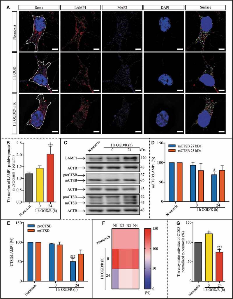
During reperfusion, lysosomal dysfunction may result from reduced CTSD activity. (A) LAMP1-positive puncta in the indicated groups were observed with SIM; scale bar: 5 μm. (B) The number of LAMP1-positive puncta per μm2 in the cytoplasm. (C-E) Representative images and analysis of western blots of LAMP1, mCTSB, proCTSD, and mCTSD in the indicated groups. (F and G) The enzymatic activity of CTSD in the neurons was evaluated with a Fluorometric Assay Kit from Abcam. N1-N4 indicated the number of replicated times. The enzymatic activities of CTSD were normalized to those of normoxia as the ratio (0–150). (B, n = 12; D and E, n = 4; G, n = 4; *p < 0.05, ***p < 0.001 vs. the normoxia group; #p < 0.05, ###p < 0.001 vs. the 1-h OGD group). Statistical comparisons were carried out with one-way ANOVA. Data are shown as the mean ± SEM. For Quantitative analysis of western blots of LAMP1, see Figure S1.
