PLOS ONE logoLink to PLOS ONE
. 2021 Jun 15;16(6):e0249948. doi: 10.1371/journal.pone.0249948

Age-preserved semantic memory and the CRUNCH effect manifested as differential semantic control networks: An fMRI study

Niobe Haitas 1,2,*, Mahnoush Amiri 1, Maximiliano Wilson 3, Yves Joanette 1,2, Jason Steffener 4
Editor: Anna Manelis5
PMCID: PMC8205163  PMID: 34129605

Abstract

Semantic memory representations are overall well-maintained in aging whereas semantic control is thought to be more affected. To explain this phenomenon, this study aims to test the predictions of the Compensation Related Utilization of Neural Circuits Hypothesis (CRUNCH) focusing on task demands in aging as a possible framework. The CRUNCH effect would manifest itself in semantic tasks through a compensatory increase in neural activation in semantic control network regions but only up to a certain threshold of task demands. This study will compare 40 young (20–35 years old) with 40 older participants (60–75 years old) in a triad-based semantic judgment task performed in an fMRI scanner while manipulating levels of task demands (low vs. high) through semantic distance. In line with the CRUNCH predictions, differences in neurofunctional activation and behavioral performance (accuracy and response times) are expected in young vs. old participants in the low- vs. high-demand conditions manifested in semantic control Regions of Interest.

Introduction

Language overall is well preserved in aging [1] and semantic memory may even improve across the lifespan [26], despite numerous neurophysiological declines in other cognitive domains that occur in the aging brain [68]. When compared with attention or memory, the relative preservation of language throughout lifetime [9] could be justified by the necessity to maintain successful communication, resulting in compensatory, flexible or atypical recruitment of neural resources [6]. Performance in terms of accuracy in semantic tasks is generally well maintained in older adults considering their more extensive experience with word use and a larger vocabulary than younger adults [2, 5, 6, 1013]. Response times (RTs) however are often longer compared to younger adults [10], possibly because older adults are slower in accessing and retrieving conceptual representations from their semantic store [1416], engaging the required executive function resources [17], and necessary motor responses [18]. Aside from behavioral performance, findings reported in the literature about the neural correlates sustaining semantic memory of older adults when compared with younger ones, are often conflicting, depending on the task utilized, inter-individual variability and the specific age group. Though several age-focused neurofunctional reorganization phenomena (e.g. Hemispheric Asymmetry Reduction in Older Adults-HAROLD [19] and Posterior-Anterior Shift in Aging-PASA [20] aim to explain how aging affects cognitive skills in general, it is still not clear how aging impacts the underlying pattern of activation sustaining semantic memory, given its relative life-course preservation. The relative preservation of semantic memory performance in older adults when compared with other cognitive fields [9, 21, 22] could be partly justified by the proposed dual nature of the semantic memory system, as expressed within the controlled semantic cognition framework [2326]. The present study focuses on the question of preservation of semantic memory in aging, defined as the ‘cognitive act of accessing stored knowledge about the world’ [27] using a semantic judgment task manipulating semantic control with two demand levels (low and high).

To account for conflicting findings in terms of brain activation during semantic memory tasks and the relative preservation of semantic memory in normal aging, a possible explanation is to consider it the result of adaptive mechanisms captured within the CRUNCH model (Compensation Related Utilization of Neural Circuits Hypothesis) [28]. This theory states that it is the level of task difficulty that impacts performance and neurofunctional activation in both younger and older individuals, whereas aging could be thought of as the expression of increasing task demands earlier than in younger adults. Accordingly, additional neural resources are recruited to attempt compensation when faced with elevated task requirements, echoing an aspect of the aging process manifestation [29, 30]. Compensation is thus defined as ‘the cognition-enhancing recruitment of neural resources in response to relatively high cognitive demand’ [29]. Alternatively, age-related reorganization phenomena refer to reduced neural efficiency, also known as dedifferentiation, resulting in reduced performance in the old [3134].

At the same time and as part of the age-related neurofunctional reorganization, neural resources may migrate from the default mode network (DMN) towards more urgent task requirements, which can be expressed as underactivation in such areas subserving ‘redundant’ tasks [28]. Indeed, the more task demands increase, the more DMN activation is expected to decrease, however this ability to ‘silence’ the DMN reduces in older adults [35]. Both over- and underactivation are relevant terms referring to comparisons with optimal patterns of activation as seen in younger adults [28]. Although the CRUNCH model describes compensatory neural mechanisms, it is not without its limits. For older adults, the overactivation benefit is thought to reach a threshold beyond which additional neural resources do not suffice, after which activation declines and performance deteriorates [28]. The relationship between task demands and fMRI activation has been described as an inverted U-shaped one, with the curve of older adults being to the left of the curve of younger ones. In other words, older adults would recruit additional neural resources at lower levels of task demands, reach a maximum and decrease in activity as task demands continue to increase earlier than younger ones (see Fig 3a in [29, 30]).

The CRUNCH hypothesis was conceived on evidence from a working memory study. Activation increased in the dorsolateral prefrontal cortex when accuracy was maintained and decreased when accuracy was compromised, depending on task load, or else, the number of items successfully retained [36, 37]. Congruent results were found in another working memory study, claiming that older adults may achieve the same outcomes using different neural circuits or strategies to achieve age-matched performance [38]. However, the CRUNCH predictions were not confirmed in recent working memory studies. In a working memory study with 3 load conditions using functional near-infrared spectroscopy (fNIRS), activation in the young progressively increased in the PFC as difficulty increased and performance was maintained [39]. However in the older adults, when performance was compromised during the most difficult condition, activation in the PFC bilaterally remained high. Similarly, in a visuospatial working memory task with 4 levels of task demands, the CRUNCH predictions were not found [40]. Instead, an increase in activation was found in a large network (premotor, prefrontal, subcortical and visual regions) however, no ‘crunch’ point after which activation decreases was found for the older group. Though older adults showed increased activation across regions at the higher task loads when compared with the younger ones, at the group level this difference was not significant, thus challenging the CRUNCH prediction of interaction between difficulty and fMRI activation.

Compatible with the CRUNCH expectations, increased activations with relatively maintained performance have been reported in frontoparietal regions in several language studies, however the results are not always consistent. More precisely, in a discourse comprehension study using fNIRS, increased activation was found in the left dorsolateral prefrontal cortex in older adults while performance was mostly equal to their younger counterparts [41]. In a sentence comprehension study, increased activation was observed in both younger and older adults during the more complex sentences in regions such as the bilateral ventral inferior frontal gyrus (IFG)/anterior insula, bilateral middle frontal gyrus (MFG), bilateral middle temporal gyrus (MTG), and left inferior parietal lobe [42]. Older adults showed increased activity compared with the young in the IFG bilaterally and the anterior insula in the difficult condition, however their performance in terms of accuracy was not maintained. Partially compatible with CRUNCH, overactivations with maintained performance have also been observed in a picture naming study manipulating for task demands/inhibition [43]. When naming difficulty increased, both younger and older adults showed increased activation in bilateral regions such as the IFG, the anterior cingulate gyri, the pre-, post-central, supramarginal and angular gyri, together with maintained performance while response times (RTs) of older adults did not significantly increase [43]. Fewer studies exist on semantic memory in light of increasing task demands, which is the focus of the current study.

Given the large volume of concepts and processes involved, semantic memory relies on a widely distributed and interconnected mainly left-lateralized core semantic network [17, 27, 4446] and bilaterally the anterior temporal lobes (ATL) proposed to act as semantic hubs [47, 48]. Semantic memory is suggested to be organized as a dual system composed of two distinctive but interacting systems, one specific to representations and one specific to cognitive-semantic control [25, 46, 4953]. In other words, it is thought to include processes related to stored concept representations with their modality-specific features which would interact with control processes in charge of selecting, retrieving, manipulating and monitoring them for relevance and the specific context, while at the same time suppressing irrelevant information [2426, 5457]. Within the controlled semantic cognition framework [26], the semantic control network would be significantly recruited during more complex tasks underpinned by left-hemisphere regions such as the prefrontal cortex (PFC), inferior frontal gyrus (IFG), posterior middle temporal gyrus (pMTG), dorsal angular gyrus (dAG), dorsal anterior cingulate (dACC), and dorsal inferior parietal cortex (dIPC) [25, 26, 45, 46, 51, 53, 58, 59], potentially extending towards the right IFG and PFC when demands further intensify [46]. One of the most up-to-date and extensive meta-analysis of 925 peaks over 126 contrasts from 87 studies on semantic control and 257 on semantic memory, found further evidence for the regions involved in semantic control, concluding them to the left-lateralized IFG, pMTG, pITG (posterior inferior temporal gyrus), and dmPFC (dorsomedial prefrontal cortex) regions [24]. Regions related to semantic control are thought to be largely overlapped by the neural correlates of the semantic network [24] but also thought to largely overlap with regions related to the ‘multiple-demand’ frontoparietal cognitive control network involved in planning and regulating cognitive processes [26, 60].

Differential recruitment has been found for easy and harder semantic tasks in young adults including recruitment of semantic control regions for the latter. In a study using transcranial magnetic stimulation (TMS) on the roles of the angular gyrus (AG) and the pMTG, participants were required to perform identity or thematic matchings that were either strongly or weakly associated, based on ratings previously collected and where RTs were used as a function of association strength. Stimulation to the AG and the pMTG confirmed their roles in more automatic and more controlled retrieval respectively [58]. An fMRI study used a triad-based semantic similarity judgment task to compare between concrete and abstract nouns (imageability) while manipulating additionally for difficulty. Difficulty was based on semantic similarity scores based on ratings of words, and for every triad, a semantic similarity score was computed to classify them as easy or hard. Increased activations were found during the hard triads and regardless of word imageability, in regions modulating attention and response monitoring such as bilaterally in the cingulate sulcus, the medial superior frontal gyrus and left dorsal inferior frontal gyrus [61]. In a triad-based synonym judgment task comparing concrete vs. abstract words, where triads were categorized as easy or difficult based on the respective response time in relation to the group mean, a main effect of difficulty was confirmed, with increased activations reported in the left temporal pole, left IFG and left MTG [62]. In a triad-based task where participants were requested to match words for colour and semantic relation to probe more automatic or controlled semantic processing respectively, greater activation was found in the IFG and IPS during the more difficult triads that were based on colour-matching. Accuracy was overall maintained equally across conditions but there were more errors and longer RTs in the ‘difficult’ colour condition, lending support to the controlled semantic cognition idea [50]. There is therefore evidence to support an increase in activation in semantic control regions when semantic processing demands increase, which could be attributed to ‘matching’ task requirements with available neural resources, in line with CRUNCH predictions. When it comes to aging, though the system related to representations is thought to be well-maintained, the system related to cognitive-semantic control is thought to be more affected [23]. This study focuses on how the relation between semantic control network activation and increasing task demands is affected by aging.

The neural correlates sustaining semantic memory are thought to be largely age-invariant, with only small differences existing in neural recruitment as a function of age [16, 22, 6366]. In a recently conducted meta-analysis of 47 neuroimaging studies comparing younger and older people, increases in activation in semantic control regions in older adults were reported when compared with younger ones, while accuracy was found to be equal between the two groups [22]. Though this increase in activation could be attributed to compensatory accounts, it could also reflect age-related loss of neuronal specificity or efficiency [22]. Several studies report activation and performance results in line with the compensatory overactivation account. In a semantic judgment task, participants had to decide whether two words share a common feature (shape or color) with their performance being categorized as better or worse based on a split from behavioral data [56]. In better performing older adults, activation was increased relative to younger adults in control regions such as the inferior parietal and bilateral premotor cortex, regions important for executive functions and object visual processing as well as relative to poorer performing older adults, in the premotor, inferior parietal and lateral occipital cortex. A further analysis for gray matter found that increased gray matter in the right precentral gyrus was associated with maintained performance [56]. In a semantic categorization study, older participants performed as accurately as the younger ones but had slower RTs. Their maintained performance was correlated with activation in a larger network than the one of younger ones, including parts of the semantic control network (such as left frontal and superior parietal cortex, left anterior cingulate, right angular gyrus and right superior temporal cortex), which was reportedly atypical and excluded the PFC [44].

Specifically to left IFG recruitment, believed to be in charge of top-down semantic control [45, 49, 51, 67], its association with the ‘difficult’ condition has been reported in several studies. In a triad-based semantic judgment task evaluating for rhyme, semantic and perceptual similarity, interaction and conjunction analyses revealed a significant interaction between age and the high-load semantic condition. Older adults overrecruited the control-related regions of the left IFG, left fusiform gyrus and posterior cingulate bilaterally, when competition demands increased while their accuracy was even better than their younger counterparts [66]. In a picture-naming task, older adults recruited overall larger frontal areas than younger ones in both hemispheres. Though the bilateral -and not the solely-left- recruitment of the IFG was beneficial to performance of older participants, the recruitment of other right-hemisphere regions was negatively correlated with accuracy [16]. The authors provided support to the finding that the neural substrates for semantic memory representations are intact in older adults whereas it is the executive aspect of language functions, including accessing and manipulating verbal information, that are most affected by aging [16]. In another study with young adults only, aiming to dissociate the role of the IFG in phonologically vs. semantically cued word retrieval, the recruitment of anterior-dorsal parts of the LIFG was associated with the high task demands condition in the semantic fluency condition, while performance was maintained [68].

Evidence therefore exists for a correlation between an increase in activation of semantic control regions when faced with increased task demands, which could be indicative of the compensation account to favor semantic memory performance in both young and older adults, and potentially reflecting the ascending part of the U-shaped relation between fMRI activation and task demands. Attributing however a causal relation between increased activation in the semantic control network and compensation is not straightforward. Distinguishing between the compensation and de-differentiation accounts can be challenging, as merely correlating brain activation with behavioral outcomes to claim compensation is methodologically incomplete [69, 70]. Many studies do not manipulate or cannot be compared for task demands and thus interpreting results that correlate neural activation with behavior can be confusing [53]. For example, in a study where task demands are lower, reorganization may be interpreted as compensatory when performance is maintained whereas when performance is more affected, it can be attributed to dedifferentiation. Numerous methodological caveats exist when attempting to allocate meaning a posteriori to age-related reorganization, given the observational nature of neuroscience, but also the need for more robust methodological designs, including longitudinal studies that measure in-person changes, between regions comparison and better analytic approaches (for a review see [70]). Correlating increased activation with improved performance at a single point in time and attributing it to compensation would require additional measures, also given that compensation may be attempted or only partly successful [30, 71].

According to the CRUNCH theory, the compensatory increase in activation of semantic control regions is thought to reach a plateau beyond which additional resources no longer benefit performance [28]. As such, reduced activation in cognitive control regions when semantic processing demands increase has also been reported. According to CRUNCH, this reduced activation could be interpreted as neural resources having already reached their maximum capacity and no longer being sufficient to successfully sustain compensation for the task [28]. Indeed, the meta-analysis of 47 neuroimaging studies comparing activation in young and older adults (mean age of young participants: 26 years (SD = 4.1) and mean age of older participants: 69.1 (SD = 4.7) during semantic processing tasks, also reported decreased activation in the older adults in typical semantic control regions in the left hemisphere (IFG, pMTG, ventral occipitotemporal regions and dIPC) together with increased activation in ‘multiple-demand network’ regions in the right hemisphere (IFG, right superior frontal and parietal cortex including the middle frontal gyrus, dIPC and dACC) especially when performance was sub-optimal [22]. In a semantic judgment task (living vs. non-living judgement of words) study with two levels of difficulty and four across-the-lifespan age groups, activation outside the core semantic network increased with age linearly and contralaterally towards the right hemisphere (right parietal cortex and middle frontal gyrus) in the easy condition, while accuracy was maintained [64]. In the difficult condition however, RTs were slower and reduced activation was observed in older participants in semantic control regions, namely the frontal, parietal and cingulate cortex regions, suggesting a declining ability of brain to respond to increasing task demands by mobilizing semantic control network resources as age increases [64].

Similarly, increased activation in right-lateralized semantic control regions was detrimental to performance in both young and old participants in a word generation study manipulating for task difficulty [72]. Indeed, activation in the ventral IFG bilaterally was correlated with difficult items as opposed to easier ones and reduced performance irrespective of age. In a verbal fluency study by the same group using correlation analysis, a strong negative correlation was found between performance and activation in the right inferior and middle frontal gyrus ROIs [73]. Older adults demonstrated a more bilateral activation than younger ones especially in the right inferior and middle frontal regions whereas their performance during the semantic task was negatively impacted. However, this right-lateralized semantic control network increase in activation together with a drop in performance has not been consistently documented. For example, in a semantic judgment task on word concreteness using magnetic encephalography (MEG), older participants overactivated the right posterior middle temporal gyrus, inferior parietal lobule, angular gyrus and the left ATL and underactivated the control-related left IPC as a result of increased task demands while their performance was equivalent to the young, thus lending support to compensatory accounts [65]. According to CRUNCH, the above findings could be interpreted within the descending part of the inverted U-shaped relation between semantic processing demands and fMRI activation [29], whereby after a certain difficulty threshold, available neural resources from the semantic or multiple-demand control network have reached their maximum capacity and further lead to reduced activations and a decline in performance [30].

In summary, it seems that depending on the semantic task used and its perceived or actual difficulty, both increased and decreased activations have been reported in the semantic control network along with variations in consequent performances. The relationship between neural activation, task difficulty and behavioral performance is not straightforward. It is possible that the neural correlates of semantic memory remain relatively invariant throughout aging when the task is perceived as easy. On the other hand, when task difficulty or the perception of it increases, activation and behavioral performance may increase or reduce depending on the nature of the task and its level of perceived or actual difficulty, in line with CRUNCH. Accordingly, maintained performance could depend on the additional recruitment of semantic control network resources but only between certain thresholds of difficulty, before which increasing activation would be unnecessary or beneficial and after which performance would decline.

Age-related reorganization phenomena alternative to CRUNCH

A number of alternative neurofunctional reorganization phenomena have been reported to account for the evolution of general cognitive skills in aging (for reviews, see ([30, 74, 75]). Such phenomena often refer to the engagement of compensatory mechanisms and redistribution of resources through overactivation or deactivation often including in the PFC [28, 30]. For example, the HAROLD neurofunctional reorganization phenomenon refers to a hemispheric asymmetry reduction in older adults with the objective of maintaining high performance [19]. To reduce the asymmetry, brain activation can increase and/or decrease in certain brain areas by recruiting additional and alternative neuronal circuits from the contralateral hemisphere. The resulting asymmetry reduction optimizes performance, whereas elderly adults who maintain a unilateral or asymmetrical activation pattern similar to the young, do not perform as well [19]. Several studies have recently challenged the accuracy of the HAROLD model [76, 77]. An alternative pattern of neurofunctional reorganization has been reported to occur intrahemispherically. The PASA (Posterior Anterior Shift in Aging) phenomenon provides a picture of such type of reorganization [78], describing an age-related shifting of activation from the occipitotemporal to the frontal cortex [20, 79]. PASA is considered to reflect a general age-related compensation phenomenon for processing sensory deficits by decreasing activation in occipitotemporal regions and increasing activation in frontal regions rather than reflect task difficulty [20]. A recent metaanalysis [80] on healthy aging provided support for the findings of the PASA phenomenon, however, others have challenged its compensatory claim [81]. Additionnally to the above intra- and inter-hemispheric reorganization phenomena is the ‘cognitive reserve’ hypothesis, which attributes successful cognitive processing in aging to complex interactions between genetic and environmental factors that influence brain reserve and the brain’s ability to compensate for age-related pathologies [82]. Cognitive reserve is proposed to depend on both neural reserve and neural compensation, a distinction reflecting inter-individual variability to use resources efficiently, flexibly or differently while performing cognitive tasks but also using alternative strategies in pathological situations. Accordingly, older adults can adapt to aging and cope with increased task demands in a flexible manner by activating regions similarly to the young or alternative ones or both.

Alternatively, neurofunctional reorganization phenomena are attributed to reduced neural efficiency, also known as dedifferentiation, resulting in reduced performance in the old [31, 32, 34, 83, 84]. According to the dedifferentiation hypothesis, aging reduces the specialization of neurons which is critical for their optimal functioning [31]. Accordingly, increased activations could be the result of randomly recruiting neurons in an attempt to meet processing demands [19], or could reflect the brain’s failure to selectively recruit specific regions [34] whereas increasing task demands may aggravate the non-specificity of neural responses [85]. Evidence exists to support the idea that neural responses are less specific in older adults when compared with younger ones, as demonstrated in the ventral visual cortex during a viewing of pictures task [83, 86], during a working memory task [87] (for a review, see [88] and in motor evoked potentials [89]). It is not clear however whether this loss of neural specificity would be the result of aging or could be attributed to larger experience of older adults in recognizing objects [83]. At the same time, it is thought that both compensation and dedifferentiation phenomena may take place in the same person simultaneously in different regions [87]. The dedifferentiation account would predict a reduction in performance together with an increase in activation, thus resembling the descending part of the inverted-U shape relation between task demands and fMRI activation, as per CRUNCH.

An additional explanation for age-related functional reorganisation is that aging selectively affects the default mode network (DMN). This network is normally activated during a situation when one is not involved in any task but instead monitors their internal and external environment [7] and deactivated when performing cognitive tasks so as to reallocate attentional resources towards them [35]. It is thought that the semantic network is largely activated at rest, as individuals would be engaged in language-supported thinking when not performing specific tasks [90]. It has been found that when the task is cognitively demanding, DMN deactivations are smaller and slower for older adults, implying that they are more easily distracted whereas their capacity to inhibit irrelevant information is compromised [28, 35, 91], in line with the inhibitory control view [92] and the cognitive theory of aging [7]. In difficult semantic tasks, maintained performance was associated with increased segregation between DMN and semantic control regions at rest, whereas reduced performance was associated with increased verbal thinking at rest [93]. It is possible that aging reduces the efficiency of transferring attention away from resting areas towards task requirements, thus probably affecting the balance between DMN and task-related activity and resulting in reduced cognitive performance [7].

The neurofunctional reorganization proposals discussed above seem to be exclusive of another as they tend to focus and attribute meaningfulness in increased or decreased activation in isolated brain regions, whereas none seems to fully capture and explain age-related reorganization [94]. Several researchers have attempted to identify the ‘common factor’ [95] in age-related brain activation patterns to explain reorganization. Cabeza (2002) [19] considers that functional reorganization is more likely to be non-intentional and neuron-originated rather than a planned change of cognitive strategies, since it is manifested in simple tasks or following unilateral brain damage, over which one has little control. On the contrary, Reuter-Lorenz and Cappell (2008) [28] consider unlikely that such a huge variability in brain activation stems from the same ‘common factor’ or is due to age-related structural changes in the brain, because then it would be consistent across all tasks. Instead, aging seems to selectively affect specific regions, mainly default-mode regions and the dorsolateral PFC [7] whereas inter-individual variabilities need to be emphasized when accounting for age-affected cognitive domains [96].

Recent studies tend to combine data on functional, structural and lifetime environmental factors to explain reorganization in a more integrative manner. In this direction, the more comprehensive Scaffolding Theory on Aging and Cognition- STAC hypothesis proposes that aging is no longer characterized by uncontrollable decline of cognitive abilities because the brain develops its own resilience, repairs its deficiencies and protects its functions [28, 97]. This idea is reflected in the aging models that emphasize the plasticity of the brain due among other factors to training interventions and their impact on neural structure, as well as functional and behavioral outcomes [98100]. The impact of short-term practice as well as long-life training would impact young and older adults differently [69]. Accordingly, engaging in intellectually challenging activities and new learnings throughout the course of a lifetime but also on a shorter-term course could stimulate plasticity of the brain. The capacity of the brain to resolve the mismatch between intellectual demands and available neurofunctional resources and its capacity to trigger behavioral adaptive strategies, would define its plasticity and affect its brain knowledge systems and processing efficiency [69]. Plasticity would demonstrate itself as increased functional activation especially in regions that are most structurally affected by aging because of atrophy, loss of grey and white matter density and cortical thinning, such as in the fronto-parietal network [99]. Aging could thus be characterized by structural loss but also neural and functional adaptation to this loss, including through the utilization of new strategies [99]. Indeed, age-related overactivations seem to be a reliable and consistent pattern observed in multiple domains regardless of whether they are more localized, contralateral or seen in the fronto-parietal multiple-demand network [101]. In summary, the more adaptable and the more dynamic the brain is, the better it would maintain its cognitive abilities [102].

Specifically to semantic memory preservation in aging, it is not clear what mechanisms are in place to account for the preservation of semantic memory in aging, supported by the intersection of both domain-general and linguistic abilities [66]. Findings in the literature about the adoption of neurofunctional activation pattern during semantic processing in aging, vary. Two additional compensatory hypotheses have been proposed: the executive hypothesis refers to the recruitment of domain-general executive processes seen as overactivation in prefrontal, inferior frontal and inferior parietal brain regions to compensate for age-related cognitive decline [6, 103], as seen for example in a semantic judgment task [56]. Indeed, the metaanalysis of semantic memory studies performing activation likelihood estimation (ALE) between young and older participants [22], found a shift in activation from semantic-specific regions to more domain-general ones, in line with the executive hypothesis. The semantic hypothesis on the other hand, also known as left anterior-posterior aging effect (LAPA), refers to the recruitment of additional semantic processes in older adults, seen as overactivation in ‘language’ regions in the left posterior temporo-parietal cortex [104, 105]. Given the larger decline in older adults of executive over language functions could justify this latter hypothesis considering that language is better maintained over executive processes [106]. Evidence for the semantic hypothesis was found in a study using semantic judgment task where participants had to decide if a word is an animal or not. Older participants had more bilateral parietal, temporal and left fusiform activations than younger ones who presented more dorsolateral activations, which the authors interpreted as older participants relying more on semantic processes whereas younger ones relying more on executive strategies [107]. However, language and executive functions are overall intertwined given that regions such as the left inferior frontal gyrus and the PFC are proposed to serve both executive and language functions, thus blurring the intersection between the semantic and executive hypothesis [53].

An alternative approach can be seen within the good-enough theory, which claims that participants tend to construct semantic representations which are ‘good-enough’ or shallow rather than more complete or detailed ones, with the aim to perform the task at hand with the least effort and save on processing resources [108110]. This theory refers to overall language processing, but it could also be applied to the semantic representation of words as inferred by the semantic judgment task used for the current study. Accordingly, participants and especially older adults at increased task demands, may resort to a more ‘shallow’ or superficial interpretation of the semantic judgment task they are required to perform and instead of analyzing thoroughly all semantic aspects of the words they are presented with (e.g. semantic features of the apple in comparison with the grape or cherry), may bypass some aspects of the task and thus resort to a quick decision. Such a shallow processing could be manifested with decreased activation overall, as well as in the semantic control network which would be in charge of selectively controlling for semantic features while ignoring others [56]. This alternative explanation is in line with the idea that at peak levels of demand, participants may become frustrated with frequent errors or difficulty to resolve competing representations, and may deploy inefficient strategies [111].

In summary, some inconsistencies are found in interpretation of results, with both increased and decreased activation reported as the result of aging [7, 112]. Neurofunctional reorganization can take the form of both inter- and intra-hemispheric changes in activation and manifests as both increased and decreased activation of specific regions [7]. When performance is compromised, reduced activation is interpreted as impairment, attributed to neural decline, inefficient inhibitory control or de-differentiation [28] whereas when performance is maintained, it is claimed to be compensatory. Most studies seem to agree on increased activation, interpreting it as compensatory and positive, whether it is understood as increased attention or as suppression of distracting elements [113]. Overactivation is also found in Alzheimer’s disease (AD) and mild cognitive impairment (MCI) patients demonstrating either its compensatory role or a progressive pathology predicting further decline [34, 35]. It seems that neurofunctional reorganisation of the aging brain is more complex and further research is still required to be able to ‘draw’ a pattern of activation that integrates the existing findings in a comprehensive model and one that can be applied to semantic memory, one of the best preserved cognitive fields in aging.

Current study

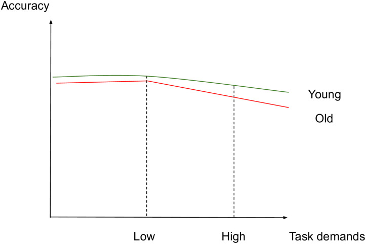
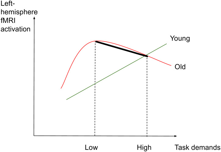
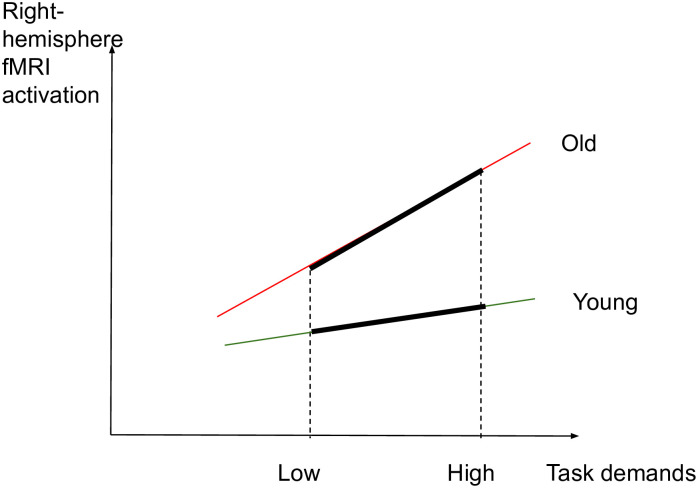
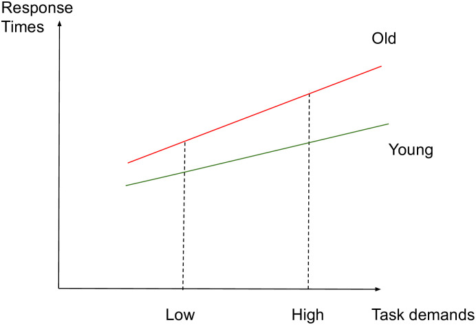
The aim of this study is to identify whether aging affects the brain activity subserving semantic memory in accordance with the CRUNCH predictions, through a semantic judgment task with two levels of demands (low and high). Task demands will be manipulated through semantic distance, which is found to influence both performance and brain activation levels [49, 61, 67, 114117]. We hypothesize that brain activity and behavioral performance (dependent variables) will support the CRUNCH model predictions when demands on semantic memory are manipulated in young and old adults (age and task demands: independent variables). More specifically, it is expected that 1) the effects of semantic distance (low vs. high-demand relations) on neurofunctional activation and behavioral performance (accuracy and RTs) during the semantic judgment task will be significantly different between younger and older participant groups, with younger adults performing with higher accuracy and faster response times than older adults. Furthermore, we predict age group differences in brain activation in semantic control regions bilaterally which are sensitive to increasing task demands [24]. This will be evident with a significant interaction effect between age group and task demands within regions of interest consisting of the core semantic control regions: IFG, pMTG, pITG and dmPFC. This will support the idea of the brain’s declining ability to respond to increasing task demands with advancing age. If this interaction is not found between task demands and age, the following are expected 2) In the low-demand (LOW) condition, both younger and older participants will perform equally in terms of accuracy and with less errors than in the high-demand condition. However, it is anticipated that older adults will present longer RTs and significant increases in activation in left-lateralized semantic control regions compared to the younger participants. 3) In the high-demand (HIGH) condition, it is expected that younger adults will perform better (higher accuracy and lower RTs) and present significant activation in the left-hemisphere semantic control regions compared to older adults. Older adults are expected to exhibit reduced performance compared to younger adults (lower accuracy and higher RTs), reduced activation in left-lateralized semantic control regions, and increased activation in right-lateralized semantic control regions compared to the younger adults. To illustrate the hypothesized relations between task demands and accuracy, RTs and activation in young and older adults, see Figs 13 below. The theoretical relations between task demands and activation are represented in the decrease in activation in the left hemisphere (cross-over interaction, Fig 3) and the increase in activation in the right hemisphere (difference in slopes interaction, Fig 4), confirming the hypothesized CRUNCH predictions. These portray the main effects of age and task demands as well as their interaction highlighted by thick lines.

Fig 1. Accuracy and task demands in younger and older adults.

Fig 1

Fig 3. Left-hemisphere activation and task demands in younger and older adults.

Fig 3

Fig 4. Right-hemisphere and task demands in younger and older adults.

Fig 4

Fig 2. RTs and task demands in younger and older adults.

Fig 2

These analyses are looking for age and load effects on task performance and on brain activation in separate analyses. Follow-up exploratory analyses within the ROIs will explicitly test how differential brain activation is related to task performance. It is hypothesized that older adults who have high levels of brain activation in left-lateralized semantic control regions during the high-demand condition, similar to the young adults, will have higher levels of task performance (reduced errors and RTs) than their counterparts whose brain activation is lower in these regions, as per the CRUNCH model, indicating that they have not yet reached their crunch point after which performance and activation decline. To accept the above hypotheses, at least one ROI from the ones mentioned is expected to be activated.

A control condition is part of the task and was designed to maximize perceptual processing requirements and minimize semantic processing ones [118, 119]. As a test of positive control, within group comparisons with the control condition are expected to show activation in the primary visual and motor cortices, which are involved with viewing of the stimuli, response preparedness and motor responses [64, 120, 121]. No CRUNCH effects are expected in the control condition. Task effects within each age group will also be tested and activation is expected to be of greater amplitude in the high vs. low condition in both young and old age groups.

This task design utilizes explicit definitions of low and high levels of task demand. However, each individual participant will experience their own subjective level of task difficulty. Perceived difficulty of triads will be measured on a difficulty 1–7 likert-scale (e.g. 1: very easy, 7: very difficult. Subsequent analyses will explore this question with heterogeneous slopes models using individualized rescaled levels of task difficulty and will compare brain activation with performance, brain activation with perceived difficulty and performance with perceived difficulty. This approach will determine how the relationship between individual task difficulty and brain activity is affected by age group.

Proposed experiment: Materials and methods

The authors comply with the Centre de Recherche Institut Universitaire de Gériatrie de Montréal (CRIUGM) Ethics Committee and the Centre intégré universitaire de santé et de services sociaux du Centre-Sud-de-l’Île-de-Montréal requirements (CÉR-VN: Comité d’Éthique de la Recherche- Vieillissement et Neuroimagerie), in line with the principles expressed in the Declaration of Helsinki. The ethics committee of CRIUGM and CÉR-VN approved this study with number CER VN 16-17-09. The approval letter is available in the OSF repository (DOI: 10.17605/OSF.IO/F2XW9). For all methodology aspects of this current study, compliance with the OHBM COBIDAS report/checklist [122] and guidelines [123] will be aimed for as much as possible. We will share the preprocessed functional datasets in MNI space publicly in Open Science Framework (https://osf.io/) with a digital object identifier (DOI: 10.17605/OSF.IO/F2XW9) to permanently identify the dataset [122], and we will index it at the Canadian Open Neuroscience Platform (https://conp.ca/) to increase findability. In addition, once these become available, we will upload our unthresholded statistical maps to neurovault (https://neurovault.org/), an online platform sharing activation data. Permanent links to the unthresholded statistical maps to be uploaded at Neurovault will be provided as part of the dataset deposited on the OSF, under the same DOI (DOI: 10.17605/OSF.IO/F2XW9). Data will be organized following the Brain Imaging Data Structure (BIDS) to maximize shareability. Supporting documentation for this study is available at DOI: 10.17605/OSF.IO/F2XW9.

Participants

A sample of 80 participants will be tested for this study: 40 in each group, Young: 20–35 years old and Older: 60–75 years old (male = female). We will recruit 86 participants assuming that some will be excluded in the process due to low task performance, excessive motion or technical issues. Participants will be recruited through the Centre de Recherche Institut Universitaire de Gériatrie de Montréal (CRIUGM) ‘Banque de Participants’, but also through poster announcements posted in Montreal and in social media. Participants will be bilingual (French and English-speaking) with French as their dominant language used on a daily basis. Multilingual participants will be excluded, as speaking many languages may influence semantic performance [124]. Participants will be matched for education level with college studies (CEGEP) as a minimum level of education, since education is a measure of cognitive reserve [82]. Participants will undergo a series of neuropsychological and health tests to determine their eligibility for the study as inclusion/exclusion criteria:

  • A health questionnaire (pre-screening to take place on the phone) to exclude participants with a history of dementia, drug addiction, major depression, stroke, aphasia, cardiovascular disease, diabetes, arterial hypertension or any drugs that could affect results. The pre-screening includes questions for bilingualism and use of French language, which needs to be the dominant one (inclusion criteria) (the complete questionnaire is available on osf.io, DOI: 10.17605/OSF.IO/F2XW9).

  • The Edinburgh Handedness Inventory scale: participants will be right-handed with minimum score for right-handedness of 80 [125].

  • The MoCA (Montreal Cognitive Assessment) test with a minimum cutoff score of 26 [126, 127].

  • The MRI-compatibility checklist (Unité Neuroimagerie Fonctionnelle/UNF) test (available at https://unf-montreal.ca/forms-documents/).

The following tests will also be performed with participants:

  • The Similarities (Similitudes) part of the Weschler Adult Intelligence Scale (WAIS-III) test [128, 129]

  • The Pyramids and Palm Trees Test (PPTT) (version images) [130] will be used as a measure of semantic performance.

  • The questionnaire Habitudes de Lecture (Reading Habits) (based on [131] as a measure of cognitive reserve [82].

Participants will provide written informed consent and will be financially compensated for their participation according to the CRIUGM and Ethics Committee policies.

Power analysis

This sample size is based on power calculation results from an age group comparison on a similar semantic task [132]. This dataset used a Boston naming semantic task and compared healthy young and old age groups. From this dataset effect size estimates were calculated from the contrasts for high versus low task demands within and between age groups. Effect sizes were extracted from the primary regions of interest for this study as defined by a recent meta-analysis of semantic control [24]. From the identified locations, a 10 mm cube was defined to identify the effect size at the published location, mean effect size and the robust maximum effect size in the ROI. Statistical power was then estimated using the G*Power tool [133]. Within group measures had robust effect sizes and demonstrated that sufficient power (alpha = 0.05, beta = 0.90) was achieved with a sample size of 40 participants in each group. The between group comparison of differential activation had sufficient power within bilateral temporal gyri and medial PFC. In addition, the proposed study will use more than twice the number of trials used in the data used for power estimations. This will decrease the within participant variation and will increase the power above that provided by the [132] study. The table of effect sizes used for the power analyses for within and between group comparisons are included as supplementary material at OSF.

Materials

Participants will be administered a task of semantic similarity judgment in French and that is suitable for the Quebec context developed for the current study, similar to the Pyramids and Palm Trees test (PPTT) [130]. The task proposed here involves triads of words resembling a pyramid where participants will need to judge within a time limit of 4 seconds which of the two words below (target or distractor) is more related to the word above (stimulus). Both target and distractor words are associated in a semantic relation with the stimulus word. Participants will thus be required to select which of the two competing words has a stronger semantic relationship to the stimulus word as measured by semantic distance between the stimulus and the distractor. Two types of triads exist: a) low-demand (distant) relations: the more distant the semantic relation between stimulus and distractor, the less demanding will be to select the correct target and b) high-demand (close) relations: the closer the semantic relation between stimulus and distractor, the more demanding will be to select the correct target as competition between the target and distractor words will be higher [61].

The task (150 triads in total) has two experimental conditions (120 triads: 60 low-demand (LOW) and 60 high-demand (HIGH) semantic relations) and one control condition (30 triads). For the control condition, the task will be to indicate which of the two consonant strings, which will be presented pseudo-randomly, are in the same case as the target strings (e.g. DKVP: RBNT-kgfc). The stimuli will look like Fig 5 below:

Fig 5. Examples of triads.
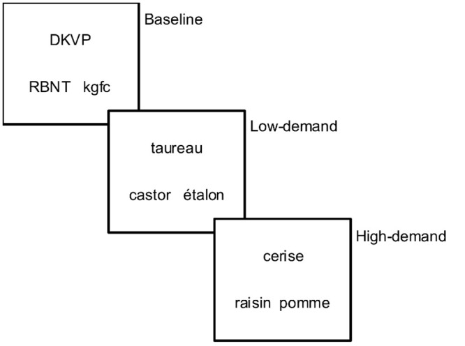
Fig 5

Stimuli description

The stimuli were developed for the current study. In every condition, the targets and distractors were matched for: a) Type of semantic relation: taxonomic and thematic. For thematic relations, the semantic distance was calculated with the help of a dictionary: ‘Dictionnaire des associations verbales (sémantiques) du français’ (http://dictaverf.nsu.ru/dict, version accessed on 2014), as a function of the number of respondents that associated two words together (i.e., the larger the number of respondents, the more closely associated the two words are, and vice versa). As such, a score of 1 means that only one person provided this answer (distant thematic relation) whereas a score of 100 means that 100 people provided this answer (close thematic relation). b) Frequency, based on the Lexique 3 database referring to films [134] c) Imageability, based on the Desrochers 3600 database [135]. Additional imageability ratings were collected from 30 participants for items without ratings in the above database. A Pearson’s correlation was performed with 30 test words from the Desrochers database to confirm that the ratings given for the new words were relevant compared to the ones that already exist. Participants rating items with a correlation value less than 0.6 were excluded, as it was deemed that they were not concentrated on the task. The final imageability rating of an item was the mean of the scores given by all included participants. ANOVA and Bonferroni corrected Tukey tests were performed to ensure the matching of a target and distractor for every condition. Finally, targets and distractors were matched on d) Word length.

The stimuli were created in a gradual process, continuously testing and evaluating its adequacy and aiming for a less than 40% error rate with pilots to test it, measure response times and gather comments. Every time, the four conditions were matched and passed an ANOVA test for mean frequency, imageability and length. Also, pilot participants were asked questions about the duration of the task and the sufficiency of time to respond. To evaluate the validity of the stimuli pertaining to low vs. high demands and younger vs. older adults, a pilot evaluation of stimuli was conducted by 28 participants (14 were older adults, age range: 67–79 years old, female = 9 and 14 were younger, age range: 21–35 years old, female = 10) for 60 triads (30 low-demand and 30 high-demand) using E-Prime. Repeated measures analysis of variance (ANOVA) was applied to the mean accuracy and median response data within each level of task demand (control, low, high) across the two age groups. The results are described below:

Accuracy. The Greenhouse-Geisser estimate for the departure from sphericity was ε = 0.63. There was not a significant interaction between age group and task demand, F(1.27, 32.94) = 0.065. p = 0.85, η2 = 0.0025. The main effect of task demand was significant, F(1.27, 32.94) = 10.36, p = 0.0015, η2 = 0.28. The estimated marginal means were: Control = 0.84, Low = 0.80 and High = 0.72. The main effect of age group was not significant, F(1, 26) = 0.34, p = 0.57, η2 = 0.013.

Response times. The Greenhouse-Geisser estimate for the departure from sphericity was ε = 0.54. There was not a significant interaction between age group and task demand, F(1.08, 28.14) = 1.14. p = 0.30, η2 = 0.042. The main effect of task demand was significant, F(1.08, 28.14) = 49.38, p < 0.0001, η2 = 0.66. The estimated marginal means were: Control = 1390ms, Low = 2230ms and High = 2292ms. The main effect of age group was significant, F(1, 26) = 4.78, p = 0.038, η2 = 0.15.

Based on the above pilot data, we confirm that our task includes a load effect that impacts task performance (accuracy and RTs) differently between younger and older adults, in the expected directions.

The following definitions were used:

  • Low-demand (distant) triads:
    • For taxonomic relations:

All items (stimulus, target, distractor) belong in the same semantic category (e.g., animals). Stimulus and target words belong in the same semantic sub-category (e.g. birds). For example, taureau: ÉTALON-castor (bull: STALLION-beaver).

  • For thematic relations:

Both the target and distractor words are thematically related to the stimulus and belong in the list of answers referred by dictaverf. To ensure the biggest distance possible, the target was the first adequate answer mentioned in dictaverf, whereas the distractor was the last or closest to the last answer, meaning that it had a score close to 1. For example, sorcier: village-BAGUETTE (wizard: village-WAND).

Alternatively, to ensure the biggest distance possible, the following criteria were used: when the distractor word is 1 (which means only 1 person provided this answer), when the distractor word is between 2–5 and the target word is above 10, and when the difference between the target and distractor words is bigger than 100.

  • High-demand (close) triads:
    • For taxonomic relations:

All items in the triad come from the same semantic sub-category (e.g. birds). The stimulus and target items share a visual or structural feature whereas the distractor word does not. For example, ‘cerise: RAISIN-pomme’ (cherry: GRAPE-apple) where cherries and grapes have a similar size and bunch structure.

  • For thematic relations:

Both the target and distractor words are thematically related to the stimulus. The target was the first adequate answer mentioned in dictaverf whereas the distractor had a score smaller or equal to half of the score of the target and was bigger or equal to 4. This criterion was used to ensure that the distractor was a more frequently mentioned answer but distant enough from the target (e.g. half of the people mentioned the distractor as opposed to mentioning the target). For example, ‘enfant: JOUET-sourire’ (child: TOY-smile).

Experimental design

Session 1: Neuropsychological tests

Participants will be recruited through the CRIUGM pool of participants and public announcements, with initial eligibility assessed through a phone interview (health questionnaire and MRI compatibility form). If eligible, the participant will partake in the first experimental session (approximately 90 minutes), during which they will sign the informed consent and MRI-compatibility forms, complete neuropsychological tests (see Participants section above) and practice with 15 practice triads (5 for every condition). Participants who qualify (meet the inclusion criteria from health questionnaire, MRI-compatibility questionnaire, MOCA and Edinburgh Handedness Inventory scale) for the fMRI scanning session following tests will proceed with the second session one week later (maximum 2 weeks later).

Session 2: fMRI scanning

For the second experimental session, the time commitment from the participant: is 90 minutes to allow for practice with triads, getting ready and leaving, following COVID-19 requirements. During this session, participants will listen to task instructions, and practice with 3 triads (1 per condition). Participants’ vision will be corrected, if necessary, with MRI-175 compatible lenses according to their prescription shared from the previous session. Additionally, pregnancy tests will be carried out when relevant, earplugs will be given to reduce machine noise and instructions will be given to remain still in the scanner while foam rubber pads in the head coil will restrict movement. Participants will then proceed with the actual task in the scanner. Stimuli will be presented with E-Prime 2.0.10.356 software run on Microsoft Windows 10 through an LCD projector projecting to a mirror over the participant’s head. Participants will select their responses using the index fingers of both hands on the MRI-compatible response box. A response on the right will be with their right hand and a response on the left with their left hand. Response data and response times (RTs) will be recorded via E-Prime for further analysis. No feedback will be shared with participants. Participant testing will alternate between young and older adults to minimize any bias due to scanner changes/upgrades.

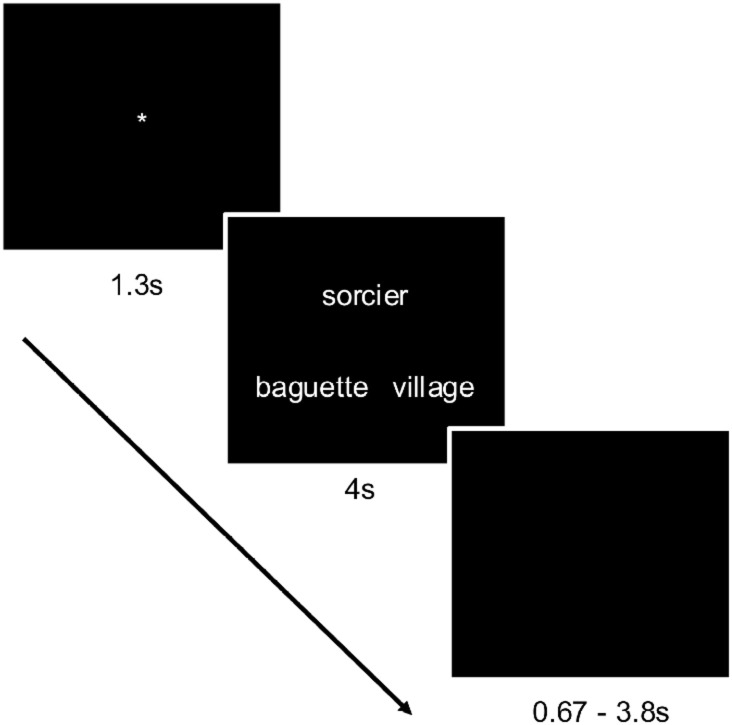
The semantic task will be event-related. Triads will be presented for 4 seconds, during which participants will need to make their choice by pressing on the left or the right button to select the word on the left or right respectively. A black screen will follow for approximately 2.2s (this interstimulus interval (ISI) will vary randomly between 0.67s and 3.8s to minimize possible correlations with the BOLD signal). A fixation point will appear for 1.3s to prepare the participant for the next trial. The whole trial will last between 5.97s and 9.10s, with an average of 7.5s. See below for a description of the methods used to determine the ISIs. Black screens were included at the beginning and the end of the runs. Information on the scanning flow is available in Fig 6 below:

Fig 6. Example of trial.

Fig 6

The task will be split in two runs with 75 triads per run (30 low-demand (LOW), 30 high-demand (HIGH) and 15 control triads), interleaved in a pseudo-random fashion. The duration for every run will be 9:45 minutes. The whole session is expected to last 45 minutes, including a 5-min break between runs 1 and 2.

Session 3

In regards to perceived task difficulty, an additional session with participants one week following the fMRI acquisitions will take place, whereby they will rate each triad on a difficulty 1–7 likert scale (eg. 1: very easy, 7: very difficult). We will further assess whether perceived difficulty correlates with actual performance scores (accuracy rates and RTs) and whether perceived difficulty correlates with levels of activation in the young and older adults (e.g. whether increased levels of perceived difficulty correlate with increased RTs and reduced accuracy, as well as levels of activation in semantic control regions).

Stimuli order and ISIs

To design the experiment in a way that maximizes design efficiency, optimal trial ordering and interstimulus intervals (ISIs) were chosen [136]. The methodology used simulated designs of random ordering of the three conditions. In addition, the ISIs were randomly drawn from Gamma distributions across a range of parameter values (shape: 0.1 to 10, scale: 0.1 to 5). This approach included expected error rates produced during the stimuli pilots to maximize design efficiency in the face of errors. A total of 800,000 simulations were performed. The ISI distribution and specific list as well as the condition order in which there was the smallest decrease in required BOLD signal response for detection as errors increased were chosen. The related ISIs are uploaded to the OSF platform.

fMRI data acquisition

Functional scans will be performed on a 3Tesla Syngo MR E11 Prisma_fit Siemens MRI machine with 32 channels at UNF (Unité de Neuroimagérie Fonctionnelle), CRIUGM. The start of the stimulus presentation software will be triggered by a pulse sent from the MRI to the stimulus laptop. To detect effects between conditions and to ensure a good fMRI signal in the brain, pilot data collected using the scanning protocol described here suggested a minimum temporal signal to noise ratio (TSNR) of 20 throughout the brain [137]. Participant data will be excluded if TSNR, assessed from every participant’s time series, is below 20. We will acquire T1-weighted MRI images for co-registration with fMRI data and atlases and to identify ROIs to be used as masks in the functional data analysis. An meMPRAGE (multi-echo MPRAGE) sequence (704 total MRI files) will be acquired with 1x1x1mm resolution, 2.2s repetition time, 256x256 acquisition matrix, a Field of View (FOV) of 256mm covering the whole head and echo times of 1.87ms, 4.11ms, 6.35ms, 8.59ms, 13ms and 15ms. The phase encoding orientation will be sagittal with a flip angle of 8 degrees.

For the functional scans (run 1 and 2), T2-weighted BOLD data will be acquired on the entire brain (including the cerebellum) using an Echo Planar Imaging (EPI) sequence with 50 slices, resolution 2.5x2.5x3mm, echo time of 20ms, repetition time of 3s and a flip angle of 90 degrees. Field of view will be 220x220mm and the acquisition matrix will be 88x88, in AC-PC direction covering 150mm in the z-direction. Slice order will be ascending-interleaved. For each run, 195 scans will be collected. The SIEMENS default double-echo FLASH sequence for field map distortion correction with the same parameters will be acquired after each sequence for inhomogeneity correction. Functional images will be reconstructed to the collected matrix size with no prospective motion correction. Two initial dummy scans will be collected and discarded by the MRI allowing for T1 saturation.

Proposed analyses

Behavioral data analysis

Response times and accuracy rates will be collected for every participant. Sex will be used as a covariate in all analyses. To account for performance, brain imaging analysis will focus on correct trials only ensuring that we are looking at brain activation related to accurate performance. Behavioral data (RT and accuracy) will be analyzed using mixed- design ANOVA with age as a between-subjects factor and condition (high vs. low demands) as within-subject factor. Accuracy rates will be transformed using Fisher logit approximation to avoid ceiling effects. Group analyses of the imaging data will be performed including behavioral covariates to investigate age group differences in the relationships between brain activity and task performance. Multiple comparisons across the 40 ROIs will be made using false discovery rate adjustments. Analyses will explicitly focus on the relationships between brain activation and task performance. These analyses will identify brain regions where age group differences in activation are dependent or independent of task performance. Time-outs (delayed responses) will be modeled and analyzed separately. Any missing or incomplete data will be excluded (the whole participant).

Imaging data analysis

Preprocessing

Preprocessing image analysis will be performed with SPM12 software. Images will be corrected for slice timing (differences in slice acquisition time), with ascending-interleaved slice order and using the acquisition time for the middle slice as the reference. We will use field map correction to correct EPI images for distortion using the Calculate VDM toolbox and the first EPI image as reference. The gradient field map images will be pre-subtracted by the scanner to provide phase and magnitude data separately. Motion correction will be applied for within-subject registration and unwarping. Motion parameters will be used later as confound variables. Data will be visually inspected for excessive motion. Participants with estimated acute motion parameters of more than 2mm, or 1-degree rotation, between scans in any direction, will be excluded. EPI functional volumes will be registered to the average anatomical volume calculated by the machine over the 4 echoes of meMPRAGE T1-weighted anatomical scan. The mean anatomical image will be used as the reference image and as quality control. Anatomical variations between subjects will be reduced by aligning the structural images to the standard space MNI template, followed by visual inspection of their overlay. An 8mm full width at half maximum (FWHM) Gaussian blur will be then applied to smooth images within each run. The final voxel size after preprocessing will be 3x3x3 mm.

fMRI data analysis

fMRI data analysis will be performed with SPM12 focusing on semantic control primary ROIs. Using files created by E-Prime during stimulus presentation, stimulus onset files will be created and regressors will be defined. For the 1st level (intrasubject) analysis, a General Linear Model (GLM) employing the canonical Hemodynamic Response Function (HRF) and its derivative both convolved with a model of the trials will be used to estimate BOLD activation for every subject as a function of condition for the fMRI task. The inclusion of the derivative term accounts for inter-individual variations in the shape of the hemodynamic response. Correct trials will be modeled separately for low demand and high demand conditions. Incorrect trials for low and high demands will be modelled together in their own regressor and not investigated further. Each participant’s fMRI time series (2 runs) will be analyzed in separate design matrices using a voxel-wise GLM (first-level models). Movement parameters obtained during preprocessing, and their first and second derivatives, will be included as covariates (regressors) of no interest to reduce the residual variance and the probability of movement-related artifacts. A high-pass filter with a temporal cut-off of 200s and a first-order autoregressive function correcting for serial autocorrelations will be applied to the data before assessing the models. Two contrasts of interest will be calculated collapsing across the two runs. These contrasts will be low-demand, correct trials > control and high-demand, correct trials > control. These contrasts will be used for second level group analyses to compare age group and effects of task demand.

The analysis will first test for an interaction between age group and task demands. A significant finding will support hypothesis one. It is expected that a significant interaction will be driven by significant post-hoc t-tests of age group within the low-demand condition, where the older age group will have significantly greater activation than the younger age group in left semantic control regions. This finding will support hypothesis two. It is also expected that there will be a significant post-hoc t-test of age group within the high demand condition where the young age group will have significantly greater activation than the old in the left semantic control regions. It is also expected that the old age group will have significant greater activation in the right semantic control regions. This finding will support hypothesis three.

To account for differences in HDR between younger and older adults, the event-related first-level statistical model of the fMRI data will include the event-chain convolved with the double-Gamma hemodynamic response function and its first derivative. The inclusion of this extra regressor will capture variance in the data due to any inter-participant or inter-group variations in the shape of the hemodynamic responses.

Defining the anatomical/functional ROIs

This study’s hypotheses depend on ROIs that include semantic control regions associated with low and high-demand conditions. To identify ROIs of the semantic control network demonstrating demand related differences in brain activation, the results of a recent meta-analysis will be used [24]. This analysis utilized data from 126 comparisons and 925 activation peaks and is the most comprehensive and up to date meta-analysis of semantic control networks. The results identified twenty highly significant peak locations throughout the inferior frontal gyrus, insula, orbitofrontal cortex, precentral gyrus, middle and inferior temporal gyri and the fusiform gyrus, see Table 1 [24] for specific x,y,z locations. Spheres of diameter of 10mm will be created at each of these locations and the corresponding contralateral locations, by flipping the sign of the x-coordinate. Participant level parameter estimates (contrast values) will be extracted using MarsBar [138]. This approach uses the methods presented in a recent analysis of the CRUNCH effect in a similar population [40]. Correction for multiple comparisons will use the false discovery rate across the forty ROIs [139]. Secondary, exploratory analyses of the more general semantic control network will use the maps of semantic control for domain general control as identified in the [24] metaanalysis.

Supporting information

S1 File

(DOC)

Acknowledgments

We wish to thank Perrine Ferré for sharing statistical maps that were used for power analysis.

Data Availability

All related data (stimuli, instructions, ethics’ approval) are in the osf.io platform (doi: 10.17605/OSF.IO/F2XW9)https://osf.io/f2xw9/?view_only=c36d4ac68e6d422ba0208ff2eda617bc. In addition, once they become available, we will upload our unthresholded statistical maps to neurovault (https://neurovault.org/), an online platform sharing activation data. Permanent links to the unthresholded statistical maps to be uploaded at Neurovault will be provided as part of the dataset deposited on the OSF, under the same DOI (DOI: 10.17605/OSF.IO/F2XW9). Though the authors intend to make their raw data publicly available, ethical regulations at our institute do not allow for sharing of raw data at the moment, due to privacy risks for the human subjects and risk of re-identification (data contain potentially identifying information). These will remain stored in a private server, accessible on demand and following ethics committee approval. Data access requests can be made at: Unité de Neuroimagerie Fonctionnelle (UNF) https://unf-montreal.ca/contact/ Centre de Recherche de l’institut Universitaire de Gériatrie de Montréal 4565 Queen-Mary Road.

Funding Statement

The study was funded by the Canadian Institutes of Health Research (CIHR).

References

  • 1.Meyer A. M. and Federmeier K. D., “Event-related potentials reveal the effects of aging on meaning selection and revision,” Psychophysiology, vol. 47, no. 4, pp. 673–686, 2010. doi: 10.1111/j.1469-8986.2010.00983.x [DOI] [PMC free article] [PubMed] [Google Scholar]
  • 2.Kavé G., Samuel-Enoch K., and Adiv S., “The association between age and the frequency of nouns selected for production.,” Psychol. Aging, vol. 24, no. 1, pp. 17–27, 2009. doi: 10.1037/a0014579 [DOI] [PubMed] [Google Scholar]
  • 3.Prinz A., Bucher D., and Marder E., “Similar network activity from disparate circuit parameters.,” Nat. Neurosci., vol. 7, no. 12, pp. 1345–52, Dec. 2004. doi: 10.1038/nn1352 [DOI] [PubMed] [Google Scholar]
  • 4.Salthouse T. A., Major Issues in Cognitive Aging. Oxford Psychology Series, 2009. [Google Scholar]
  • 5.Verhaegen C. and Poncelet M., “Changes in Naming and Semantic Abilities With Aging From 50 to 90 years,” J. Int. Neuropsychol. Soc., vol. 19, no. 2, pp. 119–126, 2013. doi: 10.1017/S1355617712001178 [DOI] [PubMed] [Google Scholar]
  • 6.Wingfield A. and Grossman M., “Language and the aging brain: patterns of neural compensation revealed by functional brain imaging.,” J. Neurophysiol., vol. 96, no. 6, pp. 2830–9, Dec. 2006. doi: 10.1152/jn.00628.2006 [DOI] [PubMed] [Google Scholar]
  • 7.Grady C. L., Springer M. V, Hongwanishkul D., McIntosh A. R., and Winocur G., “Age-related changes in brain activity across the adult lifespan.,” J. Cogn. Neurosci., vol. 18, no. 2, pp. 227–41, Feb. 2006. doi: 10.1162/089892906775783705 [DOI] [PubMed] [Google Scholar]
  • 8.Kemper S. and Anagnopoulos C., “Language and Aging,” Annu. Rev. Appl. Linguist., vol. 10, pp. 37–50, 1989. [Google Scholar]
  • 9.Salthouse T. A., “Localizing age-related individual differences in a hierarchical structure,” Intelligence, vol. 32, no. 6, pp. 541–561, Dec. 2004. [DOI] [PMC free article] [PubMed] [Google Scholar]
  • 10.Balota D. A., Cortese M. J., Sergent-Marshall S. D., Spieler D. H., and Yap M. J., “Visual word recognition of single-syllable words.,” J. Exp. Psychol. Gen., vol. 133, no. 2, pp. 283–316, Jun. 2004. doi: 10.1037/0096-3445.133.2.283 [DOI] [PubMed] [Google Scholar]
  • 11.Kahlaoui K., Di Sante G., Barbeau J., Maheux M., Lesage F., Ska B. et al. , “Contribution of NIRS to the study of prefrontal cortex for verbal fluency in aging.,” Brain Lang., vol. 121, no. 2, pp. 164–73, May 2012. doi: 10.1016/j.bandl.2011.11.002 [DOI] [PubMed] [Google Scholar]
  • 12.Laver G. D., “Adult aging effects on semantic and episodic priming in word recognition.,” Psychol. Aging, vol. 24, no. 1, pp. 28–39, 2009. doi: 10.1037/a0014642 [DOI] [PubMed] [Google Scholar]
  • 13.Methqal I., Marsolais Y., Wilson M. A., Monchi O., and Joanette Y., “More expertise for a better perspective: Task and strategy-driven adaptive neurofunctional reorganization for word production in high-performing older adults,” Aging, Neuropsychol. Cogn., 2018. doi: 10.1080/13825585.2017.1423021 [DOI] [PubMed] [Google Scholar]
  • 14.Bonner M. F., Peelle J. E., Cook P. A., and Grossman M., “Heteromodal conceptual processing in the angular gyrus,” Neuroimage, vol. 71, pp. 175–186, 2013. doi: 10.1016/j.neuroimage.2013.01.006 [DOI] [PMC free article] [PubMed] [Google Scholar]
  • 15.Huang H. W., Meyer A. M., and Federmeier K. D., “A ‘concrete view’ of aging: Event related potentials reveal age-related changes in basic integrative processes in language,” Neuropsychologia, vol. 50, no. 1, pp. 26–35, 2012. doi: 10.1016/j.neuropsychologia.2011.10.018 [DOI] [PMC free article] [PubMed] [Google Scholar]
  • 16.Wierenga C. E., Benjamin M., Gopinath K., Perlstein W. M., Leonard C. M., Rothi L. J. G., et al. , “Age-related changes in word retrieval: Role of bilateral frontal and subcortical networks,” Neurobiol. Aging, vol. 29, no. 3, pp. 436–451, 2008. doi: 10.1016/j.neurobiolaging.2006.10.024 [DOI] [PubMed] [Google Scholar]
  • 17.Diaz M. T., Johnson M. A., Burke D. M., and Madden D. J., “Age-related differences in the neural bases of phonological and semantic processes,” Cogn. Affect. Behav. Neurosci., 2018. [DOI] [PMC free article] [PubMed] [Google Scholar]
  • 18.Falkenstein M., Yordanova J., and Kolev V., “Effects of aging on slowing of motor-response generation,” Int. J. Psychophysiol., vol. 59, no. 1, pp. 22–29, 2006. doi: 10.1016/j.ijpsycho.2005.08.004 [DOI] [PubMed] [Google Scholar]
  • 19.Cabeza R., “Hemispheric asymmetry reduction in older adults: The HAROLD model.,” Psychol. Aging, vol. 17, no. 1, pp. 85–100, 2002. doi: 10.1037/0882-7974.17.1.85 [DOI] [PubMed] [Google Scholar]
  • 20.Davis S. W., Dennis N. A., Daselaar S. M., Fleck M. S., and Cabeza R., “Que PASA? The posterior-anterior shift in aging.,” Cereb. cortex, vol. 18, no. 5, pp. 1201–9, May 2008. doi: 10.1093/cercor/bhm155 [DOI] [PMC free article] [PubMed] [Google Scholar]
  • 21.Fabiani M., “It was the best of times, it was the worst of times: A psychophysiologist ‘ s view of cognitive aging,” Psychophysiology, vol. 49, pp. 283–304, 2012. doi: 10.1111/j.1469-8986.2011.01331.x [DOI] [PubMed] [Google Scholar]
  • 22.Hoffman P. and Morcom A. M., “Age-related changes in the neural networks supporting semantic cognition: A meta-analysis of 47 functional neuroimaging studies,” Neurosci. Biobehav. Rev., vol. 84, pp. 134–150, 2018. doi: 10.1016/j.neubiorev.2017.11.010 [DOI] [PubMed] [Google Scholar]
  • 23.Hoffman P., “An individual differences approach to semantic cognition: Divergent effects of age on representation, retrieval and selection,” Sci. Rep., vol. 8, no. 1, pp. 1–13, 2018. [DOI] [PMC free article] [PubMed] [Google Scholar]
  • 24.Jackson R. L., “The neural correlates of semantic control revisited,” Neuroimage, vol. 224, no. October 2020, p. 117444, 2021. [DOI] [PMC free article] [PubMed] [Google Scholar]
  • 25.Jefferies E., “The neural basis of semantic cognition: converging evidence from neuropsychology, neuroimaging and TMS.,” Cortex, vol. 49, no. 3, pp. 611–25, Mar. 2013. doi: 10.1016/j.cortex.2012.10.008 [DOI] [PubMed] [Google Scholar]
  • 26.Lambon Ralph M. A., Jefferies E., Patterson K., and Rogers T. T., “The neural and computational bases of semantic cognition,” Nat. Rev. Neurosci., vol. 18, p. 42, Nov. 2017. doi: 10.1038/nrn.2016.150 [DOI] [PubMed] [Google Scholar]
  • 27.Binder J. R., Desai R. H., Graves W. W., and Conant L. L., “Where is the semantic system? A critical review and meta-analysis of 120 functional neuroimaging studies.,” Cereb. cortex, vol. 19, pp. 2767–96, Dec. 2009. doi: 10.1093/cercor/bhp055 [DOI] [PMC free article] [PubMed] [Google Scholar]
  • 28.Reuter-Lorenz P. A. and Cappell K. A., “Neurocognitive Aging and the Compensation Hypothesis,” Curr. Dir. Psychol. Sci., vol. 17, no. 3, pp. 177–182, Jun. 2008. [Google Scholar]
  • 29.Cabeza R., Albert M., Belleville S., Craik F. I. M., Duarte A., Grady C. L. et al. , “Maintenance, reserve and compensation: the cognitive neuroscience of healthy ageing,” Nat. Rev. Neurosci., vol. 19, no. 11, pp. 701–710, 2018. doi: 10.1038/s41583-018-0068-2 [DOI] [PMC free article] [PubMed] [Google Scholar]
  • 30.Cabeza R. and Dennis N. a, “Frontal lobes and aging: Deterioration and Compensation,” in Principles of Frontal Lobe Function, no. 2, Stuss D. T. and Knight R. T., Eds. New York: Oxford University Press, 2009, pp. 628–652. [Google Scholar]
  • 31.Baltes P. B. and Lindenberger U., “Emergence of a powerful connection between sensory and cognitive functions across the adult life span: a new window to the study of cognitive aging?,” Psychol. Aging, vol. 12, no. 1, pp. 12–21, Mar. 1997. doi: 10.1037/0882-7974.12.1.12 [DOI] [PubMed] [Google Scholar]
  • 32.Jiang X., Petok J. R., Howard D. V., and Howard J. H., “Individual differences in cognitive function in older adults predicted by neuronal selectivity at corresponding brain regions,” Front. Aging Neurosci., vol. 9, no. APR, pp. 1–12, 2017. doi: 10.3389/fnagi.2017.00103 [DOI] [PMC free article] [PubMed] [Google Scholar]
  • 33.Park D. C., Polk T. A., Park R., Minear M., Savage A., and Smith M. R., “Aging reduces neural specialization in ventral visual cortex,” Proc. Natl. Acad. Sci., vol. 101, no. 35, pp. 13091–13095, 2004. doi: 10.1073/pnas.0405148101 [DOI] [PMC free article] [PubMed] [Google Scholar]
  • 34.Reuter-Lorenz P. A. and Lustig C., “Brain aging: reorganizing discoveries about the aging mind.,” Curr. Opin. Neurobiol., vol. 15, no. 2, pp. 245–51, Apr. 2005. doi: 10.1016/j.conb.2005.03.016 [DOI] [PubMed] [Google Scholar]
  • 35.Persson J., Lustig C., Nelson J. K., and Reuter-Lorenz P. A., “Age differences in Deactivation: A link to Cognitive Control?,” J. Cogn. Neurosci., vol. 19, no. 6, pp. 1021–32, 2007. doi: 10.1162/jocn.2007.19.6.1021 [DOI] [PubMed] [Google Scholar]
  • 36.Cappell K. A., Gmeindl L., and Reuter-Lorenz P. A., “Age differences in prefontal recruitment during verbal working memory maintenance depend on memory load,” Cortex, vol. 46, no. 4, pp. 462–473, 2010. doi: 10.1016/j.cortex.2009.11.009 [DOI] [PMC free article] [PubMed] [Google Scholar]
  • 37.Reuter-Lorenz P. A. and Park D. C., “Human Neuroscience and the Aging Mind: A New Look at Old Problems,” J. Gerontol. Psychol. Sci., vol. 65(B), no. 4, pp. 405–415, 2010. [DOI] [PMC free article] [PubMed] [Google Scholar]
  • 38.Schneider-Garces N. J., Gordon B. A., Brumback-Peltz C. R., Shin E., Lee Y., Sutton B. P., et al. , “Span, CRUNCH, and beyond: working memory capacity and the aging brain.,” J. Cogn. Neurosci., vol. 22, no. 4, pp. 655–69, Apr. 2010. doi: 10.1162/jocn.2009.21230 [DOI] [PMC free article] [PubMed] [Google Scholar]
  • 39.Agbangla N. F., Audiffren M., Pylouster J., and Albinet C. T., “Working Memory, Cognitive Load andCardiorespiratory Fitness: Testing the CRUNCHModel with Near-Infrared Spectroscopy,” Brain Sci., vol. 9, no. 2, p. 38, 2019. doi: 10.3390/brainsci9020038 [DOI] [PMC free article] [PubMed] [Google Scholar]
  • 40.Jamadar S. D., “The CRUNCH model does not account for load-dependent changes in visuospatial working memory in older adults,” Neuropsychologia, vol. 142, p. 107446, 2020. doi: 10.1016/j.neuropsychologia.2020.107446 [DOI] [PubMed] [Google Scholar]
  • 41.Martin C. O., Pontbriand-Drolet S., Daoust V., Yamga E., Amiri M., Hübner L. C. et al. , “Narrative discourse in young and older adults: Behavioral and NIRS analyses,” Front. Aging Neurosci., vol. 10, no. FEB, pp. 1–13, 2018. doi: 10.3389/fnagi.2018.00069 [DOI] [PMC free article] [PubMed] [Google Scholar]
  • 42.Peelle J. E., Troiani V., Wingfield A., and Grossman M., “Neural processing during older adults’ comprehension of spoken sentences: Age differences in resource allocation and connectivity,” Cereb. Cortex, vol. 20, no. 4, pp. 773–782, 2010. doi: 10.1093/cercor/bhp142 [DOI] [PMC free article] [PubMed] [Google Scholar]
  • 43.Zhang H., Eppes A., and Diaz M. T., “Task difficulty modulates age-related differences in the behavioral and neural bases of language production,” Neuropsychologia, vol. 124, no. June 2018, pp. 254–273, 2019. doi: 10.1016/j.neuropsychologia.2018.11.017 [DOI] [PMC free article] [PubMed] [Google Scholar]
  • 44.Baciu M., Boudiaf N., Cousin E., Perrone-Bertolotti M., Pichat C., Fournet N., et al. , “Functional MRI evidence for the decline of word retrieval and generation during normal aging,” Age (Omaha)., vol. 38, no. 3, 2016. doi: 10.1007/s11357-015-9857-y [DOI] [PMC free article] [PubMed] [Google Scholar]
  • 45.Badre D. and Wagner A. D., “Semantic Retrieval, Mnemonic Control and Prefrontal Cortex,” Behav. Cogn. Neurosci. Rev., vol. 1, no. 3, pp. 206–218, 2002. doi: 10.1177/1534582302001003002 [DOI] [PubMed] [Google Scholar]
  • 46.Noonan K. A., Jefferies E., Visser M., and Lambon Ralph M. A., “Going beyond Inferior Prefrontal Involvement in Semantic Control: Evidence for the Additional Contribution of Dorsal Angular Gyrus and Posterior Middle Temporal Cortex,” J. Cogn. Neurosci., vol. 25, no. 11, pp. 1824–50, 2013. doi: 10.1162/jocn_a_00442 [DOI] [PubMed] [Google Scholar]
  • 47.Patterson K., Nestor P. J., and Rogers T. T., “Where do you know what you know? The representation of semantic knowledge in the human brain.,” Nat. Rev. Neurosci., vol. 8, no. 12, pp. 976–87, Dec. 2007. doi: 10.1038/nrn2277 [DOI] [PubMed] [Google Scholar]
  • 48.Rice G. E., Ralph M. A. L., and Hoffman P., “The roles of left versus right anterior temporal lobes in conceptual knowledge: An ALE meta-analysis of 97 functional neuroimaging studies,” Cereb. Cortex, vol. 25, no. 11, pp. 4374–4391, 2015. doi: 10.1093/cercor/bhv024 [DOI] [PMC free article] [PubMed] [Google Scholar]
  • 49.Badre D., Poldrack R. A., Paré-Blagoev E. J., Insler R. Z., and Wagner A. D., “Dissociable controlled retrieval and generalized selection mechanisms in ventrolateral prefrontal cortex,” Neuron, vol. 47, no. 6, pp. 907–918, 2005. doi: 10.1016/j.neuron.2005.07.023 [DOI] [PubMed] [Google Scholar]
  • 50.Chiou R., Humphreys G. F., Jung J. Y., and Lambon Ralph M. A., “Controlled semantic cognition relies upon dynamic and flexible interactions between the executive ‘semantic control’ and hub-and-spoke ‘semantic representation’ systems,” Cortex, vol. 103, pp. 100–116, 2018. doi: 10.1016/j.cortex.2018.02.018 [DOI] [PMC free article] [PubMed] [Google Scholar]
  • 51.Davey J., Thompson H. E., Hallam G., Karapanagiotidis T., Murphy C., De Caso I., et al. , “Exploring the role of the posterior middle temporal gyrus in semantic cognition: Integration of anterior temporal lobe with executive processes,” Neuroimage, vol. 137, pp. 165–177, 2016. doi: 10.1016/j.neuroimage.2016.05.051 [DOI] [PMC free article] [PubMed] [Google Scholar]
  • 52.Davey J., Rueschemeyer S. A., Costigan A., Murphy N., Krieger-Redwood K., Hallam G. et al. , “Shared neural processes support semantic control and action understanding,” Brain Lang., vol. 142, pp. 24–35, 2015. doi: 10.1016/j.bandl.2015.01.002 [DOI] [PMC free article] [PubMed] [Google Scholar]
  • 53.Diaz M. T., Rizio A. A., and Zhuang J., “The Neural Language Systems That Support Healthy Aging: Integrating Function, Structure, and Behavior,” Lang. Linguist. Compass, vol. 10, no. 7, pp. 314–334, 2016. doi: 10.1111/lnc3.12199 [DOI] [PMC free article] [PubMed] [Google Scholar]
  • 54.Fedorenko E., Duncan J., and Kanwisher N., “Broad domain generality in focal regions of frontal and parietal cortex,” Proc. Natl. Acad. Sci., vol. 110, no. 41, pp. 16616–16621, 2013. doi: 10.1073/pnas.1315235110 [DOI] [PMC free article] [PubMed] [Google Scholar]
  • 55.Jefferies E. and Lambon Ralph M. A., “Semantic impairment in stroke aphasia versus semantic dementia: a case-series comparison,” Brain, vol. 129, pp. 2132–2147, 2006. doi: 10.1093/brain/awl153 [DOI] [PubMed] [Google Scholar]
  • 56.Peelle J. E., Chandrasekaran K., Powers J., Smith E. E., and Grossman M., “Age-related vulnerability in the neural systems supporting semantic processing,” Front. Aging Neurosci., vol. 5, no. SEP, pp. 1–11, 2013. doi: 10.3389/fnagi.2013.00046 [DOI] [PMC free article] [PubMed] [Google Scholar]
  • 57.Reilly J., Rodriguez A. D., Peelle J. E., and Grossman M., “Frontal Lobe Damage Impairs Process and Content in Semantic Memore: Evidence from Category Specific Effects in Progressive Nonfluent Aphasia,” NIH Public Access, vol. 47, no. 6, pp. 645–658, 2011. [DOI] [PMC free article] [PubMed] [Google Scholar]
  • 58.Davey J., Cornelissen P. L., Thompson H. E., Sonkusare S., Hallam G., Smallwood J., et al. , “Automatic and controlled semantic retrieval: TMS reveals distinct contributions of posterior middle temporal gyrus and angular gyrus,” J. Neurosci., vol. 35, no. 46, pp. 15230–15239, 2015. doi: 10.1523/JNEUROSCI.4705-14.2015 [DOI] [PMC free article] [PubMed] [Google Scholar]
  • 59.Duncan J., “The multiple-demand (MD) system of the primate brain: mental programs for intelligent behaviour,” Trends Cogn. Sci., vol. 14, no. 4, pp. 172–179, 2010. doi: 10.1016/j.tics.2010.01.004 [DOI] [PubMed] [Google Scholar]
  • 60.Vincent J. L., Kahn I., Snyder A. Z., Raichle M. E., and Buckner R. L., “Evidence for a frontoparietal control system revealed by intrinsic functional connectivity,” J. Neurophysiol., vol. 100, no. 6, pp. 3328–3342, 2008. doi: 10.1152/jn.90355.2008 [DOI] [PMC free article] [PubMed] [Google Scholar]
  • 61.Sabsevitz D. S., Medler D. a, Seidenberg M., and Binder J. R., “Modulation of the semantic system by word imageability.,” Neuroimage, vol. 27, pp. 188–200, Aug. 2005. doi: 10.1016/j.neuroimage.2005.04.012 [DOI] [PubMed] [Google Scholar]
  • 62.Noppeney U. and Price C. J., “Retrieval of abstract semantics.,” Neuroimage, vol. 22, no. 1, pp. 164–70, May 2004. doi: 10.1016/j.neuroimage.2003.12.010 [DOI] [PubMed] [Google Scholar]
  • 63.Federmeier K. D., “Thinking ahead: The role and roots of prediction in language comprehension,” Psychophysiology, vol. 44, no. 4, pp. 491–505, 2007. doi: 10.1111/j.1469-8986.2007.00531.x [DOI] [PMC free article] [PubMed] [Google Scholar]
  • 64.Kennedy K. M., Rodrigue K. M., Bischof G. N., Hebrank A. C., Reuter-Lorenz P. A., and Park D. C., “Age trajectories of functional activation under conditions of low and high processing demands: An adult lifespan fMRI study of the aging brain,” Neuroimage, vol. 104, pp. 21–34, 2015. doi: 10.1016/j.neuroimage.2014.09.056 [DOI] [PMC free article] [PubMed] [Google Scholar]
  • 65.Lacombe J., Jolicoeur P., Grimault S., Pineault J., and Joubert S., “Neural changes associated with semantic processing in healthy aging despite intact behavioral performance,” Brain Lang., vol. 149, pp. 118–127, 2015. doi: 10.1016/j.bandl.2015.07.003 [DOI] [PubMed] [Google Scholar]
  • 66.Zhuang J., Johnson M. A., Madden D. J., Burke D. M., and Diaz M. T., “Age-related differences in resolving semantic and phonological competition during receptive language tasks,” Neuropsychologia, vol. 93, no. October, pp. 189–199, 2016. doi: 10.1016/j.neuropsychologia.2016.10.016 [DOI] [PMC free article] [PubMed] [Google Scholar]
  • 67.Thompson-Schill S. L., D’Esposito M., Aguirre G. K., and Farah M. J., “Role of left inferior prefrontal cortex in retrieval of semantic knowledge: A reevaluation,” Neurobiology, vol. 94, no. December, pp. 14792–7, 1997. [DOI] [PMC free article] [PubMed] [Google Scholar]
  • 68.Katzev M., Tuscher O. K., Hennig J., Weiller C., and Kaller C. P., “Revisiting the Functional Specialization of Left Inferior Frontal Gyrus in Phonological and Semantic Fluency: The Crucial Role of Task Demands and Individual Ability,” J. Neurosci., vol. 33, no. 18, pp. 7837–7845, 2013. doi: 10.1523/JNEUROSCI.3147-12.2013 [DOI] [PMC free article] [PubMed] [Google Scholar]
  • 69.Lövdén M., Bäckman L., Lindenberger U., Schaefer S., and Schmiedek F., “A Theoretical Framework for the Study of Adult Cognitive Plasticity,” Psychol. Bull., vol. 136, no. 4, pp. 659–676, 2010. doi: 10.1037/a0020080 [DOI] [PubMed] [Google Scholar]
  • 70.Morcom A. M. and Johnson W., “Neural Reorganization and Compensation in Aging,” J. Cogn. Neurosci., vol. 24, no. 6, 2015. [DOI] [PubMed] [Google Scholar]
  • 71.Eyler L., Sherzai A., and Jeste D. V., “A Review of Functional Brain Imaging Correlates of Successful Cognitive Aging,” Biol. Psychiatry, vol. 70, no. 2, pp. 115–122, 2011. doi: 10.1016/j.biopsych.2010.12.032 [DOI] [PMC free article] [PubMed] [Google Scholar]
  • 72.Meinzer M., Flaisch T., Seeds L., Harnish S., Antonenko D., Witte V., et al. , “Same modulation but different starting points: Performance modulates age differences in inferior frontal cortex activity during word-retrieval,” PLoS One, vol. 7, no. 3, 2012. doi: 10.1371/journal.pone.0033631 [DOI] [PMC free article] [PubMed] [Google Scholar]
  • 73.Meinzer M., Wilser L., Flaisch T., Eulitz C., Rockstroh B., Conway T. et al. , “Neural signatures of semantic and phonemic fluency in young and old adults,” J. Cogn. Neurosci., vol. 21, no. 10, pp. 2007–2018, 2009. doi: 10.1162/jocn.2009.21219 [DOI] [PMC free article] [PubMed] [Google Scholar]
  • 74.Festini S. B., Zahodne L., Reuter-Lorenz P. A., Festini S. B., Zahodne L., and Reuter-Lorenz P. A., “Theoretical Perspectives on Age Differences in Brain Activation: HAROLD, PASA, CRUNCH—How Do They STAC Up?,” Oxford Res. Encycl. Psychol., pp. 1–24, 2018. [Google Scholar]
  • 75.Sala-Llonch R., Bartrés-Faz D., and Junqué C., “Reorganization of brain networks in aging: a review of functional,” vol. 6, no. May, pp. 1–11, 2015. [DOI] [PMC free article] [PubMed] [Google Scholar]
  • 76.Berlingeri M., Danelli L., Bottini G., Sberna M., and Paulesu E., “Reassessing the HAROLD model: Is the hemispheric asymmetry reduction in older adults a special case of compensatory-related utilisation of neural circuits?,” Exp. Brain Res., vol. 224, no. 3, pp. 393–410, 2013. doi: 10.1007/s00221-012-3319-x [DOI] [PubMed] [Google Scholar]
  • 77.Nenert R., Allendorfer J. B., Martin A. M., Banks C., Vannest J., Holland S. K., et al. , “Age-related language lateralization assessed by fMRI: The effects of sex and handedness,” Brain Res., vol. 1674, pp. 20–35, 2017. doi: 10.1016/j.brainres.2017.08.021 [DOI] [PubMed] [Google Scholar]
  • 78.Dennis N. A. and Cabeza R., “Neuroimaging of Healthy Cognitive Aging,” in The Handbook of Cognition and Aging, vol. 3, 2008, pp. 1–54. [Google Scholar]
  • 79.Grady C. L., Maisog J. M., Horwitz B., Ungerleider L. G., Mentis M. J., Salerno J. A., et al. , “Age-related Changes in Cortical Blood Flow Activation during Visual Processing of Faces and Location,” J. Neurosci., vol. 14, no. 3, pp. 1450–62, 1994. doi: 10.1523/JNEUROSCI.14-03-01450.1994 [DOI] [PMC free article] [PubMed] [Google Scholar]
  • 80.Li H. J., Hou X. H., Liu H. H., Yue C. L., Lu G. M., and Zuo X. N., “Putting age-related task activation into large-scale brain networks: A meta-analysis of 114 fMRI studies on healthy aging,” Neurosci. Biobehav. Rev., vol. 57, no. 16, pp. 156–174, 2015. doi: 10.1016/j.neubiorev.2015.08.013 [DOI] [PubMed] [Google Scholar]
  • 81.Morcom A. M. and Henson R. N. A., “Increased Prefrontal Activity with Aging Reflects Nonspecific Neural Responses Rather than Compensation,” J. Neurosci., vol. 38, no. 33, pp. 7303–7313, 2018. doi: 10.1523/JNEUROSCI.1701-17.2018 [DOI] [PMC free article] [PubMed] [Google Scholar]
  • 82.Stern Y., “Cognitive reserve.,” Neuropsychologia, vol. 47, no. 10, pp. 2015–28, Aug. 2009. doi: 10.1016/j.neuropsychologia.2009.03.004 [DOI] [PMC free article] [PubMed] [Google Scholar]
  • 83.Park D. C., Polk T. A., Park R., Minear M., Savage A., and Smith M. R., “Aging reduces neural specialization in ventral visual cortex,” Proc. Natl. Acad. Sci. U. S. A., vol. 101, no. 35, pp. 13091–13095, 2004. doi: 10.1073/pnas.0405148101 [DOI] [PMC free article] [PubMed] [Google Scholar]
  • 84.Wilson R. S., Segawa E., Hizel L. P., Boyle P. A., and Bennett D. A., “Terminal dedifferentiation of cognitive abilities,” Neurology, vol. 78, no. 15, pp. 1116–1122, 2012. doi: 10.1212/WNL.0b013e31824f7ff2 [DOI] [PMC free article] [PubMed] [Google Scholar]
  • 85.Logan J. M., Sanders A. L., Snyder A. Z., Morris J. C., and Buckner R. L., “Under-Recruitment and Nonselective Recruitment: Dissociable Neural Mechanisms Associated with Aging,” Neuron, vol. 33, no. 1, pp. 827–40, 2002. doi: 10.1016/s0896-6273(02)00612-8 [DOI] [PubMed] [Google Scholar]
  • 86.Carp J., Park J., Polk T. A., and Park D. C., “Pattern analysis,” Neuroimage, vol. 56, no. 2, pp. 736–43, 2011. doi: 10.1016/j.neuroimage.2010.04.267 [DOI] [PMC free article] [PubMed] [Google Scholar]
  • 87.Carp J., Gmeindl L., and Reuter-Lorenz P. A., “Age differences in the neural representation of working memory revealed by multi-voxel pattern analysis,” Front. Hum. Neurosci., vol. 4, no. November, pp. 1–10, 2010. doi: 10.3389/fnhum.2010.00217 [DOI] [PMC free article] [PubMed] [Google Scholar]
  • 88.Grady C. L., “The cognitive neuroscience of ageing,” Nat. Rev. Neurosci., vol. 13, no. 7, pp. 491–505, 2012. doi: 10.1038/nrn3256 [DOI] [PMC free article] [PubMed] [Google Scholar]
  • 89.Bernard J. A. and Seidler R. D., “Evidence for motor cortex dedifferentiation in older adults,” Neurobiol. Aging, vol. 33, no. 9, pp. 1890–1899, 2012. doi: 10.1016/j.neurobiolaging.2011.06.021 [DOI] [PMC free article] [PubMed] [Google Scholar]
  • 90.Humphreys G. F., Hoffman P., Visser M., Binney R. J., and Lambon Ralph M. A., “Establishing task- and modality-dependent dissociations between the semantic and default mode networks,” Proc. Natl. Acad. Sci. U. S. A., vol. 112, no. 25, pp. 7857–7862, 2015. doi: 10.1073/pnas.1422760112 [DOI] [PMC free article] [PubMed] [Google Scholar]
  • 91.Nyberg L., Cabeza R., and Tulving E., “PET studies of encoding and retrieval: The HERA model,” Psychon. Bull. Rev., vol. 3, no. 2, pp. 135–148, Jun. 1996. doi: 10.3758/BF03212412 [DOI] [PubMed] [Google Scholar]
  • 92.Zacks R. T., “Working memory, comprehension, and aging: A review and a new view.,” Psychol. Learn., vol. 22, 1989. [Google Scholar]
  • 93.Vatansever D., Bzdok D., Wang H. T., Mollo G., Sormaz M., Murphy C., et al. , “Varieties of semantic cognition revealed through simultaneous decomposition of intrinsic brain connectivity and behaviour,” Neuroimage, vol. 158, no. January, pp. 1–11, 2017. doi: 10.1016/j.neuroimage.2017.06.067 [DOI] [PubMed] [Google Scholar]
  • 94.Sala-Llonch R., Bartrés-Faz D., and Junqué C., “Reorganization of brain networks in aging: a review of functional connectivity studies,” Front. Psychol., vol. 6, no. May, pp. 1–11, 2015. doi: 10.3389/fpsyg.2015.00663 [DOI] [PMC free article] [PubMed] [Google Scholar]
  • 95.Salthouse T. A., Atkinson T. M., and Berish D. E., “Executive functioning as a potential mediator of age-related cognitive decline in normal adults,” J. Exp. Psychol. Gen., vol. 132, no. 4, pp. 566–94, Dec. 2003. doi: 10.1037/0096-3445.132.4.566 [DOI] [PubMed] [Google Scholar]
  • 96.Nyberg L., “Functional brain imaging of episodic memory decline in ageing,” J. Intern. Med., vol. 281, no. 1, pp. 65–74, 2017. doi: 10.1111/joim.12533 [DOI] [PubMed] [Google Scholar]
  • 97.Park D. C. and Reuter-Lorenz P. A., “The Adaptive Brain: Aging and Neurocognitive Scaffolding,” Annu. Rev. Psychol., pp. 173–96, 2009. doi: 10.1146/annurev.psych.59.103006.093656 [DOI] [PMC free article] [PubMed] [Google Scholar]
  • 98.Barulli D. and Stern Y., “Efficiency, capacity, compensation, maintenance, plasticity: Emerging concepts in cognitive reserve,” Trends Cogn. Sci., vol. 17, no. 10, pp. 502–509, 2013. doi: 10.1016/j.tics.2013.08.012 [DOI] [PMC free article] [PubMed] [Google Scholar]
  • 99.Greenwood P. M., “Functional plasticity in cognitive aging: Review and hypothesis.,” Neuropsychology, vol. 21, no. 6, pp. 657–673, 2007. doi: 10.1037/0894-4105.21.6.657 [DOI] [PubMed] [Google Scholar]
  • 100.Lövdén M., Bodammer N. C., Kühn S., Kaufmann J., Schütze H., Tempelmann C., et al. , “Experience-dependent plasticity of white-matter microstructure extends into old age,” Neuropsychologia, vol. 48, no. 13, pp. 3878–3883, 2010. doi: 10.1016/j.neuropsychologia.2010.08.026 [DOI] [PubMed] [Google Scholar]
  • 101.Spreng R. N., Wojtowicz M., and Grady C. L., “Reliable differences in brain activity between young and old adults: A quantitative meta-analysis across multiple cognitive domains,” Neurosci. Biobehav. Rev., vol. 34, no. 8, pp. 1178–1194, 2010. doi: 10.1016/j.neubiorev.2010.01.009 [DOI] [PubMed] [Google Scholar]
  • 102.Turner G. R. and Spreng R. N., “Executive functions and neurocognitive aging: Dissociable patterns of brain activity,” Neurobiol. Aging, vol. 33, no. 4, p. 826.e1–826.e13, 2012. doi: 10.1016/j.neurobiolaging.2011.06.005 [DOI] [PubMed] [Google Scholar]
  • 103.Helder E. J., Zuverza-Chavarria V., and Whitman R. D., “Executive functioning and lateralized semantic priming in older adults,” Cogent Psychol., vol. 3, no. 1, pp. 1–12, 2016. [Google Scholar]
  • 104.Boudiaf N., Laboissière R., Cousin É., Fournet N., Krainik A., and Baciu M., “Behavioral evidence for a differential modulation of semantic processing and lexical production by aging: a full linear mixed-effects modeling approach,” Aging, Neuropsychol. Cogn., vol. 25, no. 1, pp. 1–22, 2016. doi: 10.1080/13825585.2016.1257100 [DOI] [PubMed] [Google Scholar]
  • 105.Hoyau E., Boudiaf N., Cousin E., Pichat C., Fournet N., Krainik A., et al. , “Aging modulates the hemispheric specialization during word production,” Front. Aging Neurosci., vol. 9, no. MAY, pp. 1–13, 2017. doi: 10.3389/fnagi.2017.00125 [DOI] [PMC free article] [PubMed] [Google Scholar]
  • 106.Harada C. N., Love M. C. N., and Triebel K., “Normal Cognitive Aging,” Clin Geriatr Med, vol. 29, no. 4, pp. 737–752, 2013. doi: 10.1016/j.cger.2013.07.002 [DOI] [PMC free article] [PubMed] [Google Scholar]
  • 107.Ansado J., Marsolais Y., Methqal I., Alary F., and Joanette Y., “The adaptive aging brain: evidence from the preservation of communication abilities with age,” Eur. J. Neurosci., vol. 37, pp. 1887–95, Jun. 2013. doi: 10.1111/ejn.12252 [DOI] [PubMed] [Google Scholar]
  • 108.Ferreira F., Bailey K. G. D., Ferraro V., and Bailey K. G. D., “Good-Enough Representations Language Comprehension,” Curr. Dir. Psychol. Sci., vol. 11, no. 1, pp. 11–15, 2002. [Google Scholar]
  • 109.Ferreira F. and Patson N. D., “The ‘ Good Enough ‘ Approach to Language Comprehension,” Lang. Linguist. Compass, vol. 2, no. 1–2, pp. 71–83, 2007. [Google Scholar]
  • 110.Karimi H. and Ferreira F., “Good-enough linguistic representations and online cognitive equilibrium in language processing,” Q. J. Exp. Psychol., no. June, pp. 37–41, 2015. doi: 10.1080/17470218.2015.1053951 [DOI] [PubMed] [Google Scholar]
  • 111.Mattay V. S., Fera F., Tessitore A., Hariri A. R., Berman K. F., Das S., et al. , “Neurophysiological correlates of age-related changes in working memory capacity,” Neurosci. Lett., vol. 392, no. 1–2, pp. 32–37, 2006. doi: 10.1016/j.neulet.2005.09.025 [DOI] [PubMed] [Google Scholar]
  • 112.Stern Y., Habeck C., Moeller J., Scarmeas N., Anderson K. E., Hilton H. J., et al. , “Brain networks associated with cognitive reserve in healthy young and old adults.,” Cereb. Cortex, vol. 15, no. 4, pp. 394–402, Apr. 2005. doi: 10.1093/cercor/bhh142 [DOI] [PMC free article] [PubMed] [Google Scholar]
  • 113.Raichle M. E., MacLeod A. M., Snyder A. Z., Powers W. J., Gusnard D. A., and Shulman G. L., “A default mode of brain function.,” PNAS, vol. 98, no. 2, pp. 676–82, Jan. 2001. doi: 10.1073/pnas.98.2.676 [DOI] [PMC free article] [PubMed] [Google Scholar]
  • 114.Beeman M. J. and Chiarello C., “Complementary Right- and Left-Hemisphere Language Comprehension,” Curr. Dir. Psychol. Sci., vol. 7, no. 1, pp. 2–8, 1998. [Google Scholar]
  • 115.Joanette Y., Goulet P., and Hannequin D., “The contribution of the right hemisphere to lexical semantics,” in Right hemisphere and verbal communication, vol. 31, no. 12, Joanette Y., Goulet P., and Hannequin D., Eds. New York: Springer-Verlag, 1990, pp. 42–115. [Google Scholar]
  • 116.Kenett Y. N., Levi E., Anaki D., Faust M., Levi E., Anaki D., et al. , “The semantic distance task: Quantifying Semantic Distance With Semantic Network Path Length,” J. Exp. Psychol. Learn. Mem. Cogn., 2017. doi: 10.1037/xlm0000391 [DOI] [PubMed] [Google Scholar]
  • 117.Ramscar M., Sun C. C., Hendrix P., and Baayen H., “The Mismeasurement of Mind: Life-Span Changes in Paired-Associate-Learning Scores Reflect the ‘Cost’ of Learning, Not Cognitive Decline,” Psychol. Sci., vol. 28, no. 8, pp. 1171–1179, 2017. doi: 10.1177/0956797617706393 [DOI] [PubMed] [Google Scholar]
  • 118.Binney R. J., Hoffman P., and Ralph M. A. L., “Mapping the Multiple Graded Contributions of the Anterior Temporal Lobe Representational Hub to Abstract and Social Concepts: Evidence from Distortion-corrected fMRI,” Cereb. cortex, pp. 1–15, 2016. [DOI] [PMC free article] [PubMed] [Google Scholar]
  • 119.Gutchess A. H., Hedden T., Ketay S., Aron A., and Gabrieli J. D. E., “Neural differences in the processing of semantic relationships across cultures,” Soc. Cogn. Affect. Neurosci., vol. 5, no. 2–3, pp. 254–263, 2010. doi: 10.1093/scan/nsp059 [DOI] [PMC free article] [PubMed] [Google Scholar]
  • 120.Geng J. and Schnur T. T., “Role of features and categories in the organization of object knowledge: Evidence from adaptation fMRI,” Cortex, vol. 78, pp. 174–194, 2016. doi: 10.1016/j.cortex.2016.01.006 [DOI] [PubMed] [Google Scholar]
  • 121.Sachs O., Weis S., Krings T., Huber W., and Kircher T., “Categorical and thematic knowledge representation in the brain: neural correlates of taxonomic and thematic conceptual relations.,” Neuropsychologia, vol. 46, no. 2, pp. 409–18, Jan. 2008. doi: 10.1016/j.neuropsychologia.2007.08.015 [DOI] [PubMed] [Google Scholar]
  • 122.Nichols T., Das S., Eickhoff S. B., Evans A. C., Glatard T., Hanke M., N. et al. , “Best Practices in Data Analysis and Sharing in Neuroimaging using MRI (COBIDAS report),” 2016. [DOI] [PMC free article] [PubMed] [Google Scholar]
  • 123.Poldrack R. A., Fletcher P. C., Henson R. N., Worsley K. J., Brett M., and Nichols T. E., “Guidelines for reporting an fMRI study,” Hum. Brain Mapp. J., vol. 40, no. 2, pp. 409–414, 2008. doi: 10.1016/j.neuroimage.2007.11.048 [DOI] [PMC free article] [PubMed] [Google Scholar]
  • 124.Perquin M., Vaillant M., Schuller A. M., Pastore J., Dartigues J. F., Lair M. L., et al. , “Lifelong Exposure to Multilingualism: New Evidence to Support Cognitive Reserve Hypothesis,” PLoS One, vol. 8, no. 4, 2013. doi: 10.1371/journal.pone.0062030 [DOI] [PMC free article] [PubMed] [Google Scholar]
  • 125.Oldfield R. C., “The assessment and analysis of handedness: The Edinburgh inventory,” Neuropsychologia, vol. 9, no. 1, pp. 97–113, 1971. doi: 10.1016/0028-3932(71)90067-4 [DOI] [PubMed] [Google Scholar]
  • 126.Nasreddine Z. S., Phillips N. A., Bedirian V., Charbonneau S., Whitehead V., Collin I., et al. , “The Montreal Cognitive Assessment, MoCA: A Brief Screening,” JAGS, vol. 53, no. 4, pp. 695–699, 2005. doi: 10.1111/j.1532-5415.2005.53221.x [DOI] [PubMed] [Google Scholar]
  • 127.Waldron-Perrine B. and Axelrod B. N., “Determining an appropriate cutting score for indication of impairment on the Montreal Cognitive Assessment.,” Int. J. Geriatr. Psychiatry, vol. 27, no. 11, pp. 1189–94, Nov. 2012. doi: 10.1002/gps.3768 [DOI] [PubMed] [Google Scholar]
  • 128.Axelrod B. N., “Validity of the Wechsler Abbreviated Scale of Intelligence and Other Very Short Forms of Estimating Intellectual Functioning,” Assessment, vol. 9, no. 1, pp. 17–23, 2002. doi: 10.1177/1073191102009001003 [DOI] [PubMed] [Google Scholar]
  • 129.Schrimsher G. W., O’Bryant S. E., O’Jile J. R., and Sutker P. B., “Comparison of Tetradic WAIS-III Short Forms in Predicting Full Scale IQ Scores in Neuropsychiatric Clinic Settings,” J. Psychopathol. Behav. Assess., vol. 30, no. 3, pp. 235–240, Sep. 2007. [Google Scholar]
  • 130.Howard D. and Patterson K. E., The Pyramids and Palm Trees Test: A test of semantic access from words and pictures. Thames Valley Test Company, 1992. [Google Scholar]
  • 131.Wilson R. S., Barnes L. L., and Bennett D. A., “Assessment of lifetime participation in cognitively stimulating activities,” J. Clin. Exp. Neuropsychol., vol. 25, no. 5, pp. 634–642, 2003. doi: 10.1076/jcen.25.5.634.14572 [DOI] [PubMed] [Google Scholar]
  • 132.Ferre P., Jarret J., Brambati S. M., Bellec P., and Joanette Y., “Task-Induced Functional Connectivity of Picture Naming in Healthy Aging: The Impacts of Age and Task Complexity,” Neurobiol. Lang. Adv. Publ., pp. 1–24, 2020. [Google Scholar]
  • 133.Faul F., Erdfelder E., Lang A.-G., and Buchner A., “G*Power 3: a flexible statistical power analysis program for the social, behavioral, and biomedical sciences.,” Behav. Res. Methods, vol. 39, no. 2, pp. 175–91, May 2007. doi: 10.3758/bf03193146 [DOI] [PubMed] [Google Scholar]
  • 134.New B., Pallier C., Ferrand L., and Matos R., “Une base de données lexicales du français contemporain sur internet: LEXIQUE,” Annee. Psychol., vol. 101, no. (3), pp. 447–462, 2001. [Google Scholar]
  • 135.Desrochers A. and Thompson G. L., “Subjective frequency and imageability ratings for 3,600 French nouns.,” Behav. Res. Methods, vol. 41, no. 2, pp. 546–57, May 2009. doi: 10.3758/BRM.41.2.546 [DOI] [PubMed] [Google Scholar]
  • 136.Smith S., Jenkinson M., Beckmann C., Miller K., and Woolrich M., “Meaningful design and contrast estimability in FMRI,” Neuroimage, vol. 34, no. 1, pp. 127–136, 2006. doi: 10.1016/j.neuroimage.2006.09.019 [DOI] [PubMed] [Google Scholar]
  • 137.Binder J. R. and Desai R. H., “The neurobiology of semantic memory,” Trends Cogn. Sci., vol. 15, no. 11, pp. 527–536, 2011. doi: 10.1016/j.tics.2011.10.001 [DOI] [PMC free article] [PubMed] [Google Scholar]
  • 138.M. Brett, J.-L. Anton, R. Valabregue, and J.-B. Poline, “Region of interest analysis using an SPM toolbox,” in 8th International Conferance on Functional Mapping of the Human Brain, June 2–6, 2002, Sendai, Japan, 2002, no. June.
  • 139.Benjamini Y. and Hochberg Y., “Controlling the False Discovery Rate: A Practical and Powerful Approach to Multiple Testing,” J. R. Stat. Soc. Ser. B, vol. 57, no. 1, pp. 289–300, 1995. [Google Scholar]

Decision Letter 0

Anna Manelis

22 Dec 2020

PONE-D-20-27105

Age-preserved semantic memory and the CRUNCH effect manifested as differential semantic control networks: an fMRI study

PLOS ONE

Dear Dr. Haitas,

Thank you for submitting your manuscript to PLOS ONE. After careful consideration, we feel that it has merit but does not fully meet PLOS ONE’s publication criteria as it currently stands. Therefore, we invite you to submit a revised version of the manuscript that addresses the points raised during the review process.

The reviewers think that the proposed study may contribute to better understanding of cognitive compensation. However, they expressed multiple concerns regarding the theoretical framework, study design, statistical analysis, power calculation, and inclusion criteria. These concerns are summarized below and should be addressed before the manuscript is considered for publication

Theoretical framework:

The reviewers found it confusing that the Compensation Related Utilization of Neural Circuits Hypothesis (CRUNCH) focuses on task demands rather than age, but all proposed analyses propose to compare old and young adults for easy vs. difficult tasks that are not adjusted based on performance. From this perspective, it is unclear how ‘preservation’ of semantic memory is going to be tested in older adults because they are expected to show a decline in performance for a more difficult condition compared to young participants. Several reviewers pointed out that the differences between “perceived” and “actual” task difficulty should be examined and the implications for behavioral and neuroimaging results should be discussed in more detail. Given that the CRUNCH model explanation of the decreases in brain activation for more difficult tasks is only one possibility of several, the reviewers would appreciate you discuss alternative explanations for this phenomenon. In order to illustrate your hypotheses regarding RT, accuracy and brain activation change across levels of difficulty in younger and older adults, you might consider adding a figure that schematically illustrate these relationships.

Study design:

The ‘Stimuli description’ suggests that the task was piloted or validated already (p.14) to create the stimuli. It would help if you provided more detail on the age and other demographic factors of the sample that was tested to create the stimuli. How the semantic space associations were different for older vs. younger adults? Given that the proposed task was not validated against the CRUNCH model (at least no related information was reported in the manuscript), the reviewers were unclear about the correspondence between the U-shaped curve and task difficulty for older participants. Please provide basis for your hypothesis that older participants will be at the descending part of the U-shaped curve for difficult condition. Also please clarify how you are going to examine the U-shaped curve with only 2 levels of difficulty. Ideally, one would want at least 4 (better more) levels of difficulty to examine this kind of relationship.

Statistical Analyses and power calculation:

The reviewers pointed out that older and younger participants may have different HDR. Please indicate how you are going to account for these differences. The reviewers were also confused about whether you are going to conduct the analyses in the whole brain or in the ROIs. If you propose the ROI analysis, please give a more detailed description of how ROIs are going to be defined and how you are going to account for multiple comparisons across ROIs as well as across multiple task conditions and measures. Please describe specific statistical analyses that will be conducted for behavioral data and the fMRI-behavior relationships. It would be very helpful if you could give a definition of optimal and sub-optimal performance in the context of your specific study. For example, you propose that participants with a high error rate (outliers) will be excluded from further analysis but expect that old participants will have sub-optimal performance. How does the error rate correspond to ‘sub-optimal’ performance in the proposed study? The reviewers would like to see more detailed description of power calculation using fmripower. What pilot fMRI data were used as inputs and what were the expected effect size and alpha level?

Inclusion criteria:

Given that older participants may have a range of systemic illnesses due to age, please clarify how you are going to control for endocrine disorders, high blood pressure, medications, and other health issues in these individuals.

Please submit your revised manuscript by Feb 04 2021 11:59PM. If you will need more time than this to complete your revisions, please reply to this message or contact the journal office at plosone@plos.org. When you're ready to submit your revision, log on to https://www.editorialmanager.com/pone/ and select the 'Submissions Needing Revision' folder to locate your manuscript file.

Please include the following items when submitting your revised manuscript:

  • A rebuttal letter that responds to each point raised by the academic editor and reviewer(s). You should upload this letter as a separate file labeled 'Response to Reviewers'.

  • A marked-up copy of your manuscript that highlights changes made to the original version. You should upload this as a separate file labeled 'Revised Manuscript with Track Changes'.

  • An unmarked version of your revised paper without tracked changes. You should upload this as a separate file labeled 'Manuscript'.

If you would like to make changes to your financial disclosure, please include your updated statement in your cover letter. Guidelines for resubmitting your figure files are available below the reviewer comments at the end of this letter.

If applicable, we recommend that you deposit your laboratory protocols in protocols.io to enhance the reproducibility of your results. Protocols.io assigns your protocol its own identifier (DOI) so that it can be cited independently in the future. For instructions see: http://journals.plos.org/plosone/s/submission-guidelines#loc-laboratory-protocols

We look forward to receiving your revised manuscript.

Kind regards,

Anna Manelis, Ph.D.

Academic Editor

PLOS ONE

Journal requirements:

When submitting your revision, we need you to address these additional requirements.

1. Please ensure that your manuscript meets PLOS ONE's style requirements, including those for file naming. The PLOS ONE style templates can be found at

https://journals.plos.org/plosone/s/file?id=wjVg/PLOSOne_formatting_sample_main_body.pdf and

https://journals.plos.org/plosone/s/file?id=ba62/PLOSOne_formatting_sample_title_authors_affiliations.pdf

[Note: HTML markup is below. Please do not edit.]

Reviewers' comments:

Reviewer's Responses to Questions

Comments to the Author

1. Does the manuscript provide a valid rationale for the proposed study, with clearly identified and justified research questions?

The research question outlined is expected to address a valid academic problem or topic and contribute to the base of knowledge in the field.

Reviewer #1: Partly

Reviewer #2: Partly

Reviewer #3: Partly

Reviewer #4: Yes

**********

2. Is the protocol technically sound and planned in a manner that will lead to a meaningful outcome and allow testing the stated hypotheses?

The manuscript should describe the methods in sufficient detail to prevent undisclosed flexibility in the experimental procedure or analysis pipeline, including sufficient outcome-neutral conditions (e.g. necessary controls, absence of floor or ceiling effects) to test the proposed hypotheses and a statistical power analysis where applicable. As there may be aspects of the methodology and analysis which can only be refined once the work is undertaken, authors should outline potential assumptions and explicitly describe what aspects of the proposed analyses, if any, are exploratory.

Reviewer #1: Yes

Reviewer #2: Yes

Reviewer #3: No

Reviewer #4: Partly

**********

3. Is the methodology feasible and described in sufficient detail to allow the work to be replicable?

Reviewer #1: Yes

Reviewer #2: Yes

Reviewer #3: No

Reviewer #4: Yes

**********

4. Have the authors described where all data underlying the findings will be made available when the study is complete?

The PLOS Data policy requires authors to make all data underlying the findings described in their manuscript fully available without restriction, with rare exception, at the time of publication. The data should be provided as part of the manuscript or its supporting information, or deposited to a public repository. For example, in addition to summary statistics, the data points behind means, medians and variance measures should be available. If there are restrictions on publicly sharing data—e.g. participant privacy or use of data from a third party—those must be specified.

Reviewer #1: Yes

Reviewer #2: Yes

Reviewer #3: Yes

Reviewer #4: Yes

**********

5. Is the manuscript presented in an intelligible fashion and written in standard English?

PLOS ONE does not copyedit accepted manuscripts, so the language in submitted articles must be clear, correct, and unambiguous. Any typographical or grammatical errors should be corrected at revision, so please note any specific errors here.

Reviewer #1: Yes

Reviewer #2: Yes

Reviewer #3: Yes

Reviewer #4: Yes

**********

6. Review Comments to the Author

Please use the space provided to explain your answers to the questions above and, if applicable, provide comments about issues authors must address before this protocol can be accepted for publication. You may also include additional comments for the author, including concerns about research or publication ethics.

You may also provide optional suggestions and comments to authors that they might find helpful in planning their study.

(Please upload your review as an attachment if it exceeds 20,000 characters)

Reviewer #1: Haitas et al. proposed to investigate the preservation of semantic memory in old age using a semantic judgment paradigm during fMRI. The primary objective is to test the compensation-related Utilization of Neural Circuits Hypothesis (CRUNCH) by comparing the differential effects of changing task demands on neural activation in young and older participants. Though testing of the popular theory in regard to semantic memory could be a valuable addition to the existing literature, I have two major and few minor concerns, including some clarification questions.

Major concerns:

1). Though the topic and aim of the study claim to explain the ‘preservation’ of semantic memory in old age but at the same time, the authors propose to do that according to CRUNCH. This is contradictory, as CRUNCH indicates a decline in performance in the face of a difficult task, in older adults, secondary to the exhaustion of compensatory neural activity. More specifically, the authors hypothesize that younger adults will surpass the older ones in performance when presented with a task of high difficulty (page 9, line 196). If the decrease in performance is expected, how could you justify the idea of ‘preservation’ in older folks?

2). In the abstract (page 3, lines 22-24), it says, “this study aims to test the Compensation Related Utilization of Neural Circuits Hypothesis (CRUNCH) focusing on task demands rather than age as a possible framework” but all the hypotheses of the study are based on condition-wise comparisons between the two age groups (page 9). It is suggested to make the objective of the study congruous with its research plan.

Minor concerns:

1). Page 3, line 64; The statement is inconsistent with the cited literature. The concept of CRUNCH proposed by Reuter-Lorenz et al. was not irrelevant to aging. The proper reference supporting the statement could be a seminal study by Schneider-Garces et al., 2010

Schneider-Garces, N.J., Gordon, B.A., Brumback-Peltz, C.R., Shin, E., Lee, Y., Sutton, B.P., Maclin, E.L., Gratton, G., Fabiani, M., 2010. Span, CRUNCH, and beyond: working memory capacity and the aging brain. J Cogn Neurosci 22, 655-669.

2). Throughout the paper, the authors use the term “CRUNCH-like”. CRUNCH is a hypothesis, and such wording is not accurate. Use of a better word (e.g., compensatory) is suggested.

3). Page 4, lines 76-80; The mentioned figure in the cited paper does not show a relationship between task demands and compensation but between task demand and fMRI activity to illustrate the concept of compensation. Please correct the wording throughout the paper.

4). Page 4, line 79, did the authors mean “a decrease in activity at higher task demands level”? a typo, maybe?

5). Pages 5&6, lines 107-109; Please clarify what age-range participants are being referred to.

6). Page 6, lines 116-119; This is not correct. The cited meta-analysis did not analyze the accuracy, they did measure performance in terms of reaction time, and the overall findings were more inclined to a decrease in performance in older adults as compared to the younger ones.

7). Page 7, lines 134-138; Please mention the age-range of the population of interest in the cited paper, as that information is highly relevant to the current proposal.

8). Page 8, lines 157-160; Here again, the age-range of the population studied in the cited literature needs to be mentioned.

9). Page 8, line 165; It's not clear what the authors mean by ‘stable’. Previously the authors supported the idea of compensatory neural activity in old age when performing simple tasks, but now here they are speculating that neural correlate of semantic memory may remain ‘stable’ with aging when performing simple tasks and only change when faced with a difficult one? This discrepancy in statements needs to be addressed.

10). Page 9, lines 187-188; Consider mentioning if the left/right lateralized or bilateral effects are expected in the regions of interest.

11) Page 10, lines 204-208; The hypothesis stated is not in line with CRUNCH. According to CRUNCH, in high demand condition, the compensatory activity exhausts in older adults. Secondly, please clarify what is meant by ‘higher level of task performance? Increased accuracy? decreased reaction time? or both?

12). Page 12; As the proposal suggests that the cohort of older people would have an age-range of 60-75 years. How would the common comorbidities like diabetes and hypertension be controlled? As both of these conditions are known to cause cognitive deficits. Understandably, the exclusion of people with such common comorbidities may make a goal of 40 participants challenging to reach. In such a case, is there any consideration for collecting the relevant information and controlling the effect of such confounders at the back end?

13). Page 12; Would any other relevant neuropsychological tests (e.g., semantic and episodic memory test, verbal fluency, etc.) be administered besides MoCA?

14). What is the statistical plan for behavioral effects? Also, need to mention statistical tests that would be used to probe the relation between behavioral and neural findings. Though the authors say that an exploratory analysis would be performed to investigate that, a little statistical detail would be helpful for the clarity of the planned analyses.

Reviewer #2: This proposal aims at investigating whether semantic control in younger and older adults follows the pattern predicted by the CRUNCH model. Participants will be performing a semantic decision task whereby they should pick one of two words that they think is more closely related to a target word. The semantic distance between the two choice words and the target word will either be great or small, producing “easy” and “difficult” conditions, respectively. The prediction is that in the easy condition, older adults will exhibit compensatory brain activity with maintained behavioral performance. However, in the “difficult” condition, brain activity will decline and behavioral performance will suffer.

I think this research has good potential, and I’m generally in favor of supporting this project. However, I also do have some concerns and recommendations that should be taken care of before the main project is initiated. I outline these below:

1. Theoretical rigor: Although the CRUNCH models provides a reasonable explanation for compensatory brain activity at low levels of task difficulty, its explanation for high levels of task difficulty is not very convincing (at least to me). Why should brain activity decline for difficult tasks? Why not just plateau? If an increase in brain activity is supposed to compensate for weakened cognitive ability, one would expect brain activity to increase up to a ceiling level and stay at that level. Behavioral performance cannot further improve when the ceiling brain activity is reached. One alternative possibility for why brain activity (and behavior) declines for difficult tasks is that processing becomes “good-enough” or “shallow” to save processing resources (e.g., Ferreira et al., 2002; Ferreira & Patson, 2007; Karimi & Ferreira, 2016). In other words, at high levels of task difficulty, older adults may resort to strategic performance. Such strategic processing may lead to a decrease in brain activity because participants may essentially bypass some aspects of the task. This is more likely for older adults because they don’t have the neural reserve to attend to all aspects of the task. In the current proposal, this may translate into making a quick decision based on whatever information becomes available first, rather than evaluating all the semantic features of the word before making a decision. I recommend that the authors go beyond CRUNCH to explain the expected pattern of results, especially for the “difficult” condition.

2. Confirmatory data via eye-tracking: This is just a suggestion and I don’t know if the authors have an MRI-compatible eye-tracker. But just in case they do, it would be nice to show that participants fixate on the two choice words more often (by going back and forth between them more often) in the difficult relative to the easy condition. Then, if older adults do any good-enough/shallow processing, their eye-movements should reveal this; they should look at the choice words less often in the difficult relative to the easy condition, but younger adults may show the opposite pattern.

3. Apparent vs. actual difficulty: It is not currently clear how “apparent” and “actual” difficulty may affect the results. If eye-tracking is not possible, the authors can explicitly ask the participants how difficult the current item was after each trial. This way, they can run another analysis on the “participant-rated” easy and difficult conditions and see if the results differ.

4. Neuropsychological tests: Currently, the neuropsychological tests are very limited. How can we ensure that the results are not caused by cognitive abilities other than semantic control, including inhibition, attention, speed of processing, working memory capacity, language knowledge etc.? I recommend that that author collect data on these individual differences and analyze the data accordingly.

5. Feature vs. association: It is currently unclear if the semantic distance between words is affected by semantic features or semantic associations. Because associations may give rise to stronger automatic cognitive processes such as semantic priming, and because such automatic processing are largely preserved in older adults, I recommend the author try to minimize strongly-associated words and focus more on shared semantic features to increase power.

Reviewer #3: This study investigates the influences of task demands on the brain activity serving semantic memory, based on the CRUNCH model. To conduct the study, the authors plan to manipulate the levels of task demands and compare the corresponding differences in neurofunctional activation in young and old age groups. Several expectations are made by the authors: 1) the activation in semantic control regions will be significantly different between young and old participant groups. Specifically, in the low-demand condition, older adults will present significant increases in activation in left-hemisphere control regions compared to the younger participants, reflecting the compensation for higher perceived task difficulty. 2) In the high-demand condition, younger adults will present higher activation in the semantic control regions than older adults, reflecting the compensational overactivation in the older group has reached a threshold beyond which additional neural resources do not suffice. 3) the behavioral performance will also follow the inverted U-shaped relation between compensation and task demands.

Comments

The motivation of this study is clear and the relevant background literature is covered at an appropriate level of detail. It has potential to make some contribution to the understanding of the neural basis of cognitive aging in semantics. However, this paper is difficult to follow in places and the logic and rationale for the authors’ hypotheses are unclear, so I feel some clarification should be made. Additionally, I have a number of concerns about the details of the experimental design and data analysis which need to be better articulated to understand what the authors are proposing.

First of all, the logic and rationale for the authors’ three hypotheses are unclear (Page 9). Based on the CRUNCH model, semantic control regions increase their activations to compensate for increased task demands, whereby after a certain difficulty threshold, available neural resources have reached their maximum capacity and further demand increases lead to reduced activations. However, it’s not clear how they’ve translated this verbally described theory into experimental predictions in hypotheses 1 to 3. Why do they believe, for example, that their high-demand condition will catch older participants at the descending part of the CRUNCH curve and not, for example, on the ascent or at the plateau? It’s not clear to me how they can be confident about this. Do they, for example, have some behavioral data indicating that older people are at ceiling in the low-demand condition but are more error-prone in the high-demand condition? This would help to reassure me that older people will have exceeded their processing limits in the high-demand condition, particularly if young people showed a different pattern.

The concept of “perceived task difficulty” in different age groups is introduced to help establish the authors’ hypotheses, but the relation between “perceived difficulty” and actual task demand/difficulty and “perceived difficulty” fits in the CRUNCH theory are not well discussed. Additionally, the authors assume that by manipulating the two levels of task demands (low and high) they can precisely locate the “perceived difficulties” in the increasing and descending part of the inverted U-shaped model, I feel this is not that easy to achieve.

I also found the methods of data analysis unsatisfactory and lacking in detail.

First, hypotheses seem under-specified:

Hypothesis 1 predicts a demand x age interaction in accuracy and RT and in BOLD activation in 6 separate ROIs (pg. 9). These requires 8 statistical tests. To conclude that the hypothesis is upheld, do all of these effects need to be significant? Or only some of them? If so, which ones? Confusingly, in the data analysis section it’s stated that this hypothesis will be tested at the whole brain level, contradicting the description provided earlier in the paper.

Hypothesis 2 predicts that in the low-demand condition, participants will make minimal errors. How will this be tested? What is the definition of minimal errors? It is also hypothesized the older people will present longer RTs and more activation in control regions. Again, how many tests have to be significant to accept this hypothesis?

Hypothesis 3 predicts that in the high-demand condition, only young adults will perform optimally (again, defined how?), that they will perform better than older people in accuracy and RT, that older people will show less activation in left-hemisphere semantic control regions (all 6? Or is just 1 enough?) and more activation in right-hemisphere homologues (again, 6 tests here?).

Second, critical information is lacking about the ROIs. There are 12 ROIs in total in this study (Page 22, 23), but very little information is provided about how they are defined and how they will treated statistically. The Neuromorphometrics atlas that the authors want to use to define the ROIs does not include some of the specific regions they discuss. For example, this atlas does not have a single IFG area. How will the various IFG subregions in this atlas be combined to make a single ROI? It does not have an area labelled dorsal inferior parietal cortex. How will this be defined? It does not divide the MTG into anterior and posterior parts, so how will pMTG be defined? And so on.

In addition, as stated earlier, the same hypotheses are being tested in multiple ROIs. The authors should state how they are handling issues of multiple comparisons as some sort of correction seems necessary here.

Finally, more information should be given on the discussion of power calculations (Page 11, 12). The authors mention that “power estimates calculated that a sample of 38 participants per age group would be sufficient to detect age group differences bilaterally in the IFG, AG, dACC, dIPC, and large areas of the PFC”. What are the expected effect sizes in each of these regions, what alpha level has been used to estimate power (and does it account for multiple comparisons?) and what power is achieved with this sample size?

Minor comments

Page 9. The sentences of the three hypotheses are long and difficult to understand. For example, the paper says: “If this interaction is not found between task demands and age, it is expected that … (Line 190)”. The causal relationship between hypothesis 1 and 2/3 that the authors are claiming here is not clear.

Page 16. The estimation of the duration of the fMRI experiment does not add up. There are two runs in total in this experiment, and the duration for each run is 9:45 minutes. The authors suggest that the fMRI session will take 90 minutes, which is much longer than I expected even considering all the related preparation procedures.

Reviewer #4: Haitas et al. present a protocol for a registered report for an fMRI experiment testing the CRUNCH model of cognitive compensation. They propose to compare 40 young and 40 older adults on a semantic judgement task. In general, the experiment is well described, and the manuscript is clearly written. I am a little concerned that perhaps the design requires some additional consideration, and I outline this below. I welcome this registered report examining the CRUNCH model as numerous studies in this area are under-powered and poorly designed; as such I believe this work may have a substantial impact in this area.

MAJOR COMMENTS

1. I am surprised that the experimental design only seems to include 2 levels of task difficulty, when the literature states that 3-4 levels of difficulty (Cappell et al.; Reuter-Lorenz & Cappell; Fabiani et al.) is required to determine the hypothesised non-linear relationship between task difficulty and brain activity. The authors refer to this non-linear relationship in the introduction (‘inverted U-shaped one’, page 4). How can this experimental design adequately test the CRUNCH model with only 2 levels of difficulty? I would recommend adding at least a third level of difficulty to the experimental design.

2. A few points about the fMRI data analysis:

a. it is becoming much more common to include derivatives of the motion parameters in the GLM as well as the motion parameters themselves.

b. Also, is there a reason why HDR derivatives are not included in the model? Older participants may have different HDR shape/timing compared to younger people.

c. I also strongly suggest accounting for confounds in the model – at the very least, accounting for grey matter/cortical thickness differences between the groups, if not other physiological parameters like cardiovascular parameters, sex, haemoglobin, etc. Differences in grey matter volume between the groups should be explicitly tested for and controlled in the analysis.

d. Also, is the ‘baseline’ implicit baseline or the control task? Is the post-hoc tests of age group (pg 22 ln 469) a t-test? How does this control for potential cardiovascular confounds between the age-groups?

e. I am a little confused about whole brain vs. ROI analysis. Most of the analysis section seems to be referring to a whole brain approach but then the hypotheses are focused on the ROIs. Which parameters will be calculated from the ROIs? How will they be analysed?

f. I think that if the analysis approach is modified (e.g., including derivatives of HDR & motion parameters, grey matter, etc.) then the power calculation should be revisited.

3. One of the reasons why I welcome this registered report is the recent study by Jamadar et al. in Neuropsychologica. These authors suggest that there may be publication bias in the reporting of the CRUNCH effect, and perhaps the effect is not as robust as believed. It would be worthwhile mentioning this criticism in the manuscript, maybe in the introduction.

4. “Power estimates calculated that a sample of 38 participants per group would be sufficient to detect age group differences bilaterally …” does this account for multiple comparisons correction? Could the authors provide an alpha level here, and note how they will correct for multiple comparisons across the 10 (?) ROIs. Also, what does ‘large areas of the PFC’ mean? Is this a large ROI across subsections of the PFC?

5. Stimuli description – I didn’t quite follow this section. Have the authors already run a pilot in 100 people to collect the semantic relationship data? Are these people similarly aged as the experimental groups? I could imagine that a semantic relationship might exists for one age group and not another (e.g., stream – music), have the authors assessed if the semantic relationships are maintained across age groups? How many participants were excluded on the basis of the correlation < 0.6 threshold? Is there behavioural data to confirm that ‘hard’ trials are actually difficult, and ‘easy’ trials are actually easy – and comparable between the age groups? One of the challenges of the CRUNCH model is the definition of ‘difficulty’ is not as clear cut in all cognitive domains as it is in the memory domain, and I think that it is worthwhile confirming that task difficulty is working as expected, and similarly across age groups.

MINOR COMMENTS

1. A figure would be useful to illustrate the hypotheses.

2. The motion exclusion strategy is fairly conservative. Are the parameters given (2mm/1deg) acute motion or drift across time? Perhaps there may be more data loss for the older than younger participants with this criterion.

3. The TR is quite long – is there a reason for this? And is there a reason why the EPI resolution is not isotropic?

4. Participants respond with ‘any of their fingers’ – it is typical to standardise the response keys across subjects.

5. I think that the activation height threshold noted on pg 22 ln 478 needs ‘uncorrected’ specified.

6. “Participants who qualify for the fMRI scanning session…” do participants need to meet a certain threshold of performance in the 15 practice triads to qualify? Does ‘qualify’ here mainly mean ‘meet inclusion criteria’? This is unclear. Also will session 2 always be one week later, or will there be a window of opportunity.

**********

7. PLOS authors have the option to publish the peer review history of their article (what does this mean?). If published, this will include your full peer review and any attached files.

If you choose “no”, your identity will remain anonymous but your review may still be made public.

Do you want your identity to be public for this peer review? For information about this choice, including consent withdrawal, please see our Privacy Policy.

Reviewer #1: No

Reviewer #2: Yes: Hossein Karimi

Reviewer #3: No

Reviewer #4: No

[NOTE: If reviewer comments were submitted as an attachment file, they will be attached to this email and accessible via the submission site. Please log into your account, locate the manuscript record, and check for the action link "View Attachments". If this link does not appear, there are no attachment files.]

While revising your submission, please upload your figure files to the Preflight Analysis and Conversion Engine (PACE) digital diagnostic tool, https://pacev2.apexcovantage.com/. PACE helps ensure that figures meet PLOS requirements. To use PACE, you must first register as a user. Registration is free. Then, login and navigate to the UPLOAD tab, where you will find detailed instructions on how to use the tool. If you encounter any issues or have any questions when using PACE, please email PLOS at figures@plos.org. Please note that Supporting Information files do not need this step.

PLoS One. 2021 Jun 15;16(6):e0249948. doi: 10.1371/journal.pone.0249948.r002

Author response to Decision Letter 0


5 Mar 2021

PONE-D-20-27105

Age-preserved semantic memory and the CRUNCH effect manifested as differential semantic control networks: an fMRI study

PLOS ONE

Dear Dr. Haitas,

Thank you for submitting your manuscript to PLOS ONE. After careful consideration, we feel that it has merit but does not fully meet PLOS ONE’s publication criteria as it currently stands. Therefore, we invite you to submit a revised version of the manuscript that addresses the points raised during the review process.

The reviewers think that the proposed study may contribute to better understanding of cognitive compensation. However, they expressed multiple concerns regarding the theoretical framework, study design, statistical analysis, power calculation, and inclusion criteria. These concerns are summarized below and should be addressed before the manuscript is considered for publication

Theoretical framework:

The reviewers found it confusing that the Compensation Related Utilization of Neural Circuits Hypothesis (CRUNCH) focuses on task demands rather than age, but all proposed analyses propose to compare old and young adults for easy vs. difficult tasks that are not adjusted based on performance.

Thank you for your critical and constructive comments.

To account for performance, brain imaging analysis will focus on correct trials only ensuring that we are looking at brain activation related to accurate performance. Analyses will explicitly focus on the relationships between brain activation and task performance. These analyses will identify brain regions where age group differences in activation are dependent or independent of task performance.

From this perspective, it is unclear how ‘preservation’ of semantic memory is going to be tested in older adults because they are expected to show a decline in performance for a more difficult condition compared to young participants.

Indeed, there seems to be a contradiction when we refer to well-maintained semantic memory and then aim to test a hypothesis that suggests that older adults will perform worse than younger ones in a semantic judgment task. It is generally thought that in healthy aging, performance in tasks that require attention and control decrease, whereas tasks that depend on lifelong learning (such as semantic memory) are typically well maintained (Fabiani, 2012). Thus, when compared with other cognitive functions in aging (e.g. attention, memory), semantic memory is well preserved (Hoffman & Morcom, 2018; Salthouse, 2004) (see e.g. (St-Laurent, Abdi, Burianova, & Grady, 2011) comparing autobiographical, episodic and semantic memory in young vs. older adults). When comparing semantic memory of older with younger adults, the literature has yielded numerous results in regards to neural activation increases or decreases, and performance (accuracy and response times), depending on the task utilized, inter-individual variability and the specific age group. For example, older adults are found to be performing equally to younger adults in semantic priming tasks (Allen, Madden, Weber, & Groth, 1993; Laver, 2009; Lustig & Buckner, 2004) or tasks assessing vocabulary size (Krieger-Redwood et al., 2019). However, more ‘tip of the tongue’ phenomena are reported for older adults (Diaz, Rizio, & Zhuang, 2016), reduced performance in older adults in a naming task (Verhaegen & Poncelet, 2013) and increased response times when tasks involve semantic selection (Hoffman & Morcom, 2018). The answer may lie within the system of semantic memory which is thought to comprise of 2 or 3 sub-systems, and with each one of them differentially affected by aging. More specifically, a) semantic representations are thought to be well-maintained in aging, b) retrieval processes are thought to be age-invariant whereas c) semantic control processes are thought to be negatively impacted by aging (Hoffman 2018).

In the manuscript, it is reported: Performance in terms of accuracy in semantic tasks is generally well maintained in older adults considering their more extensive experience with word use and a larger vocabulary than younger adults (Balota, Cortese, Sergent-Marshall, Spieler, & Yap, 2004; Kahlaoui et al., 2012; Kavé, Samuel-Enoch, & Adiv, 2009; Laver, 2009; Methqal, Marsolais, Wilson, Monchi, & Joanette, 2018; Verhaegen & Poncelet, 2013; Wingfield & Grossman, 2006). Response times (RTs) however are often longer compared to younger adults (Balota et al., 2004), possibly because older adults are slower in accessing and retrieving conceptual representations from their semantic store (Bonner, Peelle, Cook, & Grossman, 2013; Huang, Meyer, & Federmeier, 2012; Wierenga et al., 2008), engaging the required executive function resources (Diaz, Johnson, Burke, & Madden, 2018), and necessary motor responses (Falkenstein, Yordanova, & Kolev, 2006).

In terms of brain activation, semantic memory is overall well-maintained throughout aging, as the neural correlates sustaining it are reported to be largely age-invariant, with only small differences existing in neural recruitment as a function of age (Hoffman & Morcom, 2018; Kennedy114 et al., 2015; Lacombe, Jolicoeur, Grimault, Pineault, & Joubert, 2015). Other studies report that even if semantic memory performance is equivalent between younger and older, the neural circuits that support it are different between the two groups (Wierenga 2008, Federmeier 2007). Similar findings of different neural circuits despite age-matched performance were found on working memory tasks, indicating that older adults may achieve the same outcomes using different neural circuits or strategies for example (Cappell, Gmeindl, & Reuter-Lorenz, 2010). However, despite semantic memory performance (e.g. accuracy) being well preserved in aging when compared to other cognitive functions, this preservation can be potentially affected by increased task demands, as demonstrated for example in studies that have manipulated levels of demand in semantic memory tasks (Chiou, Humphreys, Jung, & Lambon Ralph, 2018; Davey et al., 2015; Kennedy et al., 2015; Noppeney & Price, 2004; Sabsevitz, Medler, Seidenberg, & Binder, 2005; Zhuang, Johnson, Madden, Burke, & Diaz, 2016). Semantic memory of younger adults as well is affected by increasing task demands (Badre, Poldrack, Paré-Blagoev, Insler, & Wagner, 2005; Krieger-Redwood, Teige, Davey, Hymers, & Jefferies, 2015; Noonan, Jefferies, Visser, & Lambon Ralph, 2013). To reflect the above, we have edited some parts of the manuscript to emphasize the contradiction of well-maintained semantic memory e.g. maintained representations but affected semantic control in aging, as well as performance being affected by increasing task demands.

Several reviewers pointed out that the differences between “perceived” and “actual” task difficulty should be examined and the implications for behavioral and neuroimaging results should be discussed in more detail.

In regards to perceived task difficulty, an additional session with participants one week following the fMRI acquisitions will take place, whereby they will rate each triad on a difficulty 1-7 likert scale (eg. 1: very easy, 7: very difficult). We will further assess whether perceived difficulty correlates with actual performance scores (accuracy rates and RTs) and whether perceived difficulty correlates with levels of activation in the young and older adults e.g. whether increased levels of perceived difficulty correlate with increased RTs and reduced accuracy, as well as levels of activation in semantic control regions.

Given that the CRUNCH model explanation of the decreases in brain activation for more difficult tasks is only one possibility of several, the reviewers would appreciate you discuss alternative explanations for this phenomenon.

Additional theoretical frameworks were introduced as an alternative to the CRUNCH framework to justify neurofunctional differences between older and younger participants (page 4). These include the good-enough model suggested by reviewer #2 (Karimi & Ferreira, 2015), the incapacity to deactivate the default mode network (Humphreys 2015, Vatansever 2017, Persson 2007), the HAROLD (Cabeza, 2002), PASA (Davis, Dennis, Daselaar, Fleck, & Cabeza, 2008) and cognitive reserve (Stern, 2009) hypotheses. We included also critiques that have been voiced for the above models, (Berlingeri, Danelli, Bottini, Sberna, & Paulesu, 2013, Li et al. 2015, Festini 2018) and also for CRUNCH (Jamadar, 2020). As the neuroimaging and aging literature is quite vast, we chose to focus on the CRUNCH compensatory framework to test it on semantic memory since it is more general, it accounts for individual differences and makes predictions within the individual, when compared with HAROLD or PASA for example (Festini 2018).

In order to illustrate your hypotheses regarding RT, accuracy and brain activation change across levels of difficulty in younger and older adults, you might consider adding a figure that schematically illustrate these relationships.

Three figures were added to illustrate our hypotheses on brain activation, accuracy and RTs differences between young and older across different levels of task difficulty.

Study design:

The ‘Stimuli description’ suggests that the task was piloted or validated already (p.14) to create the stimuli. It would help if you provided more detail on the age and other demographic factors of the sample that was tested to create the stimuli. How the semantic space associations were different for older vs. younger adults?

Information from the stimuli pilots was added in the section ‘Stimuli description’ page 36 of the manuscript, showing that there was a significant main effect of task for accuracy and RTs and a significant effect of age group for RTs.

Given that the proposed task was not validated against the CRUNCH model (at least no related information was reported in the manuscript), the reviewers were unclear about the correspondence between the U-shaped curve and task difficulty for older participants. Please provide basis for your hypothesis that older participants will be at the descending part of the U-shaped curve for difficult condition. Also please clarify how you are going to examine the U-shaped curve with only 2 levels of difficulty. Ideally, one would want at least 4 (better more) levels of difficulty to examine this kind of relationship.

Several papers in the literature have referred to the inverted U-shape relation between task demands and fMRI activation (Rypma 2007, Cabeza et al., 2018; Reuter-Lorenz & Cappell, 2008), and this has inspired our research aims. However, our aim is not to test the validity of the CRUNCH model or the validity of the inverted U-shape itself. This would indeed require more task demand level points. Semantic memory on the other hand is context-dependent and creating task demand levels for semantic memory would be complex. Instead, we aim to test the predictions of the CRUNCH model regarding differences between age groups. These are expressed as: 1) age related over-activation at low loads 2) age-related under-activation at high loads (Cappell et al., 2010). These predictions can be evaluated with 2 levels of task demands only. Our second focus is to test these predictions as manifested specifically in the semantic control network (ROIs).

More specifically, the CRUNCH model was conceived on a working memory task of a quantifiable nature (number of items to retain) (Cappell et al., 2010). Studies that have tested CRUNCH with more than 2 difficulty levels, have done so on working memory ((Fabiani, 2012; Rypma, Eldreth, & Rebbechi, 2007; Schneider-Garces et al., 2010), including recently an evaluation of the model with 4 demand levels on working memory (Jamadar, 2020). Our focus is on semantic memory which is of a different nature, more context-dependent and more difficult to quantify or manipulate for task demands (see for example Patterson et al. 2007 on the salience of semantic features and Badre et al. 2002 on the variability of strength between semantic representations). There is no previous study in our knowledge which has evaluated the CRUNCH model in the context of semantic memory with more than two levels of task demands (see however the behavioral study of Fu et al. 2017). Studies on semantic memory that have examined the impact of differing task demands, have done so with 2 levels (Chiou et al., 2018; Davey et al., 2015; Kennedy et al., 2015; Noppeney & Price, 2004; Sabsevitz et al., 2005; Zhuang et al., 2016).

Testing the validity of the CRUNCH inverted-U shape on semantic memory with 4 levels of task demands would require a very complex methodology. For example, Jamadar (2020) states: ‘the CRUNCH model is challenging to test because it must be possible to manipulate task difficulty, parametrically, across 3–4 levels. Not all cognitive constructs/tasks are amenable to these requirements’. The stimuli that was developed for the current study (360 words overall) was developed following numerous iterations, evaluations from pilots and peer-reviewed by a team of linguists. Words have numerous connotations and can elicit varying semantic representations. Factors such as frequency, imageability, length, age of acquisition, familiarity, emotional valence, concreteness (among many other factors) interplay with task demands and can thus influence processing of these representations (see for example, Sabsevitz, Binder 2011, Meteyard 2012, Barsalou 2008).

In regards to our stimuli and the required difficulty level to provoke a differential activation between the 2 task demand levels, 1) by design, there is an added level of complexity, provided within the definition of conditions (low-high) and 2) our behavioral data from pilots with 28 participants provide evidence of a significant main effect of task for both accuracy and RTs. Additionally, to account for individual differences of difficulty between participants, we have included in the manuscript: ‘Subsequent analyses will explore this question with heterogeneous slopes models using individualized rescaled levels of task difficulty and will compare brain activation with performance, brain activation with perceived difficulty and performance with perceived difficulty. using a throughput measure (Schneider-Garces et al., 2010). This approach will determine how the relationship between individual task difficulty and brain activity is affected by age group’.

As such, our goal is to test the main predictions of the CRUNCH framework as first evidence of its applicability to semantic memory, manifested with differential activation in the semantic control network. We have rephrased these sentences in our hypotheses to better explain that we focus on the predictions rather than the validity of the whole CRUNCH model.

Statistical Analyses and power calculation:

The reviewers pointed out that older and younger participants may have different HDR. Please indicate how you are going to account for these differences.

We will account for differences in HDR between younger and older adults as such (p. 45): The event-related first-level statistical model of the fMRI data will include the event-chain convolved with the double-Gamma hemodynamic response function and its first derivative. The inclusion of this extra regressor will capture variance in the data due to any inter-participant or inter-group variations in the shape of the hemodynamic responses.

The reviewers were also confused about whether you are going to conduct the analyses in the whole brain or in the ROIs. If you propose the ROI analysis, please give a more detailed description of how ROIs are going to be defined and how you are going to account for multiple comparisons across ROIs as well as across multiple task conditions and measures.

The analyses to address the primary hypotheses of this study will be done within ROIs. The description of the ROI definition and analyses, as well as correction for multiple comparisons are now revisited in the ‘Defining the anatomical/functional ROIs’ section p.46.

Please describe specific statistical analyses that will be conducted for behavioral data and the fMRI-behavior relationships.

Behavioral data (RT and accuracy) will be analyzed using mixed- design ANOVA with age as a between-subjects factor and condition (high vs. low demands) as within-subject factor. Accuracy rates will be transformed using Fisher logit approximation to avoid ceiling effects.

Group analyses of the imaging data will be performed including behavioral covariates to investigate age group differences in the relationships between brain activity and task performance. Multiple comparisons across the 40 ROIs will be made using false discovery rate adjustments.

It would be very helpful if you could give a definition of optimal and sub-optimal performance in the context of your specific study. For example, you propose that participants with a high error rate (outliers) will be excluded from further analysis but expect that old participants will have sub-optimal performance. How does the error rate correspond to ‘sub-optimal’ performance in the proposed study?

The terms ‘optimal’ and ‘sub-optimal’ are confusing. We replaced them with ‘better’ and ‘worse’ respectively, as performance is planned to be defined from the comparison between the young and the older adults. In regards to outliers, these are defined as two standard deviations higher than group average, and will be excluded from the analysis.

The reviewers would like to see more detailed description of power calculation using fmripower. What pilot fMRI data were used as inputs and what were the expected effect size and alpha level?

The power analysis section was completely redone. The expected effect size is 90% and alpha level is 0.05. We uploaded the table of effect sizes of the Ferre et al. (2020) used as input for our power analysis at osf.io.

Inclusion criteria:

Given that older participants may have a range of systemic illnesses due to age, please clarify how you are going to control for endocrine disorders, high blood pressure, medications, and other health issues in these individuals.

In the participants’ section (page 15), information had been shared on a health questionnaire /pre-screening to take place on the phone, with the aim to exclude participants with a history of illness, drug use and other health issues (including endocrine disorders, high blood pressure and medication use), as well as multilingualism. We uploaded this questionnaire at the osf.io platform (currently in French language).

Please submit your revised manuscript by Feb 04 2021 11:59PM. If you will need more time than this to complete your revisions, please reply to this message or contact the journal office at plosone@plos.org. When you're ready to submit your revision, log on to https://www.editorialmanager.com/pone/ and select the 'Submissions Needing Revision' folder to locate your manuscript file.

Please include the following items when submitting your revised manuscript:

● A rebuttal letter that responds to each point raised by the academic editor and reviewer(s). You should upload this letter as a separate file labeled 'Response to Reviewers'.

● A marked-up copy of your manuscript that highlights changes made to the original version. You should upload this as a separate file labeled 'Revised Manuscript with Track Changes'.

● An unmarked version of your revised paper without tracked changes. You should upload this as a separate file labeled 'Manuscript'.

If you would like to make changes to your financial disclosure, please include your updated statement in your cover letter. Guidelines for resubmitting your figure files are available below the reviewer comments at the end of this letter.

If applicable, we recommend that you deposit your laboratory protocols in protocols.io to enhance the reproducibility of your results. Protocols.io assigns your protocol its own identifier (DOI) so that it can be cited independently in the future. For instructions see: http://journals.plos.org/plosone/s/submission-guidelines#loc-laboratory-protocols

We look forward to receiving your revised manuscript.

Kind regards,

Anna Manelis, Ph.D.

Academic Editor

PLOS ONE

Journal requirements:

When submitting your revision, we need you to address these additional requirements.

1. Please ensure that your manuscript meets PLOS ONE's style requirements, including those for file naming. The PLOS ONE style templates can be found at

https://journals.plos.org/plosone/s/file?id=wjVg/PLOSOne_formatting_sample_main_body.pdf and

https://journals.plos.org/plosone/s/file?id=ba62/PLOSOne_formatting_sample_title_authors_affiliations.pdf

[Note: HTML markup is below. Please do not edit.]

Reviewers' comments:

Reviewer's Responses to Questions

Comments to the Author

1. Does the manuscript provide a valid rationale for the proposed study, with clearly identified and justified research questions?

The research question outlined is expected to address a valid academic problem or topic and contribute to the base of knowledge in the field.

Reviewer #1: Partly

Reviewer #2: Partly

Reviewer #3: Partly

Reviewer #4: Yes

________________________________________

2. Is the protocol technically sound and planned in a manner that will lead to a meaningful outcome and allow testing the stated hypotheses?

The manuscript should describe the methods in sufficient detail to prevent undisclosed flexibility in the experimental procedure or analysis pipeline, including sufficient outcome-neutral conditions (e.g. necessary controls, absence of floor or ceiling effects) to test the proposed hypotheses and a statistical power analysis where applicable. As there may be aspects of the methodology and analysis which can only be refined once the work is undertaken, authors should outline potential assumptions and explicitly describe what aspects of the proposed analyses, if any, are exploratory.

Reviewer #1: Yes

Reviewer #2: Yes

Reviewer #3: No

Reviewer #4: Partly

________________________________________

3. Is the methodology feasible and described in sufficient detail to allow the work to be replicable?

Reviewer #1: Yes

Reviewer #2: Yes

Reviewer #3: No

Reviewer #4: Yes

________________________________________

4. Have the authors described where all data underlying the findings will be made available when the study is complete?

The PLOS Data policy requires authors to make all data underlying the findings described in their manuscript fully available without restriction, with rare exception, at the time of publication. The data should be provided as part of the manuscript or its supporting information, or deposited to a public repository. For example, in addition to summary statistics, the data points behind means, medians and variance measures should be available. If there are restrictions on publicly sharing data—e.g. participant privacy or use of data from a third party—those must be specified.

Reviewer #1: Yes

Reviewer #2: Yes

Reviewer #3: Yes

Reviewer #4: Yes

________________________________________

5. Is the manuscript presented in an intelligible fashion and written in standard English?

PLOS ONE does not copyedit accepted manuscripts, so the language in submitted articles must be clear, correct, and unambiguous. Any typographical or grammatical errors should be corrected at revision, so please note any specific errors here.

Reviewer #1: Yes

Reviewer #2: Yes

Reviewer #3: Yes

Reviewer #4: Yes

________________________________________

6. Review Comments to the Author

Please use the space provided to explain your answers to the questions above and, if applicable, provide comments about issues authors must address before this protocol can be accepted for publication. You may also include additional comments for the author, including concerns about research or publication ethics.

You may also provide optional suggestions and comments to authors that they might find helpful in planning their study.

(Please upload your review as an attachment if it exceeds 20,000 characters)

Reviewer #1: Haitas et al. proposed to investigate the preservation of semantic memory in old age using a semantic judgment paradigm during fMRI. The primary objective is to test the compensation-related Utilization of Neural Circuits Hypothesis (CRUNCH) by comparing the differential effects of changing task demands on neural activation in young and older participants. Though testing of the popular theory in regard to semantic memory could be a valuable addition to the existing literature, I have two major and few minor concerns, including some clarification questions.

Major concerns:

1). Though the topic and aim of the study claim to explain the ‘preservation’ of semantic memory in old age but at the same time, the authors propose to do that according to CRUNCH. This is contradictory, as CRUNCH indicates a decline in performance in the face of a difficult task, in older adults, secondary to the exhaustion of compensatory neural activity. More specifically, the authors hypothesize that younger adults will surpass the older ones in performance when presented with a task of high difficulty (page 9, line 196). If the decrease in performance is expected, how could you justify the idea of ‘preservation’ in older folks?

(see paragraph above -response to the editor):

2). In the abstract (page 3, lines 22-24), it says, “this study aims to test the Compensation Related Utilization of Neural Circuits Hypothesis (CRUNCH) focusing on task demands rather than age as a possible framework” but all the hypotheses of the study are based on condition-wise comparisons between the two age groups (page 9). It is suggested to make the objective of the study congruous with its research plan.

The incongruence was corrected in the abstract as such: ‘…focusing on task demands and aging as a possible framework…’ in order to more accurately depict the CRUNCH framework.

Minor concerns:

1). Page 3, line 64; The statement is inconsistent with the cited literature. The concept of CRUNCH proposed by Reuter-Lorenz et al. was not irrelevant to aging. The proper reference supporting the statement could be a seminal study by Schneider-Garces et al., 2010

Schneider-Garces, N.J., Gordon, B.A., Brumback-Peltz, C.R., Shin, E., Lee, Y., Sutton, B.P., Maclin, E.L., Gratton, G., Fabiani, M., 2010. Span, CRUNCH, and beyond: working memory capacity and the aging brain. J Cogn Neurosci 22, 655-669.

Thank you for your comment and recommending the reference. Indeed, CRUNCH is not irrelevant to aging, it emphasizes however that is the level of task demands that impacts the level of brain activation and this, at any age (Reuter-Lorenz & Cappell, 2008). The defining feature of the CRUNCH model is task demands whereas predictions are made within-person as a function of task demands and availability of neural resources (Festini 2018). The authors also suggest that ‘We also show that this age-related over- activation occurs in a region… that was also recruited by young adults at higher memory loads suggesting that increased recruitment of this regions is not an aberrant sign of aging, but may instead be a typical compensatory neural response to increased cognitive demand’ (Cappell et al., 2010). When we put the emphasis on task demands rather than aging we were aiming at ‘paralleling’ aging to increased task demands and emphasizing the resilience of the brain to face its own deficits, as expressed by CRUNCH, and later on elaborated by the same authors in the STAC model (Park & Reuter-Lorenz, 2009): ‘Scaffolding is a process that characterizes neural dynamics across the lifespan. It is not merely the brain’s response to normal aging; it is the brain’s normal response to challenge’, as well as in (Cabeza et al., 2018) where aging is parallelized to brain injuries, lesions or disorders: ‘That is, individuals with a neurological disease or disorder may compensate for their disorder-related deficits in ways similar to those described here for healthy older adults’. The CRUNCH framework is thought to apply to young adults as well, who would demonstrate increased compensatory activations once task demands exceed a certain level. To reflect the CRUNCH framework more accurately, the manuscript has been modified as per your recommendation and every part referring to ‘task-demands only’ was replaced by ‘task-demands and aging’.

2). Throughout the paper, the authors use the term “CRUNCH-like”. CRUNCH is a hypothesis, and such wording is not accurate. Use of a better word (e.g., compensatory) is suggested.

This was corrected accordingly throughout the manuscript such as ‘compensatory’ or ‘compatible with CRUNCH’.

3). Page 4, lines 76-80; The mentioned figure in the cited paper does not show a relationship between task demands and compensation but between task demand and fMRI activity to illustrate the concept of compensation. Please correct the wording throughout the paper.

This was corrected accordingly throughout the manuscript (compensation was replaced by fMRI activation in the context of the inverted-U shaped discussion).

4). Page 4, line 79, did the authors mean “a decrease in activity at higher task demands level”? a typo, maybe?

Indeed a typo, thank you for pointing out, this was corrected.

5). Pages 5&6, lines 107-109; Please clarify what age-range participants are being referred to.

This was added in the revised manuscript.

6). Page 6, lines 116-119; This is not correct. The cited meta-analysis did not analyze the accuracy, they did measure performance in terms of reaction time, and the overall findings were more inclined to a decrease in performance in older adults as compared to the younger ones.

Thank you for the diligent reading of the cited literature. However, we have a slightly different interpretation of the conclusions of the cited article, (Hoffman & Morcom, 2018) where it is stated: ‘Effect sizes were computed from number of correct responses/errors but not from reaction times, since older people exhibit general reductions in processing speed that may not reflect changes in semantic processing per se’.

7). Page 7, lines 134-138; Please mention the age-range of the population of interest in the cited paper, as that information is highly relevant to the current proposal.

The specific article mentioned (Reuter-Lorenz & Cappell, 2008) is a review article and does not specify age ranges of the younger-older populations. Previous work of the authors (Reuter-Lorenz, Stanczak, & Miller, 1999) mentions ‘Twenty-four healthy older adults (age 65-75 years) and 24 younger adults (age 18-25 years) participated…’. In the study (Cappell, Gmeindl, & Reuter-Lorenz, 2010) used for the 2008 paper mean age of young was 20.8 years old and of older 68.4 years old (Cappell, K., Gmeindl, L.,&Reuter-Lorenz, P.A. (2006, November) (Age differences in DLPFC recruitment during verbal working memory maintenance depend on memory load. Paper presented at the annual meeting of the Society for Neuroscience, Atlanta, GA)

8). Page 8, lines 157-160; Here again, the age-range of the population studied in the cited literature needs to be mentioned.

The (Cabeza et al., 2018) and (Cabeza & Dennis, 2009) are review rather than experimental articles and thus do not focus on specific populations but comment on previous research comparing younger with older adults. The (Hoffman & Morcom, 2018) metaanalysis states: The mean age of young participants was 26.0 years (SD=4.1) and the mean age of older participants was 69.1 (SD=4.7), which was added in the manuscript.

9). Page 8, line 165; It's not clear what the authors mean by ‘stable’. Previously the authors supported the idea of compensatory neural activity in old age when performing simple tasks, but now here they are speculating that neural correlate of semantic memory may remain ‘stable’ with aging when performing simple tasks and only change when faced with a difficult one? This discrepancy in statements needs to be addressed.

Thank you for your comment. The response is related to the one above about maintained semantic memory. Some studies report that the neural correlates sustaining semantic memory are largely age-invariant, with only small differences existing in neural recruitment as a function of age (Hoffman & Morcom, 2018; Kennedy114 et al., 2015; Lacombe, Jolicoeur, Grimault, Pineault, & Joubert, 2015). Other studies report that even if semantic memory performance is equivalent between younger and older, the neural circuits that support it are different between the two groups (Wierenga 2008). Similar findings of different neural circuits despite age-matched performance were found on working memory tasks, indicating that older adults may achieve the same outcomes using different neural circuits or strategies for example (Cappell, Gmeindl, & Reuter-Lorenz, 2010). However, despite semantic memory performance (e.g. accuracy) being well-preserved in aging when compared to other cognitive functions, this preservation can be potentially affected by increased task demands, as demonstrated in studies that have manipulated semantic memory task demands (Chiou, Humphreys, Jung, & Lambon Ralph, 2018; Davey et al., 2015; Kennedy et al., 2015; Noppeney & Price, 2004; Sabsevitz, Medler, Seidenberg, & Binder, 2005; Zhuang, Johnson, Madden, Burke, & Diaz, 2016). Semantic memory of younger adults as well is affected by increasing task demands (Badre, Poldrack, Paré-Blagoev, Insler, & Wagner, 2005; Krieger-Redwood, Teige, Davey, Hymers, & Jefferies, 2015; Noonan, Jefferies, Visser, & Lambon Ralph, 2013). To resolve the contradiction, the term ‘stable’ was replaced by ‘relatively age-invariant’, to reflect small only changes in activation.

10). Page 9, lines 187-188; Consider mentioning if the left/right lateralized or bilateral effects are expected in the regions of interest.

Thank you for your comment. There is a bias in semantic processing to recruit mainly left-lateralized regions. However, when task demands increase, activation tends to become more bilateral. Hence, the primary ROIs are bilateral and this was added in the manuscript.

11) Page 10, lines 204-208; The hypothesis stated is not in line with CRUNCH. According to CRUNCH, in high demand condition, the compensatory activity exhausts in older adults. Secondly, please clarify what is meant by ‘higher level of task performance? Increased accuracy? decreased reaction time? or both?

Thank you for your comment. Indeed, according to CRUNCH we would expect that compensatory activity exhausts in older adults in the high-demand condition. However, the CRUNCH model predicts variability in older adults’ performance and level at which they reach their personal ‘crunch’ point (peak level of the inverted-U curve), depending on cognitive reserve and perceived task difficulty (Lustig et al. 2009). We expect that if there are adults who perform better than their counterparts (reduced errors and RTs) they may not have exhausted their compensatory activity during the high-demand condition, in which case we could expect them to show activation similar to the young. The definition of higher level of task performance was clarified in the manuscript.

12). Page 12; As the proposal suggests that the cohort of older people would have an age-range of 60-75 years. How would the common comorbidities like diabetes and hypertension be controlled? As both of these conditions are known to cause cognitive deficits. Understandably, the exclusion of people with such common comorbidities may make a goal of 40 participants challenging to reach. In such a case, is there any consideration for collecting the relevant information and controlling the effect of such confounders at the back end?

(see paragraph above - response to the editor): In the participants’ section (page 15), information had been shared on a health questionnaire /pre-screening to take place on the phone, with the aim to exclude participants with a history of illness, drug use and other health issues (including endocrine disorders, high blood pressure and medication use), as well as multilingualism. We uploaded this questionnaire at the osf.io platform (currently in French language).

13). Page 12; Would any other relevant neuropsychological tests (e.g., semantic and episodic memory test, verbal fluency, etc.) be administered besides MoCA?

The following neuropsychological tests will also be administered during the 1st session to describe participants’ individual differences:

● The Similarities (Similitudes) part of the Weschler Adult Intelligence Scale (WAIS-III) test (Axelrod, 2002; Schrimsher, O’Bryant, O’Jile, & Sutker, 2007).

● The Pyramids and Palm Trees Test (PPTT) (version images) (Howard & Patterson, 1992) will be used as a measure of semantic performance.

● The questionnaire Habitudes de Lecture (Reading Habits) (based on (Wilson, Barnes, & Bennett, 2003) as a measure of cognitive reserve (Stern, 2009).

14). What is the statistical plan for behavioral effects? Also, need to mention statistical tests that would be used to probe the relation between behavioral and neural findings. Though the authors say that an exploratory analysis would be performed to investigate that, a little statistical detail would be helpful for the clarity of the planned analyses.

Response times and accuracy rates will be collected for every participant. Participants with a high error rate (outliers) will be excluded from further analysis following data collection to make sure participants do not answer by chance but remain concentrated to the task. Outliers are defined as two standard deviations higher than group average. Trials with errors will be collapsed with correct trials and analyzed in the same single process, given that errors in trials still reflect semantic processing. To account for performance, brain imaging analysis will focus on correct trials only. Behavioral data (RT and accuracy) will be analyzed using mixed- design ANOVA with age as a between-subjects factor and condition (high vs. low demands) as within-subject factor. Accuracy rates will be transformed using Fisher logit approximation to avoid ceiling effects. Group analyses of the imaging data will be performed including behavioral covariates to investigate age group differences in the relationships between brain activity and task performance. Multiple comparisons across the 40 ROIs will be made using false discovery rate adjustments. Analyses will explicitly focus on the relationships between brain activation and task performance. These analyses will identify brain regions where age group differences in activation are dependent or independent of task performance. Time-outs (delayed responses) will be modeled and analyzed separately. Any missing or incomplete data will be excluded (the whole participant).

Reviewer #2: This proposal aims at investigating whether semantic control in younger and older adults follows the pattern predicted by the CRUNCH model. Participants will be performing a semantic decision task whereby they should pick one of two words that they think is more closely related to a target word. The semantic distance between the two choice words and the target word will either be great or small, producing “easy” and “difficult” conditions, respectively. The prediction is that in the easy condition, older adults will exhibit compensatory brain activity with maintained behavioral performance. However, in the “difficult” condition, brain activity will decline and behavioral performance will suffer.

I think this research has good potential, and I’m generally in favor of supporting this project. However, I also do have some concerns and recommendations that should be taken care of before the main project is initiated. I outline these below:

1. Theoretical rigor: Although the CRUNCH models provides a reasonable explanation for compensatory brain activity at low levels of task difficulty, its explanation for high levels of task difficulty is not very convincing (at least to me). Why should brain activity decline for difficult tasks? Why not just plateau? If an increase in brain activity is supposed to compensate for weakened cognitive ability, one would expect brain activity to increase up to a ceiling level and stay at that level. Behavioral performance cannot further improve when the ceiling brain activity is reached. One alternative possibility for why brain activity (and behavior) declines for difficult tasks is that processing becomes “good-enough” or “shallow” to save processing resources (e.g., Ferreira et al., 2002; Ferreira & Patson, 2007; Karimi & Ferreira, 2016). In other words, at high levels of task difficulty, older adults may resort to strategic performance. Such strategic processing may lead to a decrease in brain activity because participants may essentially bypass some aspects of the task. This is more likely for older adults because they don’t have the neural reserve to attend to all aspects of the task. In the current proposal, this may translate into making a quick decision based on whatever information becomes available first, rather than evaluating all the semantic features of the word before making a decision.

The authors agree that the CRUNCH framework as expressed is interesting and original in its conception. The phenomenon was observed by (Reuter-Lorenz & Cappell, 2008) with a working memory task whereby task demands refers to memory load (the number of items that need to be remembered). Additionally, this phenomenon is referred to by (Cabeza, 2002; Cappell et al., 2010; Fabiani, 2012; Rypma et al., 2007; Schneider-Garces et al., 2010). There is evidence to support the observed increases in activation, plateau and consequent decreases in activation. For example, (Reuter-Lorenz & Cappell, 2008) state: ‘According to CRUNCH, processing inefficiencies cause the aging brain to recruit more neural resources to achieve computational output equivalent to that of a younger brain. The resulting compensatory activation is effective at lower levels of task demand, but as demand increases, a resource ceiling is reached, leading to insufficient processing and age-related decrements for harder tasks’. Also, the authors (Reuter-Lorenz & Park, 2010) state: ‘Therefore, older adults are likely to show overactivation, including frontal or bilateral recruitment, at lower levels of cognitive demand where younger adults show more focal activations. However, as load increases, younger adults may shift to an overactive or bilateral pattern to address task demands, whereas older adults, who may have already maxed out their neural resources at the lower load, show underactivation and performance decline. The predictions of the CRUNCH model have been upheld in several studies of working memory (Cappell et al., 2010; Mattay et al., 2006; Schneider-Garces et al., 2010). This model meshes well with the notion of “reserve” in that individuals with more reserve may reach their resource limit at higher load levels (Stern 2009), making overactivation less likely at lower loads (e.g., Nagel et al., 2009; Smith et al., 2001; Rypma et al., 2007). This may be why longitudinal work has found that overactivation predicted subsequent cognitive decline (Persson et al., 2006). Additional evidence on the increase in activation, plateau and consequent decrease and decline in performance is shared by (Cabeza et al., 2018). We aim to test the CRUNCH model predictions in the field of semantic memory.

I recommend that the authors go beyond CRUNCH to explain the expected pattern of results, especially for the “difficult” condition.

(See paragraph-response to editor)

2. Confirmatory data via eye-tracking: This is just a suggestion and I don’t know if the authors have an MRI-compatible eye-tracker. But just in case they do, it would be nice to show that participants fixate on the two choice words more often (by going back and forth between them more often) in the difficult relative to the easy condition. Then, if older adults do any good-enough/shallow processing, their eye-movements should reveal this; they should look at the choice words less often in the difficult relative to the easy condition, but younger adults may show the opposite pattern.

Thank you for your recommendation, which would add enormous value to the task. Unfortunately, this methodology is not supported currently at the neuroimaging unit where MRI acquisitions will take place. However, the authors will keep it in mind as a powerful tool for future studies.

3. Apparent vs. actual difficulty: It is not currently clear how “apparent” and “actual” difficulty may affect the results. If eye-tracking is not possible, the authors can explicitly ask the participants how difficult the current item was after each trial. This way, they can run another analysis on the “participant-rated” easy and difficult conditions and see if the results differ.

(See paragraph above- response to editor)

4. Neuropsychological tests: Currently, the neuropsychological tests are very limited. How can we ensure that the results are not caused by cognitive abilities other than semantic control, including inhibition, attention, speed of processing, working memory capacity, language knowledge etc.? I recommend that that author collect data on these individual differences and analyze the data accordingly.

The following neuropsychological tests will also be administered in the 1st session to describe participants’ individual differences:

● The Similarities (Similitudes) part of the Weschler Adult Intelligence Scale (WAIS-III) test (Axelrod, 2002; Schrimsher et al., 2007).

● The Pyramids and Palm Trees Test (PPTT) (version images) (Howard & Patterson, 1992) will be used as a measure of semantic performance.

● The questionnaire Habitudes de Lecture (Reading Habits) (based on (Wilson et al., 2003) as a measure of cognitive reserve (Stern, 2009).

5. Feature vs. association: It is currently unclear if the semantic distance between words is affected by semantic features or semantic associations. Because associations may give rise to stronger automatic cognitive processes such as semantic priming, and because such automatic processing are largely preserved in older adults, I recommend the author try to minimize strongly-associated words and focus more on shared semantic features to increase power.

Indeed, sharing semantic features and belonging to the same taxonomy (e.g. taxonomic relations) vs. being in a semantic association (e.g. thematic relations) may affect semantic processing. We have aimed to construct stimuli that is controlled for as much as possible for several factors given time constraints but also availability of databases. For example, we have matched stimuli for frequency, imageability and length, according to databases mentioned in the manuscript. Also, we have matched stimuli for taxonomic (sharing semantic features) and thematic (semantic associations) relations (see stimuli section). In regards to semantic priming, indeed it has been shown to be spared by aging (Allen et al., 1993; Laver, 2009; Lustig & Buckner, 2004). Given however that semantic priming refers to short stimulus onset asynchrony (eg. SOA < 400ms) (Sass, Krach, Sachs, & Kircher, 2009) whereas our task uses a longer stimulus onset asynchrony (approximately 3.5s), we believe that this is not going to be a confounding factor in our study.

Reviewer #3: This study investigates the influences of task demands on the brain activity serving semantic memory, based on the CRUNCH model. To conduct the study, the authors plan to manipulate the levels of task demands and compare the corresponding differences in neurofunctional activation in young and old age groups. Several expectations are made by the authors: 1) the activation in semantic control regions will be significantly different between young and old participant groups. Specifically, in the low-demand condition, older adults will present significant increases in activation in left-hemisphere control regions compared to the younger participants, reflecting the compensation for higher perceived task difficulty. 2) In the high-demand condition, younger adults will present higher activation in the semantic control regions than older adults, reflecting the compensational overactivation in the older group has reached a threshold beyond which additional neural resources do not suffice. 3) the behavioral performance will also follow the inverted U-shaped relation between compensation and task demands.

Comments

The motivation of this study is clear and the relevant background literature is covered at an appropriate level of detail. It has potential to make some contribution to the understanding of the neural basis of cognitive aging in semantics. However, this paper is difficult to follow in places and the logic and rationale for the authors’ hypotheses are unclear, so I feel some clarification should be made. Additionally, I have a number of concerns about the details of the experimental design and data analysis which need to be better articulated to understand what the authors are proposing.

First of all, the logic and rationale for the authors’ three hypotheses are unclear (Page 9). Based on the CRUNCH model, semantic control regions increase their activations to compensate for increased task demands, whereby after a certain difficulty threshold, available neural resources have reached their maximum capacity and further demand increases lead to reduced activations. However, it’s not clear how they’ve translated this verbally described theory into experimental predictions in hypotheses 1 to 3. Why do they believe, for example, that their high-demand condition will catch older participants at the descending part of the CRUNCH curve and not, for example, on the ascent or at the plateau? It’s not clear to me how they can be confident about this.

(See paragraph-response to editor): Several papers in the literature have referred to the inverted U-shape relation between task demands and fMRI activation (Rypma 2007, Cabeza et al., 2018; Reuter-Lorenz & Cappell, 2008), and this has inspired our research aims. However, our aim is not to test the validity of the CRUNCH model or the validity of the inverted U-shape itself. This would indeed require more task demand level points. Semantic memory on the other hand is context-dependent and creating task demand levels for semantic memory would be complex. Instead, we aim to test the predictions of the CRUNCH model regarding differences between age groups. These are expressed as: 1) age related over-activation at low loads 2) age-related under-activation at high loads (Cappell et al., 2010). These predictions can be evaluated with 2 levels of task demands only. Our second focus is to test these predictions as manifested specifically in the semantic control network (ROIs).

More specifically, the CRUNCH model was conceived on a working memory task of a quantifiable nature (number of items to retain) (Cappell et al., 2010). Studies that have tested CRUNCH with more than 2 difficulty levels, have done so on working memory ((Fabiani, 2012; Rypma, Eldreth, & Rebbechi, 2007; Schneider-Garces et al., 2010), including recently an evaluation of the model with 4 demand levels on working memory (Jamadar, 2020). Our focus is on semantic memory which is of a different nature, more context-dependent and more difficult to quantify or manipulate for task demands (see for example Patterson et al. 2007 on the salience of semantic features and Badre et al. 2002 on the variability of strength between semantic representations). There is no previous study in our knowledge which has evaluated the CRUNCH model in the context of semantic memory with more than two levels of task demands (see however the behavioral study of Fu et al. 2017). Studies on semantic memory that have examined the impact of differing task demands, have done so with 2 levels (Chiou et al., 2018; Davey et al., 2015; Kennedy et al., 2015; Noppeney & Price, 2004; Sabsevitz et al., 2005; Zhuang et al., 2016).

Testing the validity of the CRUNCH inverted-U shape on semantic memory with 4 levels of task demands would require a very complex methodology. For example, Jamadar (2020) states: ‘the CRUNCH model is challenging to test because it must be possible to manipulate task difficulty, parametrically, across 3–4 levels. Not all cognitive constructs/tasks are amenable to these requirements’. The stimuli that was developed for the current study (360 words overall) was developed following numerous iterations, evaluations from pilots and peer-reviewed by a team of linguists. Words have numerous connotations and can elicit varying semantic representations. Factors such as frequency, imageability, length, age of acquisition, familiarity, emotional valence, concreteness (among many other factors) interplay with task demands and can thus influence processing of these representations (see for example, Sabsevitz, Binder 2011, Meteyard 2012, Barsalou 2008).

In regards to our stimuli and the required difficulty level to provoke a differential activation between the 2 task demand levels, 1) by design, there is an added level of complexity, provided within the definition of conditions (low-high) and 2) our behavioral data from pilots with 28 participants provide evidence of a significant main effect of task for both accuracy and RTs. Additionally, to account for individual differences of difficulty between participants, we have included in the manuscript: ‘Subsequent analyses will explore this question with heterogeneous slopes models using individualized rescaled levels of task difficulty and will compare brain activation with performance, brain activation with perceived difficulty and performance with perceived difficulty. using a throughput measure (Schneider-Garces et al., 2010). This approach will determine how the relationship between individual task difficulty and brain activity is affected by age group’.

As such, our goal is to test the main predictions of the CRUNCH framework as first evidence of its applicability to semantic memory, manifested with differential activation in the semantic control network. We have rephrased these sentences in our hypotheses to better explain that we focus on the predictions rather than the validity of the whole CRUNCH model.

Do they, for example, have some behavioral data indicating that older people are at ceiling in the low-demand condition but are more error-prone in the high-demand condition? This would help to reassure me that older people will have exceeded their processing limits in the high-demand condition, particularly if young people showed a different pattern.

(See paragraph-response to editor about behavioral data).

The concept of “perceived task difficulty” in different age groups is introduced to help establish the authors’ hypotheses, but the relation between “perceived difficulty” and actual task demand/difficulty and “perceived difficulty” fits in the CRUNCH theory are not well discussed. Additionally, the authors assume that by manipulating the two levels of task demands (low and high) they can precisely locate the “perceived difficulties” in the increasing and descending part of the inverted U-shaped model, I feel this is not that easy to achieve.

(See paragraph/response to editor about actual vs. perceived difficulty).

I also found the methods of data analysis unsatisfactory and lacking in detail.

First, hypotheses seem under-specified:

Hypothesis 1 predicts a demand x age interaction in accuracy and RT and in BOLD activation in 6 separate ROIs (pg. 9). These requires 8 statistical tests. To conclude that the hypothesis is upheld, do all of these effects need to be significant? Or only some of them? If so, which ones? Confusingly, in the data analysis section it’s stated that this hypothesis will be tested at the whole brain level, contradicting the description provided earlier in the paper.

Thank you for your comments. The ‘Defining the anatomical/functional ROIs’ section was rewritten. The hypotheses will be tested in 40 ROIs identified from a recent comprehensive meta-analysis of semantic control (Jackson 2021). To conclude that a hypothesis is upheld, we expect at least one region of interest (ROI) to demonstrate significant activation. We will use false discovery rate correction for multiple comparisons across the ROIs as described in the manuscript (section: Defining the anatomical/functional ROIs). The manuscript has been edited to clarify and expanded to address these important points. In addition, many other details have been added throughout the methods section to improve clarification and replication based on the excellent reviews we have received.

Hypothesis 2 predicts that in the low-demand condition, participants will make minimal errors. How will this be tested? What is the definition of minimal errors? It is also hypothesized the older people will present longer RTs and more activation in control regions. Again, how many tests have to be significant to accept this hypothesis?

Indeed the term ‘minimal errors’ is confusing. We replaced this with ‘both young and older participants will perform equally in terms of accuracy and with less errors than in the high-demand condition’. To support hypothesis 2, at least one brain region needs to show significant activation.

Hypothesis 3 predicts that in the high-demand condition, only young adults will perform optimally (again, defined how?), that they will perform better than older people in accuracy and RT, that older people will show less activation in left-hemisphere semantic control regions (all 6? Or is just 1 enough?) and more activation in right-hemisphere homologues (again, 6 tests here?).

The term optimally was replaced with ‘better (than older adults)’. In regards to brain activation, we expect that at least one ROI will show a significant decrease in left-hemisphere ROIs and at least one ROI will show a significant increase in right-hemisphere ROI. We are interested to see if the same regions will be activated for all 3 hypotheses. This would demonstrate which regions activate consistently as part of the ROIs and are thus important for semantic control.

Second, critical information is lacking about the ROIs. There are 12 ROIs in total in this study (Page 22, 23), but very little information is provided about how they are defined and how they will treated statistically. The Neuromorphometrics atlas that the authors want to use to define the ROIs does not include some of the specific regions they discuss. For example, this atlas does not have a single IFG area. How will the various IFG subregions in this atlas be combined to make a single ROI? It does not have an area labelled dorsal inferior parietal cortex. How will this be defined? It does not divide the MTG into anterior and posterior parts, so how will pMTG be defined? And so on.

The description of the region of interest definition has been completely rewritten. We will use the 20 identified locations (40, bilaterally) resulting from a recent meta-analysis on semantic control tasks combining 126 comparisons and 925 brain activation peak locations (Jackson 2021). This is the most up to date and comprehensive study of semantic control. Using these 40 locations as centers, spheres of activation will be extracted from our data to serve as ROI data, following the recent protocol for a similar study of aging and the CRUNCH model (Jamadar 2020).

In addition, as stated earlier, the same hypotheses are being tested in multiple ROIs. The authors should state how they are handling issues of multiple comparisons as some sort of correction seems necessary here.

There will be forty ROIs and the false discovery rate method will be used to correct for multiple comparisons (see section ‘Defining the anatomical/functional ROIs’).

Finally, more information should be given on the discussion of power calculations (Page 11, 12). The authors mention that “power estimates calculated that a sample of 38 participants per age group would be sufficient to detect age group differences bilaterally in the IFG, AG, dACC, dIPC, and large areas of the PFC”. What are the expected effect sizes in each of these regions, what alpha level has been used to estimate power (and does it account for multiple comparisons?) and what power is achieved with this sample size?

The power analysis section was rewritten (p.32). The expected effect size is 90% and alpha level is 0.05. We uploaded the table of effect sizes of the Ferre et al. (2020) used as input for our power analysis at osf.io.

Minor comments

Page 9. The sentences of the three hypotheses are long and difficult to understand. For example, the paper says: “If this interaction is not found between task demands and age, it is expected that … (Line 190)”. The causal relationship between hypothesis 1 and 2/3 that the authors are claiming here is not clear.

This was addressed in the manuscript.

Page 16. The estimation of the duration of the fMRI experiment does not add up. There are two runs in total in this experiment, and the duration for each run is 9:45 minutes. The authors suggest that the fMRI session will take 90 minutes, which is much longer than I expected even considering all the related preparation procedures.

Indeed, the actual scanning session will last approximately 20min. We estimate a generous 90min for the whole participant appointment, assuming extra time for participant’s eventual delay, preparation (e.g. receiving instructions and practicing with practice trials in a different room, walking to the MRI room, getting dressed adequately, putting on MRI-compatible glasses if the participant wears, pregnancy tests if necessary etc). Especially given the COVID-19 situation, extra time is required to disinfect the areas as needed and since not more than 2 participants are allowed in the testing room. The participant is not obliged to stay for 90min if the session is finished in less time. This was clarified in the manuscript.

Reviewer #4: Haitas et al. present a protocol for a registered report for an fMRI experiment testing the CRUNCH model of cognitive compensation. They propose to compare 40 young and 40 older adults on a semantic judgement task. In general, the experiment is well described, and the manuscript is clearly written. I am a little concerned that perhaps the design requires some additional consideration, and I outline this below. I welcome this registered report examining the CRUNCH model as numerous studies in this area are under-powered and poorly designed; as such I believe this work may have a substantial impact in this area.

MAJOR COMMENTS

1. I am surprised that the experimental design only seems to include 2 levels of task difficulty, when the literature states that 3-4 levels of difficulty (Cappell et al.; Reuter-Lorenz & Cappell; Fabiani et al.) is required to determine the hypothesised non-linear relationship between task difficulty and brain activity. The authors refer to this non-linear relationship in the introduction (‘inverted U-shaped one’, page 4). How can this experimental design adequately test the CRUNCH model with only 2 levels of difficulty? I would recommend adding at least a third level of difficulty to the experimental design.

(see paragraph above-response to editor)

2. A few points about the fMRI data analysis:

a. it is becoming much more common to include derivatives of the motion parameters in the GLM as well as the motion parameters themselves.

Thank you for your comments.

This was corrected in the manuscript ‘fMRI data analysis’ section (p.44): For the 1st level (intrasubject) analysis, a General Linear Model (GLM) employing the canonical Hemodynamic Response Function (HRF) and its derivative both convolved with a model of the trials will be used to estimate BOLD activation for every subject as a function of condition for the fMRI task. The inclusion of the derivative term accounts for inter-individual variations in the shape of the hemodynamic response. Each participant’s fMRI time series (2 runs) will be analyzed in separate design matrices using a voxel-wise GLM (first-level models). Movement parameters obtained during preprocessing, and their first and second derivatives, will be included as covariates (regressors) of no interest to reduce the residual variance and the probability of movement-related artifacts.

b. Also, is there a reason why HDR derivatives are not included in the model? Older participants may have different HDR shape/timing compared to younger people.

(See paragraph above-response to the editor)

c. I also strongly suggest accounting for confounds in the model – at the very least, accounting for grey matter/cortical thickness differences between the groups, if not other physiological parameters like cardiovascular parameters, sex, haemoglobin, etc. Differences in grey matter volume between the groups should be explicitly tested for and controlled in the analysis.

We agree and will aim to control for sex (male=female) and will be used as a covariate in all analyses. Controlling for global cortical thickness or volume would be ideal, but will be problematic. There are expected to be large age-related grey matter volume differences. Therefore, the introduction of a highly collinear covariate into the model will decrease the statistical power to detect age-related differences. First-level fMRI statistical analyses will include motion parameters and their derivatives to account for additional variance in the data.

d. Also, is the ‘baseline’ implicit baseline or the control task?

It is a control condition, and we corrected for this in the manuscript.

Is the post-hoc tests of age group (pg 22 ln 469) a t-test?

Yes, it is a t-test and this was corrected in the manuscript.

How does this control for potential cardiovascular confounds between the age-groups?

Though some studies have found that differences in BOLD activity may be attributed to cardiovascular differences between young and old (Tsvetanov et al. 2015), other studies have shown that neurovascular coupling does not significantly change with age (Grinband et al. 2017, Kannurpatti et al. 2010). The inclusion of the HRF derivative will account for inter-participant and inter-group variations in the hemodynamic response to stimuli. It is also reported that comparing differences between individuals accounts for potential cardiovascular differences (d’Esposito et al. 2003).

e. I am a little confused about whole brain vs. ROI analysis. Most of the analysis section seems to be referring to a whole brain approach but then the hypotheses are focused on the ROIs. Which parameters will be calculated from the ROIs? How will they be analysed?

The description of the region of interest definition has been completely rewritten (see ‘Defining the anatomical/functional ROIs’ section p51). We will use the 20 identified locations (40 bilaterally) resulting from a recent meta-analysis on semantic control tasks combining 126 comparisons and 925 brain activation peak locations (Jackson 2021). This is the most up to date and comprehensive study of semantic control. Using these 40 locations as centers, spheres of activation will be extracted from our data to serve as ROI data, following the recent protocol for a similar study of aging and the CRUNCH model (Jamadar 2020). We will therefore utilize the MarsBar (Brett et al. 2002) tool to extract data from spheres of a radii of 10mm around the 40 locations identified in the metaanalysis for analyses.

f. I think that if the analysis approach is modified (e.g., including derivatives of HDR & motion parameters, grey matter, etc.) then the power calculation should be revisited.

The data we used for our power analysis (Ferre 2020) already included motion parameters in their statistical models: ‘A motion-censoring procedure was applied to remove unwanted motion, physiological and other artifactual effects from the BOLD signal. An ART-based functional outlier detection method was used, as implemented in the CONN toolbox (Mazaika, Whitfield & Cooper 2005). The threshold was established using the maximum voxel displacement with a scrubbing criterion established at 0.9 mm scan-to-scan head motion or global signal intensity of 5 SD above the mean signal for the session (Mazaika, Hoeft, Glover & Reisse 2009, Whitfield-Gabrieli & Nieto-Castanon 2012).’ The task of Ferre et al. (2020) consisted of a block design (12 blocks lasting 17.5s). Due to their use of a block design, inclusion of HDR derivatives in their designs offers little benefits and was not done. Our power analysis based on their work has been improved and rewritten. Based on their results the current study will have sufficient power.

3. One of the reasons why I welcome this registered report is the recent study by Jamadar et al. in Neuropsychologica. These authors suggest that there may be publication bias in the reporting of the CRUNCH effect, and perhaps the effect is not as robust as believed. It would be worthwhile mentioning this criticism in the manuscript, maybe in the introduction.

Thank you for sharing this very reference, very important for the CRUNCH model and the reproducibility of science in general. We added it in our manuscript.

4. “Power estimates calculated that a sample of 38 participants per group would be sufficient to detect age group differences bilaterally …” does this account for multiple comparisons correction? Could the authors provide an alpha level here, and note how they will correct for multiple comparisons across the 10 (?) ROIs.

In regards to power analysis, the expected effect size was 90% and alpha level were 0.05. Correction for multiple comparisons will use the false discovery rate across the 40 ROIs (Benjamini and Hochberg 1995).

Also, what does ‘large areas of the PFC’ mean? Is this a large ROI across subsections of the PFC?

This part was deleted/rewritten.

5. Stimuli description – I didn’t quite follow this section. Have the authors already run a pilot in 100 people to collect the semantic relationship data? Are these people similarly aged as the experimental groups? I could imagine that a semantic relationship might exist for one age group and not another (e.g., stream – music), have the authors assessed if the semantic relationships are maintained across age groups?

The 100 people mentioned here refers to people who participated in the survey conducted for the ‘Dictionnaire des associations verbales (sémantiques) du français’ (http://dictaverf.nsu.ru/dict). This is an online database whereby respondents provided the first item that came to their mind when provided with a given target. This database was accessed and used to construct our stimuli, but is not part of the current project. There is no information available on the age of respondents of this online dictionary. To our knowledge, there is no database available in French that has controlled for words’ association according to age groups. We have controlled our stimuli for frequency and imageability, thus making it equally ‘accessible’ and understandable to both age groups. Pilot evaluations of the stimuli were conducted by both younger and older adults and any consequent changes were made (e.g. for unpopular words).

How many participants were excluded on the basis of the correlation < 0.6 threshold?

In regards to participants who were excluded on the basis of the <0.6 threshold, these participants did not participate in our stimuli pilots, but were participants who evaluated imageability of word stimuli in order to give these items a score (since these word stimuli were not included in the DesRochers imageability database). As such, 31 participants (age range 23-74) were requested to score 307 words for imageability in a scale from 1 (very low) to 7 (very high). Among these 307 words, 30 were ‘test’ words (already had a score in the imageability database to be able to compare and correlate their scoring) whereas the remaining ones did not (they were the actual word stimuli of our interest). We used Pearson’s correlation to correlate the 2 scores provided by the participants. We excluded 6 participants for giving a score to the 30 test words which had a correlation value of less than 0.6 from the one available in the database, as it was deemed they were not concentrated on the task

Is there behavioural data to confirm that ‘hard’ trials are actually difficult, and ‘easy’ trials are actually easy – and comparable between the age groups? One of the challenges of the CRUNCH model is the definition of ‘difficulty’ is not as clear cut in all cognitive domains as it is in the memory domain, and I think that it is worthwhile confirming that task difficulty is working as expected, and similarly across age groups.

(See paragraph-response to editor on behavioural data).

MINOR COMMENTS

1. A figure would be useful to illustrate the hypotheses.

Three figures were added to illustrate our hypotheses on brain activation, accuracy and RTs differences between young and older across different levels of task difficulty (see page 13).

2. The motion exclusion strategy is fairly conservative. Are the parameters given (2mm/1deg) acute motion or drift across time? Perhaps there may be more data loss for the older than younger participants with this criterion.

It concerns acute motion. The sentence now reads “Participants with estimated acute motion parameters of more than 2mm, or 1-degree rotation, between scans in any direction, will be excluded.”

3. The TR is quite long – is there a reason for this?

Yes, the TR is relatively long. This duration is used because it maximizes the signal to noise ratio for our scanner and allows for full brain coverage of 150mm in the z-direction and a voxel size of 2.5x2.5mm in-plane resolution.

And is there a reason why the EPI resolution is not isotropic?

The relatively long TR and isotropic voxels are specifically chosen to correct for the non-homogeneity of gradients on the temporal regions and to minimize signal loss in these regions.

4. Participants respond with ‘any of their fingers’ – it is typical to standardise the response keys across subjects.

That sentence now reads: “Participants will select their responses using the index fingers of both hands on the MRI-compatible response box. A response on the right will be with their right hand and a response on the left with their left hand.”

5. I think that the activation height threshold noted on pg 22 ln 478 needs ‘uncorrected’ specified.

This part was deleted, and the’ fMRI data analysis’ section was rewritten.

6. “Participants who qualify for the fMRI scanning session…” do participants need to meet a certain threshold of performance in the 15 practice triads to qualify? Does ‘qualify’ here mainly mean ‘meet inclusion criteria’? This is unclear.

Qualify for fMRI scanning session refers to the inclusion/exclusion criteria presented in section ‘Participants’, namely who meet the inclusion criteria from health questionnaire, MRI-compatibility questionnaire, MOCA and Edinburgh Handedness Inventory scale. There will be no performance threshold to achieve from the 15 practice triads.

Also will session 2 always be one week later, or will there be a window of opportunity.

The 2nd session will take place one week later (maximum within 2 weeks), depending on participant availability.

________________________________________

7. PLOS authors have the option to publish the peer review history of their article (what does this mean?). If published, this will include your full peer review and any attached files.

If you choose “no”, your identity will remain anonymous but your review may still be made public.

Do you want your identity to be public for this peer review? For information about this choice, including consent withdrawal, please see our Privacy Policy.

Reviewer #1: No

Reviewer #2: Yes: Hossein Karimi

Reviewer #3: No

Reviewer #4: No

[NOTE: If reviewer comments were submitted as an attachment file, they will be attached to this email and accessible via the submission site. Please log into your account, locate the manuscript record, and check for the action link "View Attachments". If this link does not appear, there are no attachment files.]

While revising your submission, please upload your figure files to the Preflight Analysis and Conversion Engine (PACE) digital diagnostic tool, https://pacev2.apexcovantage.com/. PACE helps ensure that figures meet PLOS requirements. To use PACE, you must first register as a user. Registration is free. Then, login and navigate to the UPLOAD tab, where you will find detailed instructions on how to use the tool. If you encounter any issues or have any questions when using PACE, please email PLOS at figures@plos.org. Please note that Supporting Information files do not need this step.

Reviewer #5

It was a pleasure to review this fMRI registered report manuscript. I like the study idea and general design, and the authors’ commitment to COBIDAS guidelines, power analysis and preregistration. And this is a good study to do to test CRUNCH. The operationalisation of (objective) task difficulty is also meaningful and well-motivated. So there are many positives – but there are some quite deep theoretical issues in this literature on ageing and possible compensation that should be taken into account in the Introduction and methods, mainly by more explicitly considering alternative hypotheses, and by more clearly defining key terms and the statistical tests that are needed to support interpretations.

Below are my main points in chronological order through the manuscript. Points 2, 3 (particularly a-c) and 7 are the most important. More minor issues are listed at the end.

1. In the first part of Introduction, the explanatory emphasis is on age-related preservation (eg., line xx ) or even improvement (eg line 35) of semantic cognition with age but there is only a bare mention of semantic control, the focus of this study. This is given a good introduction later, but it would help the reader unfamiliar with the concept, or the existing studies, to preview this right at the start.

Thank you for your thorough review and constructive comments.

We addressed this issue in the introduction, adding the phrase: ‘The relative preservation of semantic memory performance in older adults when compared with other cognitive fields (Hoffman & Morcom, 2018; Salthouse, 2004, Fabiani 2018) could be partly justified by the proposed dual nature of the semantic memory system, as expressed within the controlled semantic cognition framework (Jefferies 2013, Lambon Ralph et al. 2017, Jackson 2021, Hoffman 2018)’.

2. The CRUNCH theory is complex (as you imply) and is probably interpreted in more than one way, with potentially critical differences in operationalisation at least. The Introduction could be clarified in the following ways:

a. A figure of the inverted U to explain the predictions for this task would be really helpful.

As we will not be testing the inverted-U shape relation between task demands and fMRI activation (see response to editor above), but we will be testing instead the CRUNCH predictions, we included 3 figures related to the hypotheses, namely on the relation between task demands with: RTs, accuracy and activation.

b. Please specify what is meant by ‘urgent’ task requirements – if you don’t find this convincing, you might use examples from the original papers.

This was further elaborated in the manuscript in the paragraph mentioned, but also in an added section on alternative to CRUNCH explanations.

c. In introducing evidence for CRUNCH, please specify explicitly the pattern/s results referred to as ‘CRUNCH-like overactivations’.

We elaborated this paragraph with more precise details and renaming CRUNCH-like as ‘Compatible with CRUNCH, increased overactivations with maintained performance...’.

d. Is it your interpretation of the CRUNCH theory that CRUNCH-like patterns of brain activity will be found for tasks tapping cognitive functions that show age effects, but not for tasks that do not? this is never explicitly stated but is implicit in the different predictions for the baseline task.

We hope we understood your comment well. The CRUNCH framework is thought to apply not only to older but to young adults as well, who would demonstrate increased compensatory activations once task demands exceed a certain level. The defining feature of the CRUNCH model is thus task demands whereas predictions are made within-person as a function of task demands and availability of neural resources (Festini 2018). Similarly, ‘individuals with a neurological disease or disorder may compensate for their disorder-related deficits in ways similar to those described here for healthy older adults’ (Cabeza et al., 2018). Semantic memory of younger adults is affected by increasing task demands (Badre, Poldrack, Paré-Blagoev, Insler, & Wagner, 2005; Krieger-Redwood, Teige, Davey, Hymers, & Jefferies, 2015; Noonan, Jefferies, Visser, & Lambon Ralph, 2013). As load increases, younger adults may shift to an overactive or bilateral pattern to address task demands, whereas older adults, who may have already maxed out their neural resources at the lower load, show underactivation and performance decline (Reuter-Lorenz & Park, 2010). The manuscript mentions: ‘Task effects within each age group will be tested and activation is expected to be of greater amplitude in the high vs. low condition in both young and old age groups’.

e. The 2008 CRUNCH paper specified that compensatory overactivation might compensate *for* either a region’s own declining efficiency, or for deficiencies elsewhere. Which is the case for semantic control? It seems to me that you might expect additional activations in older people in task-general (executive control as opposed to semantic control) regions. It might be worth testing for this and asking whether these age differences are also more pronounced in the harder condition (see Hoffman and Morcom 2018 for related observations).

Indeed, during the more high-demand condition of the semantic task, increased activations could also be expected in the task-general or, multiple-demand network and not exclusively in the semantic control network. Regions related to semantic control are thought to be largely overlapped by the neural correlates of the semantic network (Jackson 2021) but also thought to largely overlap with regions related to the ‘multiple-demand’ frontoparietal cognitive control network (Lambon Ralph, Jefferies, Patterson, & Rogers, 2017; Vincent, Kahn, Snyder, Raichle, & Buckner, 2008) and as observed in (Hoffman & Morcom, 2018). Language and executive functions are overall intertwined given that regions such as the left inferior frontal gyrus and the PFC are proposed to serve both executive and language functions (Diaz, Rizio, & Zhuang, 2016). Given the complexity of the issue and the sometimes conflicting findings, we decided to focus on the stated ROIs with followup exploratory analyses that will explore how differential brain activation is related to task performance.

3. The CRUNCH theory deliberately combines a set of observations about activation patterns with the assumption that they are compensatory. This is absolutely fine for a theory, but does not mean that the theory is the only one that could explain such activation patterns in relation to load. I suspect that you are testing here for a CRUNCH activation pattern but not the compensatory interpretation. Or, perhaps there is a way in which the proposed study can adjudicate between compensatory and non-compensatory theories. Or, are there different forms that CRUNCH might take?

a. The concept of compensation is muddy in the literature, therefore needs explicit definition in this context. Different definitions are used in the literature, or none. For example, the results of our (Hoffman & Morcom 2018) meta-analysis are described (line 119) as showing ‘CRUNCH-like compensatory increases in activation’. Assuming that you mean that the additional activation is helping participants to perform better than they would have done without it, this was not our sole interpretation, but one possible explanation (see next point). Moreover, without data from manipulations of load, CRUNCH was not directly tested in that meta-analysis (which is why your study is needed).

Indeed, determining if neurofunctional differences between young and older adults have a purpose or are the result of aging, is complex. The following definition for compensation was added in the manuscript: ‘the cognition-enhancing recruitment of neural resources in response to relatively high cognitive demand’ (Cabeza et al. 2018). We are sorry for misinterpreting your metaanalysis. We rectified this by replacing at this point (and the whole of the manuscript) the term CRUNCH-like with compensatory. Also, we rephrased the sentence as ‘...the authors reported increases in activation in semantic control regions in older adults …’. Throughout the manuscript we refer to the de-differentiation account more explicitly as an equally valid interpretation.

b. Without a definition of compensation and consideration of alternative accounts, the following statement is currently not supported (line 128):” Evidence therefore exists that a significant increase in activation of semantic control regions may compensate for increased task demands and favor semantic memory performance in both old and younger adults”.

We corrected this inaccuracy by replacing the term with: ‘Evidence therefore exists for a correlation between significant increase in activation of semantic control regions when faced with increased task demands, which could be indicative of the compensation account, favoring semantic memory performance in both young and older adults’.

c. An alternative set of theories assume that brain activation becomes less specific or efficient with age (eg Park et al., 2004). Please consider these in your interpretations of the literature and predictions for the proposed study. for example, nonspecific responses might follow a non-compensatory CRUNCH-type pattern in that the overactivation would track difficulty and interact with age, but would not actually help people perform better at the task (compared to how they would perform without the overactivation).

We elaborated more on the de-differentiation account in the introduction.

d. Another relevant theory is that of cognitive flexibility, closer to but distinct from other ideas about compensation (see Lovden et al. 2010) – the proposal that overactivation with age reflects greater engagement of task-general processes (see also 2e above).

We elaborated more on the plasticity account in the introduction.

e. A related point: you refer to ‘attempts at compensation’ (line 153, re. Meinzer et al. 2009). Would there be any evidence that could convince you that nonspecific activity is just that, rather than an attempt at compensation? It seems to me that the answer is no, and therefore this is not a helpful term. If I’m wrong, please specify how this could be tested meaningfully.

The ‘attempt at compensation’ was removed and will not be tested. Distinguishing between the de-differentiation accounts and the attempted (but failed or partly) compensation as discussed by Cabeza et al. (2009), would be complicated, since both would present overactivations with reduced performance.

f. When referring to literature that might support CRUNCH, please specify how it’s established that brain activity is ‘beneficial’ or ‘detrimental’ to performance – e.g. in the Meinzer et al study referred to in the last point. There are different ways of trying to do this, none very satisfactory in cross-sectional brain imaging studies (see Morcom and Johnson, 2015; no need to cite us, we also cite others who have made similar points in this synthesis & review).

Thank you for pointing out this very relevant article and critique. We further elaborated on the methodologies used in the cited literature.

4. I’m concerned about the assumption that CRUNCH relates to ‘perceived’ task difficulty (lines 115,166).

a. Is there anything to support the assertion that perceived task difficulty tracks *between group* as opposed to within-participants increases in response times? I would strongly recommend restricting your CRUNCH predictions so they relate to difficulty that can be measured in some way. If you want to address perceived as opposed to objective difficulty, this should be measured too.

In regards to perceived task difficulty, we added an additional session with participants one week following the fMRI acquisitions, whereby they will rate each triad on a difficulty likert scale 1-7 (1: very easy, 7: very difficult). We will further correlate perceived (participant-rated) difficulty with performance (accuracy and RTs) as well as fMRI activation, to evaluate if the results differ. Rates of perceived difficulty will be collected in a basic descriptive manner for this study whereas no hypotheses will be formulated according to them, as its analysis would deserve its own study and is considered as a future direction. As such, the average perceived difficulty per participant will be used as a measure to compare between groups. The behavioral data we collected demonstrate that there are group differences in task performance.

b. I appreciate that there is an idea to do exploratory analyses of rescaled task difficulty (lines 218 on) but this should either be explained in more detail or omitted as it is too general for preregistration to be meaningful. What do you expect to find that is different and what will it signify?

The relationships between brain and behavior will be done within the ROIs and no brain wide exploratory analyses will be done. Analyses will be a difference in slopes analysis to determine if and where in the brain brain-behavior relationships differ between age groups.

5. Regarding the task, is there any benchmark to decide whether the ‘low’ demand condition is low enough, and the ‘high’ demand condition is high enough? should this be calibrated within Subjects? (this probably relates back to point 4b).

Please see the section on behavioral data from stimuli pilots added in the manuscript. For within-subject calibration based on performance, triads represent low-demand and high-demand conditions by design based on dictaverf for the thematic condition e.g. biggest distance possible between distractor and target for thematic relations based on dictaverf for the low-demand condition, and smallest distance possible based on dictaverf for the high-demand condition. Calibrating at the participant group level would be great, but would require a full other study.

6. Please clarify the predictions about the baseline task (line 209 onwards) – do you mean that you expect no CRUNCH patterns in this task compared to an implicit (fixation?) baseline? If so I agree this is a good control comparison but could be spelt out more clearly.

We added in the manuscript: ‘No CRUNCH effects are expected in the control condition’. For an explanation of the choice of control condition which is a high-level active one vs. a low-level passive one, see (Binney, Hoffman, & Ralph, 2016) and (Sachs 2008) for this type of letter categorization task. In the manuscript it is stated: ‘A control baseline condition was designed to maximize perceptual processing requirements and minimize semantic processing ones (Binney, Hoffman, & Ralph, 2016; Gutchess, Hedden, Ketay, Aron, & Gabrieli, 2010). As a positive control, within group comparisons with the control baseline condition are expected to show activation in the primary visual and motor cortices, which are involved with viewing of the stimuli, response preparedness and motor responses (Geng & Schnur, 2016; Kennedy et al., 2015; Sachs, Weis, Krings, Huber, & Kircher, 2008)’.

7. It’s great to see a power analysis was done but a lot more detail is needed. I have not used fMRIPower myself but believe it requires pilot data, which are not mentioned here although they are mentioned on the linked OSF site. The procedure should be specified, and it is essential at least to state the criteria entered into the power analysis, usually the required power, the alpha, the effect size, and the contrast and the model that the effect size pertains to. Also, does the Boston naming task show similar age effects to the proposed experimental task?

The power analyses have been redone using the same ROIs as for the proposed study (Ferre et al. 2020) and rewritten in the section Participants (manuscript, p. 32). In regards to power analysis, the expected effect size was 90% and alpha level are 0.05. We uploaded the table of effect sizes of the Ferre et al. (2020) used as input for our power analysis in the osf.io. The Boston Naming Task is a picture naming task measuring word retrieval, whereas our task is based on the word version of the Pyramids and Palm Trees test and measures semantic memory and the ability to access detailed conceptual information to form associations between them. Though the tasks are not perfectly similar, they both access semantic memory and activate broadly similar regions. Our choice was largely guided by the availability -or lack of- statistical maps following our request from authors of studies similar to ours.

8. Behavioural data analysis should include tests for age effects according to difficulty, please add. How does group-level performance on the task relate to the CRUNCH predictions and what do you expect to find? Some are given in the Intro, but they should be summarised in the methods too more formally, as for the fMRI data. Its also not mentioned whether age diffs in performance are expected under low demand?

To account for performance, brain imaging analysis will focus on correct trials only. This important point is now addressed in the manuscript. Analyses are now described which will explicitly focus on the relationships between brain activation and task performance. These analyses will identify brain regions where age group differences in activation are dependent or independent of task performance. Pilot behavior data, included in the manuscript, demonstrate task demand differences in both age groups.

9. fMRI hypothesis tests are for the most part clearly specified. But hypothesis three, regarding age-related activity increases in right semantic control regions, needs a bit more detail. Is the hypothesis of localised additional activation or of a change in lateralisation? If the latter, a laterality comparison would be both more sensitive and more specific to the hypothesis.

The hypothesis concerns a localized additional activation in the left and right hemispheres with aging.

More minor points

10. Please clarify what is meant by general skills in this sentence on p.3: “Though several age-focused neurofunctional reorganization phenomena (e.g. HAROLD (Cabeza, 2002) and PASA (Davis, Dennis, Daselaar, Fleck, & Cabeza, 2008)) aim to explain how aging affects general cognitive skills”.

HAROLD and PASA claim to be a general aging re-organization phenomenon that are applicable to all cognitive fields. For HAROLD: ‘The model is supported by functional neuroimaging and other evidence in the domains of episodic memory, semantic memory, working memory, perception, and inhibitory control’ (Cabeza 2008), and for PASA: ‘ Taken together, these findings demonstrate the validity, function, and generalizability of PASA, as well as its importance for the cognitive neuroscience of aging (Davis et al. 2008). To be more accurate, the sentence was restated as : ‘..aim to explain how aging affects cognitive skills in general’.

11. Please specify the interaction to be found in the Introduction briefly, ~line 190, as well as in the Methods.

This was addressed in the manuscript.

12. Regarding the task, has this task been time-limited in other studies? This may differentially impact older people. It’s not a problem if this is usual practice but might be if it is not.

The task was developed explicitly for this study, so it has not been tested elsewhere. Data from our pilots were added in the manuscript in the section stimuli description, including how the conditions differ between young and older, as well as between low and high-demand conditions. This pilot data also demonstrated that out of the 4350 trials administered to the 29 participants, 4123 of the trials were responded to within the 4 second response window. Therefore, the timing of this study will capture 95% of the responses. The instructions and training on the task with participants will also stress the need to respond within a four second window.

13. Thanks for sharing the stimuli. Some further information would be helpful in the eventual online material: i) who were the participants in the imageability task, and what was the procedure, and ii) how many triads were taxonomically and how many thematically related, with any per stimulus metrics.

i) Imageability ratings were collected for words that did not have a value in the DesRochers imageability database. As such, 31 participants (age range 23-74) were requested to score 307 words for imageability in a scale from 1 (very low) to 7 (very high). Among these 307 words, 30 were ‘test’ words (already had a score in the imageability DesRochers database to be able to compare and correlate their scoring) whereas the remaining ones did not (they were the actual word stimuli of our interest). We used Pearson’s correlation to correlate the 2 scores provided by the participants. We excluded 6 participants for giving a score to the 30 test words which had a correlation value of less than 0.6 from the one available in the database, as it was deemed they were not concentrated on the task. ii) Half of the triads are taxonomically and half are thematically related. As such, there are 30 taxonomic triads and 30 thematic triads in the low-demand condition, as well as 30 taxonomic and 30 thematic in the high-demand condition. The triads are available at OSF, we will also add the related metrics.

14. Anyone wanting to replicate this study will also need the eventual stimulus lists with ISIs – these may be in the EPrime files (I don’t have access) but please also share them in an open format like csv.

The intertrial times are now included in a CSV file which has been uploaded to the OSF sharing platform.

15. Why not use BIDS format for the imaging data to maximise shareability?

We will organize our data according to BIDS at the conclusion of the study. The manuscript has been updated to state this.

16. Preprocessing (line 430): do you mean ascending-interleaved rather than ascending (as mentioned in the previous section)?

Yes, we do and this has been fixed in the manuscript.

17. fMRI analysis (line 456) do you mean to specify a high-pass of 200s?

A high pass filter with a temporal cutoff of 200 seconds will be used. This is equivalent to a cutoff of 0.005 Hz. The software SPM specifies their filters with the temporal period.

18. For multiple comparisons correction, I appreciate that you are specifying the updated version of 3dClusterSim but please give a number if you can.

Multiple comparison correction will use the false discovery method across the multiple ROIs used in the study.

Attachment

Submitted filename: Response to reviewers.docx

Decision Letter 1

Anna Manelis

29 Mar 2021

Age-preserved semantic memory and the CRUNCH effect manifested as differential semantic control networks: an fMRI study

PONE-D-20-27105R1

Dear Dr. Haitas,

We’re pleased to inform you that your manuscript has been judged scientifically suitable for publication and will be formally accepted for publication once it meets all outstanding technical requirements.

Within one week, you’ll receive an e-mail detailing the required amendments. When these have been addressed, you’ll receive a formal acceptance letter and your manuscript will be scheduled for publication.

An invoice for payment will follow shortly after the formal acceptance. To ensure an efficient process, please log into Editorial Manager at http://www.editorialmanager.com/pone/, click the 'Update My Information' link at the top of the page, and double check that your user information is up-to-date. If you have any billing related questions, please contact our Author Billing department directly at authorbilling@plos.org.

If your institution or institutions have a press office, please notify them about your upcoming paper to help maximize its impact. If they’ll be preparing press materials, please inform our press team as soon as possible -- no later than 48 hours after receiving the formal acceptance. Your manuscript will remain under strict press embargo until 2 pm Eastern Time on the date of publication. For more information, please contact onepress@plos.org.

Kind regards,

Anna Manelis, Ph.D.

Academic Editor

PLOS ONE

Additional Editor Comments (optional):

Reviewers' comments:

Reviewer's Responses to Questions

Comments to the Author

1. Does the manuscript provide a valid rationale for the proposed study, with clearly identified and justified research questions?

The research question outlined is expected to address a valid academic problem or topic and contribute to the base of knowledge in the field.

Reviewer #1: Yes

Reviewer #2: Yes

**********

2. Is the protocol technically sound and planned in a manner that will lead to a meaningful outcome and allow testing the stated hypotheses?

The manuscript should describe the methods in sufficient detail to prevent undisclosed flexibility in the experimental procedure or analysis pipeline, including sufficient outcome-neutral conditions (e.g. necessary controls, absence of floor or ceiling effects) to test the proposed hypotheses and a statistical power analysis where applicable. As there may be aspects of the methodology and analysis which can only be refined once the work is undertaken, authors should outline potential assumptions and explicitly describe what aspects of the proposed analyses, if any, are exploratory.

Reviewer #1: Yes

Reviewer #2: Yes

**********

3. Is the methodology feasible and described in sufficient detail to allow the work to be replicable?

Reviewer #1: Yes

Reviewer #2: Yes

**********

4. Have the authors described where all data underlying the findings will be made available when the study is complete?

The PLOS Data policy requires authors to make all data underlying the findings described in their manuscript fully available without restriction, with rare exception, at the time of publication. The data should be provided as part of the manuscript or its supporting information, or deposited to a public repository. For example, in addition to summary statistics, the data points behind means, medians and variance measures should be available. If there are restrictions on publicly sharing data—e.g. participant privacy or use of data from a third party—those must be specified.

Reviewer #1: Yes

Reviewer #2: Yes

**********

5. Is the manuscript presented in an intelligible fashion and written in standard English?

PLOS ONE does not copyedit accepted manuscripts, so the language in submitted articles must be clear, correct, and unambiguous. Any typographical or grammatical errors should be corrected at revision, so please note any specific errors here.

Reviewer #1: Yes

Reviewer #2: Yes

**********

6. Review Comments to the Author

Please use the space provided to explain your answers to the questions above and, if applicable, provide comments about issues authors must address before this protocol can be accepted for publication. You may also include additional comments for the author, including concerns about research or publication ethics.

You may also provide optional suggestions and comments to authors that they might find helpful in planning their study.

(Please upload your review as an attachment if it exceeds 20,000 characters)

Reviewer #1: I don't have any additional suggestions/clarification requests. The authors have satisfactorily addressed all my comments.

Reviewer #2: I read revised research proposal as well as the authors’ response. I think the revised version of the proposal has immensely improved in terms of clarity, comprehensiveness of the relevant theories and experimental rigor. The authors have addressed all of my methodological and theoretical concerns. I therefore recommend an accept decision.

I have to last recommendations for the authors before they launch the study:

1.In relation to semantic distance, it is not clear from the paper if the norms come from younger adult respondents only. (My guess is that they do?). If yes, maybe norming the stimuli with older adults may help gain a better understanding of the degree of semantic relatedness for each group (separately).

2.I would make sure there’s no phonological overlap between the triads. We know that older adults are more susceptible to phonological interference and might therefore be disproportionately affected by such overlap.

**********

7. PLOS authors have the option to publish the peer review history of their article (what does this mean?). If published, this will include your full peer review and any attached files.

If you choose “no”, your identity will remain anonymous but your review may still be made public.

Do you want your identity to be public for this peer review? For information about this choice, including consent withdrawal, please see our Privacy Policy.

Reviewer #1: No

Reviewer #2: Yes: Hossein Karimi

Acceptance letter

Anna Manelis

14 May 2021

PONE-D-20-27105R1

Age-preserved semantic memory and the CRUNCH effect manifested as differential semantic control networks: an fMRI study

Dear Dr. Haitas:

I'm pleased to inform you that your manuscript has been deemed suitable for publication in PLOS ONE. Congratulations! Your manuscript is now with our production department.

If your institution or institutions have a press office, please let them know about your upcoming paper now to help maximize its impact. If they'll be preparing press materials, please inform our press team within the next 48 hours. Your manuscript will remain under strict press embargo until 2 pm Eastern Time on the date of publication. For more information please contact onepress@plos.org.

If we can help with anything else, please email us at plosone@plos.org.

Thank you for submitting your work to PLOS ONE and supporting open access.

Kind regards,

PLOS ONE Editorial Office Staff

on behalf of

Dr. Anna Manelis

Academic Editor

PLOS ONE

Associated Data

    This section collects any data citations, data availability statements, or supplementary materials included in this article.

    Supplementary Materials

    S1 File

    (DOC)

    Attachment

    Submitted filename: Response to reviewers.docx

    Data Availability Statement

    All related data (stimuli, instructions, ethics’ approval) are in the osf.io platform (doi: 10.17605/OSF.IO/F2XW9)https://osf.io/f2xw9/?view_only=c36d4ac68e6d422ba0208ff2eda617bc. In addition, once they become available, we will upload our unthresholded statistical maps to neurovault (https://neurovault.org/), an online platform sharing activation data. Permanent links to the unthresholded statistical maps to be uploaded at Neurovault will be provided as part of the dataset deposited on the OSF, under the same DOI (DOI: 10.17605/OSF.IO/F2XW9). Though the authors intend to make their raw data publicly available, ethical regulations at our institute do not allow for sharing of raw data at the moment, due to privacy risks for the human subjects and risk of re-identification (data contain potentially identifying information). These will remain stored in a private server, accessible on demand and following ethics committee approval. Data access requests can be made at: Unité de Neuroimagerie Fonctionnelle (UNF) https://unf-montreal.ca/contact/ Centre de Recherche de l’institut Universitaire de Gériatrie de Montréal 4565 Queen-Mary Road.


    Articles from PLoS ONE are provided here courtesy of PLOS

    RESOURCES