Figure 3.

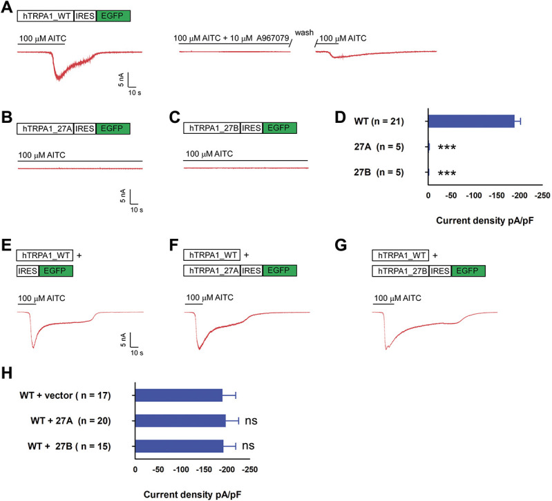
hTRPA1_27A and 27B splice variant displayed loss of function and did not affect WT current density on coexpression. (A) Top, hTRPA1 WT coding sequence was cloned into pIRES2-eGFP. On transfection, green fluorescent cells were selected for recording. Bottom, the cells were held at −60 mV, and TRPA1 current was evoked by perfusion of external solution containing 100 μM AITC as shown in the exemplar trace in red. Cotreatment of 10 μM A967079 and 100 μM AITC blocked activation of TRPA1 channels which could be reactivated on wash-out of the inhibitor. (B and C) Exemplar current of 27A and 27B on perfusion of 100 μM AITC, format as in A. (D) Bar chart reporting the peak current density of WT, 27A, and 27B singly transfected cells. *** P < 0.001 (Student unpaired t test) as compared with WT transfected cells. Values shown are mean ± SEM. (E) Exemplar current recorded from cell cotransfected with hTRPA1_WT expressed from nonfluorescent vector and empty pIRES2-eGFP vector, format as in A. (F) Exemplar current recorded from cell cotransfected with WT and 27A, format as in E. (G) Exemplar current recorded from cell cotransfected with WT and 27B, format as in E. (H) Bar chart reporting the peak current density of for conditions in F to G ns, nonsignificant (Student unpaired t test)) as compared with WT and empty vector transfected cells. Values shown are mean ± SEM. TRPA1, transient receptor potential ankyrin 1.
