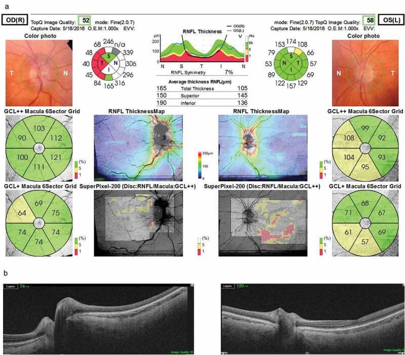
Figure 1.

Comprehensive report that includes a colour image of the optic nerve head obtained simultaneously with the OCT scanning. The right eye (upper left) shows optic disc swelling, increased brightness of the retinal nerve fibre layer, and increased vascular tortuosity and dilation. The left eye (upper right) presents mild irregularity of the optic disc borders and pallor. The circumpapillary retinal nerve fibre layer (RNFL) thickness average and sectors and a normative profile graph where the left and right eye thicknesses are plotted (top middle), the sectorial thickness of the ganglion-cell inner plexiform layer (GCL+) as well as the former with the macular RNFL added (GCL++), and an RNFL thickness map and a superpixel panoramic map merging the macular GCL++ map and the disc RNFL map are also depicted. B-scans (from the 12 × 9 mm cube) showing, on the lower left side, greater protrusion of the prelaminar tissue of the right eye compatible with the clinical findings of optic disc swelling
