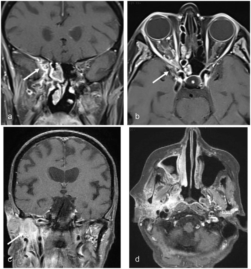
Figure 8.

MRI of case 2
(a) Gadolinium-enhanced coronal T1-weighted image with fat suppression showing acute inflammation within the right sphenoid sinus cell and orbital apex (arrow); (b) Gadolinium-enhanced axial T1-weighted image with fat suppression showing similar findings as in 8a; (c) Gadolinium-enhanced coronal T1-weighted image with fat suppression showing parapharyngeal extension of inflammatory changes (arrow); (d) Gadolinium-enhanced axial T1-weighted image with fat suppression showing similar findings as in 8c.
