



Abstract
Human adenoviruses (HAdVs) were previously detected at high prevalence by real-time reverse transcription-polymerase chain reaction (rRT-PCR) in the upper respiratory tract of residents of two Kenyan refugee camps under surveillance for acute respiratory infection (ARI) between October 2006 and April 2008. We sought to confirm this finding and characterize the HAdVs detected. Of 2148 respiratory specimens originally tested, 511 (23.8%) screened positive for HAdV and 510 were available for retesting. Of these, 421 (82.4%) were confirmed positive by repeat rRT-PCR or PCR and sequencing. Other respiratory viruses were codetected in 55.8% of confirmed HAdV-positive specimens. Species B and C viruses predominated at 82.8%, and HAdV-C1, -C2, and -B3 were the most commonly identified types. Species A, D, and F HAdVs, which are rarely associated with ARI, comprised the remainder. Viral loads were highest among species B HAdVs, particularly HAdV-B3. Species C showed the widest range of viral loads, and species A, D, and F were most often present at low loads and more often with codetections. These findings suggest that many HAdV detections were incidental and not a primary cause of ARI among camp patients. Species/type, codetections, and viral load determinations may permit more accurate HAdV disease burden estimates in these populations.
Keywords: adenovirus, Kenya, real-time reverse transcription-polymerase chain reaction (rRT-PCR), respiratory infections
1 ∣. INTRODUCTION
Acute respiratory infections (ARIs) are the leading cause of morbidity, hospitalization, and mortality among children in resource-poor countries.1 The most common etiologic agents of childhood ARI are viruses,2 and among the most frequently implicated viruses are the human adenoviruses (HAdVs). Premolecular studies using less sensitive culture and antigen-based diagnostic methods attributed 5% to 10% of symptomatic upper respiratory tract infections in young children to HAdVs.3,4 HAdV infections are most often subclinical or present with mild respiratory symptoms but can be severe, particularly in infants and the immunocompromised.5 Outbreaks of HAdV pneumonia with fatal outcomes in otherwise healthy adults have also been reported.6,7
Seven HAdV species (A-G) and over 80 genotypes have been described that can vary in geographic prevalence, disease syndrome, and severity.8,9 HAdV ARIs are most often associated with (i) species C viruses (predominately types 1, 2, and 5), endemic worldwide with sporadic ARI cases occurring primarily among young children and in whom persistent infections and subclinical shedding are also common; (ii) species B viruses (predominantly types 3, 7, 14, 21, and 55), more often associated with more severe ARI and may appear irregularly in case clusters or community-wide outbreaks that can involve older children and adults; and (iii) species E (type 4), more commonly identified in adults with acute febrile respiratory illness, particularly in unvaccinated US military recruits.10 Species A, D, and F HAdVs are uncommonly associated with ARI.
Although HAdV-associated ARI has been well described in low-, middle-, and high-income countries, few studies have been from resource-poor regions of sub-Saharan Africa and focused specifically on refugee populations. A recent study of residents of two refugee camps in Kenya under surveillance for ARI used sensitive molecular assays for multiple respiratory pathogens, and found an unexpectedly high prevalence of HAdV, exceeding most other respiratory viruses tested.11 To confirm this finding, we revisited a subset of the HAdV-positive samples to assess virus loads, codetections with other respiratory pathogens, and species/type-specific identity to gauge etiologic association with ARI.
2 ∣. MATERIALS AND METHODS
2.1 ∣. Study subjects and clinical specimens
Study subjects were enrolled from October 2006 to April 2008 at health care centers serving the Dadaab and Kakuma refugee camps located respectively in the northeast and northwest of Kenya. These centers were part of a national influenza sentinel surveillance system run jointly by the Kenya Ministry of Public Health and Sanitation and the Kenya Medical Research Institute/US Centers for Disease Control and Prevention-Kenya (KEMRI/CDC-K).11 During this surveillance period, Dadaab housed approximately 300 000 residents among three camps, over 95% were ethnic Somali, and Kakuma had approximately 70 000 residents, mostly from South Sudan. Pediatric and adult patients visiting inpatient and outpatient facilities with respiratory symptoms were classified by clinical presentation following World Health Organization definitions for influenza-like illness (ILI) or severe ARI (SARI).12 Among patients enrolled during the study period, approximately a third were classified as ILI and two-thirds as SARI. Study participants completed a questionnaire requesting demographic and clinical information, and nasopharyngeal and oropharyngeal swab specimens were collected together in commercial viral transport media. Swabs were refrigerated at 4°C to 8°C for up to 72 hours and then shipped to the KEMRI/CDC-Kenya laboratory where they were aliquoted and held at −80°C until testing. Specimens were tested at the KEMRI/CDC-Kenya laboratory for influenza A and B viruses and specimen aliquots were shipped to CDC (Atlanta) for testing for other respiratory viruses, including respiratory syncytial virus (RSV), human metapneumovirus, parainfluenza viruses 1 to 3, rhinovirus, and HAdV using in-house real-time reverse transcription-polymerase chain reaction (rRT-PCR) assays developed by CDC (Atlanta).
2.2 ∣. HAdV molecular detection and identification
2.2.1 ∣. HAdV rRT-PCR assay
Total nucleic acid was extracted from 100 to 200 μL of the swab specimens at CDC (Atlanta) using the MagMAX-96 Viral RNA Isolation Kit (Applied Biosystems, Carlsbad, CA) on a Kingfisher 96 Magnetic Particle Processor (Thermo Fisher Scientific, Waltham, MA). Extracts were tested by a pan-HAdV rRT-PCR assay modified from Heim et al13 and positive test results were assigned to samples with a well-defined amplification curve that crossed the cycle threshold (Ct) within 45 cycles. For this study, specimens were considered to be true positive if HAdV sequence could be obtained or if confirmed positive by sequencing or repeat rRT-PCR with the same assay. Positive and negative template controls were included in all runs and all specimens were also tested by rRT-PCR for the human RNAse P (RNP) gene to assess specimen quality.
2.2.2 ∣. HAdV sequence-based typing
HAdV species/type determinations of rRT-PCR positive samples were made by PCR amplification and Sanger sequencing of the hexon gene hypervariable regions 1 to 6 of loop 1 of the hexon capsid protein that have been shown to successfully predict most HAdV serotypes.14 Sample sequences were aligned with current reference sequences available on the GenBank for HAdV types 1 to 79 and 81 to 85 and species/types assigned based on the highest sequence similarity score and phylogenetic clustering analysis using the maximum parsimony method implemented in PAUP* 4.0.
2.3 ∣. Statistical analysis
Categorical data were compared using Pearson’s chi-squared test or Fisher’s exact test when the sample size was small. The Kruskal-Wallis test was used for virus load (Ct values) comparisons of all groups, and the Mann-Whitney U test was used for individual comparisons. P values < 0.01 were considered significant. Linear regression was performed to assess the relationship between virus load (Ct values) and the proportions of HAdV-positive specimens with (i) species B/C viruses and (ii) other codetected respiratory viruses. Mean Ct values were calculated for each of seven 4-Ct value bins covering the range of Ct values for all confirmed HAdV positive specimens. Simple regression lines were fitted to these data and correlation coefficients of determination (R2) were calculated to measure data fit.
2.4 ∣. Ethics
All participants or participant guardians provided informed consent. Ethical approval for the surveillance activities for respiratory diseases and specimen retention was obtained from the KEMRI Ethical Review Committee (protocol no. 1161). Surveillance activity was determined to be nonresearch that did not require CDC Institutional Review Board approval.
3 ∣. RESULTS
3.1 ∣. HAdV detection and confirmation
Respiratory swab specimens collected from 2158 camp patients with ILI or SARI during the surveillance period were tested for HAdV and other respiratory viruses by rRT-PCR. Eight specimens that were negative for RNP and two specimens with insufficient quantity were excluded from the analysis. Of the remaining 2148 specimens, 511 (23.8%) had tested positive for HAdV. One sample extract had insufficient quantity, leaving 510 sample extracts available for this study. Of these, 421 (82.4%) were confirmed positive by repeat rRT-PCR or PCR and partial hexon gene sequencing for a corrected prevalence estimate of 19.5%.
3.2 ∣. HAdV species/types identified
HAdV species and type identification was successful with 377 (89.5%) and 325 (77.2%) of the 421 confirmed positive specimens, respectively. Five HAdV species (A, B, C, D, and F) and at least 21 types were identified; 5 of the 21 type assignments could not distinguish between 2 or more reference types (Table 1). Species C (53.6%) and B (29.2%) viruses predominated, with HAdV-B3, -C1, and -C2 accounting for over 60% of identified types. HAdV-B16, an uncommonly reported type, was identified in specimens from 13 patients. Ct values differed significantly among HAdV species (P < 0.01); species B viruses (and especially HAdV-B3) and C viruses had generally lower and broader range of Ct values than species A, D, and F or samples where species/type could not be determined (Figure 1). The proportion species B/C HAdV detections among all species varied inversely with the Ct value (R2 = 0.91; Figure 2).
Table 1. Human adenovirus (HAdV) species/type determinations for 421 confirmed positive camp patients.
| Speciesa | Typea | n (%)b |
|---|---|---|
| A | 12 | 2 (0.53) |
| 18 | 3 (0.80) | |
| 31 | 2 (0.53) | |
| TND | 8 (2.12) | |
| Subtotal: | 15 (3.98) | |
| B | 3 | 74 (19.63) |
| 7/66 | 6 (1.59) | |
| 16 | 13 (3.45) | |
| 21 | 2 (0.53) | |
| 50 | 2 (0.53) | |
| TND | 13 (3.45) | |
| Subtotal: | 110 (29.18) | |
| C | 1 | 55 (14.58) |
| 2 | 73 (19.4) | |
| 5 | 37 (9.8) | |
| 6 | 15 (4.0) | |
| TND | 22 (5.8) | |
| Subtotal: | 202 (53.6) | |
| D | 10/65 | 1 (0.3) |
| 17/84 | 1 (0.3) | |
| 25/59 | 2 (0.53) | |
| 27 | 1 (0.3) | |
| 15/29/69 | 1 (0.3) | |
| 37 | 1 (0.3) | |
| 42 | 1 (0.3) | |
| TND | 9 (2.4) | |
| Subtotal: | 17 (4.5) | |
| F | 40 | 22 (5.8) |
| 41 | 11 (2.9) | |
| TND | 0 | |
| Subtotal: | 33 (8.8) | |
| STND | 44 (10.5) | |
| Total: | 421 |
Abbreviations: STND, species and type not determined; TND, type not determined.
Species/type assignment based on sequence of hexon gene hypervariable regions 1-6 (14). Indistinguishable types noted by “/.”
Denominator used for % calculations, 377 (421 confirmed HAdV detectons −44 STND).
FIGURE 1.
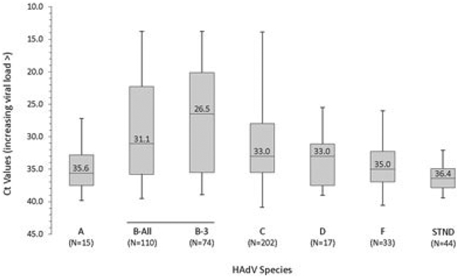
Box-and-whisker plots showing median and interquartile distributions of Ct values obtained with confirmed positive specimens by HAdV species/type and where species/type could not be determined (STND)
FIGURE 2.
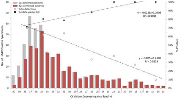
Distribution by Ct values (left axis) of 511 specimens that screened positive (gray bars) for HAdV by rRT-PCR and 421 specimens that were subsequently confirmed positive (red bars) by repeat rRT-PCR or PCR and sequencing. Percentages of HAdV-positive specimens (right axis) with codetected respiratory viruses (open circles) or with species B or C HAdVs identified (filled circles). Data averaged over 4-Ct value bins and fitted with linear regression lines. Linear regression equations and correlation coefficients of determination (R2) presented adjacent to the respective lines. Ct, cycle threshold; HAdVs, human adenoviruses; rRT-PCR, real-time reverse transcription-polymerase chain reaction
3.3 ∣. HAdV detections by campsite, demographics, and clinical case definition
Of the 2148 patients tested for HAdV who were included in the analysis, 770 (35.8%) were from Dadaab and 1378 (64.2%) were from Kakuma (Table 2). The proportions of the 421 HAdV-positive patients were similar for the two camps (Dadaab, 170/770 [22.1%]; Kakuma, 251/1378 [18.2%]) as was specimen quality based on RNP Ct values (Dadaab [median Ct, 25.5; interquartile range (IQR), 19.8-34.6]; Kakuma [median Ct, 25.7; interquartile range (IQR), 21.1-34.6]). Of the 403 HAdV-positive patients with available gender data, 59.3% were male. Of the 404 HAdV-positive patients with available age data, the median age was 2.6 years (ranging from 2 months to 56 years); overall, 84.5% were children <5 years old and 1.7% were adults ≥18 years old. Age and sex data were not available for HAdV-negative patients. Of 2073 patients with an available clinical case definition, 702 (33.9%) were classified as ILI and 1371 (66.1%) as SARI. HAdV-positive patients were equally distributed between the two case definitions (ILI, 136/702 [19.4%]; SARI, 268/1371 [19.5%]; Table 2). However, patients with species B detections had proportionately more ILI and those with species F had proportionately more SARI than other HAdV species, a finding that was consistent across age categories.
TABLE 2.
Virologic, demographic, and clincal category of 421 refugee camp patients with confirmed human adenovirus (HAdV) detections
| Respiratory HAdVa |
Nonrespiratory HAdVa |
|||||
|---|---|---|---|---|---|---|
| Categoryb | HAdV positive n = 421 (%)c |
HAdV-B n = 110 (%) |
HAdV-C n = 202 (%) |
HAdV-A n = 15 (%) |
HAdV-D n = 17 (%) |
HAdV-F n = 33 (%) |
| Location | n = 421 | n = 110 | n = 202 | n = 15 | n = 17 | n = 33 |
| Dadaab, 770 (35.8) | 170 (40.4) | 26 (23.6) | 93 (46.0) | 4 (26.7) | 6 (35.3) | 16 (48.5) |
| Kakuma, 1378 (64.2) | 251 (59.6) | 84 (76.4)d | 109 (54.0) | 11 (73.3) | 11 (64.7) | 17 (51.5) |
| Sex | n = 403 | n = 108e | n = 195e | n = 14e | n = 17 | n = 32e |
| Male | 239 (59.3) | 62 (57.4) | 116 (59.5) | 10 (71.4) | 11 (64.7) | 16 (50.0) |
| Female | 164 (40.7) | 46 (42.6) | 79 (40.5) | 4 (28.6) | 6 (35.3) | 16 (50.0) |
| Age group, y | n = 404e | n = 108e | n = 195e | n = 14e | n = 17 | n = 32e |
| <1 | 151 (37.4) | 45 (41.7) | 70 (35.9) | 6 (40.0) | 9 (52.9) | 11 (34.4) |
| 1-<5 | 202 (50.0) | 46 (42.6) | 100 (51.3) | 8 (53.3) | 8 (47.1) | 18 (54.5) |
| ≥5 | 51 (12.6) | 17 (15.7) | 25 (12.8) | 1 (6.7) | 0 | 3 (9.4) |
| Codetections | n = 421 | n = 110 | n = 202 | n = 15 | n = 17 | n = 33 |
| 235 (55.8) | 54 (49.1) | 105 (51.9) | 9 (60.0) | 10 (58.8) | 24 (72.7) | |
| Clinical case definitionf | n = 404e | n = 108e | n = 195e | n = 15 | n = 17 | n = 32e |
| ILI, 702 (33.9) | 136 (33.7) | 50 (46.3)d | 60 (30.8) | 4 (26.7) | 4 (23.5) | 3 (9.4) |
| SARI, 1371 (66.1) | 268 (66.3) | 58 (53.7) | 135 (69.2) | 11 (73.3) | 13 (76.5) | 29 (90.6)d |
Abbreviation: ILI, influenza-like illness; SARI, severe acute respiratory infection.
Respiratory HAdVs: species commonly associated with ARI; Non-respiratory HAdVs: species uncommonly associated with ARI.
Camp location data available for 2148 study patients; clinical case definition data available for 2073 patients; gender, age group, and codetections data not available for HAdV-negative patients.
HAdV species determined for 377 of 421 HAdV confirmed positive patients.
χ2 test for significance, P < 0.01.
Number of specimens in some cells differ from the column heading number due to missing data.
Clinical case definition.
3.4 ∣. Codetections with other respiratory viruses
Of the 421 specimens confirmed positive for HAdV, 235 (55.8%) had one or more of eight other respiratory viruses detected (Table 2); 184 (43.7%) were positive for one other virus, 47 (11.2%) for two other viruses, and 4 (0.95%) for three other viruses. Codetection data were not available for HAdV-negative patients. Codetections with rhinovirus were the most common (134, 31.8%) followed by RSV (40, 9.5%) and influenza A (28, 7.6%). HAdV-positive patients with ILI had significantly fewer codetections than those with SARI (44.9% vs 61.9%; P < 0.01). Whereas rhinovirus codetections were similar for both clinical groups (ILI, 57.4% vs SARI, 56.6%), influenza A codetections were more common among patients with ILI (24.6% vs 6.0%) and RSV codetections was more common among patients with SARI (20.5% vs 8.2%). Of the 377 HAdV-positive specimens identified to species, codetections were more common with HAdV species A, D, and F viruses (ranging from 58.8% to 72.7%) and least common with species B (49.1%), particularly HAdV-B3 (38.4%; Table 2). Median Ct values were significantly lower (P < 0.01) for single HAdV detections (31.8; IQR, 25.3-35.7) than with codetections (34.5; IQR, 31.5-36.8) and the proportion of codetections varied inversely with Ct values (R2 = 0.94; Figure 2).
3.5 ∣. Temporal variation of HAdV detections
HAdVs were continually detected over an 11-month period from May 2007 to March 2008 when surveillance data were available from both camps. All five species and most types were identified at both camps (Figure 3). Species C viruses were detected in every month and predominated in most months surveyed. Although species C viruses were continuously abundant at both camps, the proportion of species B viruses, and particularly HAdV-B3, was highest at Kakuma (P < 0.01). A temporal increase in HAdV-B3 detections was observed in Kakuma, beginning in June 2007, peaking in July/Aug and declining through October. Of the 74 total HAdV-B3 detections from both camps, over half occurred in Kakuma during this 3-month period; no similar increase in HAdV-B3 was detected in Dadaab during the same period.
FIGURE 3.
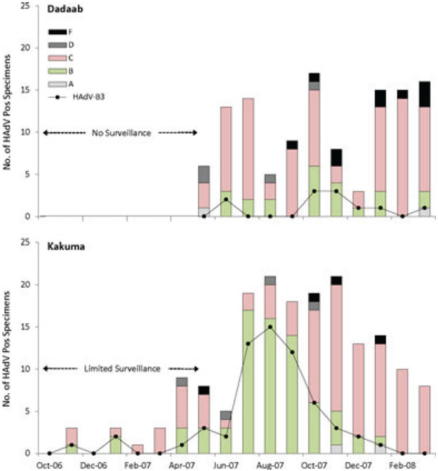
Temporal distribution of HAdV species A, B, C, D, and F (bars) and type 3 (line) identified at Dadaab and Kakuma refugee camps. Limited surveillance began in October 2006 in Kakuma and in May 2007 in Dadaab
4 ∣. DISCUSSION
We investigated an unexpected high prevalence of HAdV detected by rRT-PCR in respiratory specimens from mostly young children presenting with ILI or SARI at health care centers in the Dadaab and Kakuma refugee camps from October 2006 to April 2008. On initial screening, respiratory specimens from 23.8% of patients tested positive for HAdV. However, a high proportion of samples were found to have low virus loads and only 82.4% could be confirmed positive on retesting. Specimens that could not be confirmed had significantly higher Ct values (median, 37.6; IQR, 36.3-38.3) than those that were confirmed (median, 33.5; IQR, 28.7-36.4; P <0.01), which would be expected from the Poisson effect on assay reproducibility at lower detection limits. Although the potential for amplicon contamination during initial testing could not be excluded, rigorous quality control practices were implemented to minimize this possibility. Viral loads of many confirmed HAdV-positive specimens were still low and made it difficult in some cases to obtain high-quality sequence for species/type assignment. The presence of heterozygous base-calling with some samples suggested coamplification of mixed HAdV species/types, but limited available sample volume precluded further analyses.
Overall, five HAdV species and at least 21 distinct genotypes were identified over the study period. Species C and B viruses predominated, mirroring findings of other studies of childhood ARI in both developed and resource limited countries of North4,15,16 and South America,17,18 Europe,19 Africa,20 and Asia.21-23 Species C HAdVs comprised over half of the detections among camp patients, with most detections present at low virus loads. Although a recognized cause of ARI in young children, species C viruses are also commonly isolated from asymptomatic children and can persist in a latent state in adenoidal and tonsillar lymphocytes in up to half of infected children.24 A recent study by Song et al25 of HAdV-positive nasopharyngeal specimens from 483 children found an overabundance of low burden culture negative species C viruses with documented co-pathogens that they attributed to incidental detections. Similarly, we found species C viruses to be disproportionately associated with codetections and low virus loads, although the distribution of Ct values was skewed toward higher virus loads, perhaps reflecting the subset of infections responsible for the acute illness.
Species B HAdVs were the next most common species identified, with HAdV-B3 the most common type. HAdV-B3 is frequently associated with mild ARI and conjunctivitis,5 but is also capable of more serious lower respiratory tract infections and has been associated with community-wide ARI outbreaks.26 These findings of generally higher virus loads, lower proportions of codetections with other respiratory pathogens, and the temporal increase in HAdV-B3 detections observed in Kakuma are consistent with its reputation as a definitive respiratory pathogen. An unexpected finding was the detection of the rare HAdV-B16, the second most common species B virus identified. HAdV-B16 was originally isolated in Saudi Arabia in 1955 from a conjunctivitis case27 and has been isolated from a cluster of hospitalized patients with Reye’s-syndrome-like illness and pneumonia in Arkansas in 198128 and recovered from military recruits with ARI in Egypt between 1999 and 2002.29 Otherwise, large surveys of HAdV ARI from multiple countries in Europe, Asia, and the Americas failed to identify this virus.3,4,15-23
Species A, D, and F viruses comprised approximately 17% of HAdV detections, most often present at low virus loads. These viruses predominately infect the gastrointestinal tract with or without symptoms of acute gastroenteritis. Species A viruses have been associated with acute diarrhea in young children and have been detected in persons with underlying immune deficiencies.30,31 The higher numbered species D HAdVs were originally identified from the stool of persons with human immune-deficiency virus infections but without clear links to gastroenteritis.32 The enteric species F HAdVs are an important cause of childhood diarrhea that is uncommonly identified from respiratory tracts of children with ARI.33 Recent studies in Sub-Saharan Africa found a high prevalence of species A, D, and F HAdVs in stool from mostly children with and without diarrhea.34,35 Consequently, fecal contamination or transient infection of the oropharynx might account for their low load detections here.
Unlike definitive respiratory pathogens like RSV and influenza, HAdV detections are often incidental, as demonstrated in controlled multiple-pathogen studies.36-40 A recent etiologic study of SARI in children aged <5 years old living in a densely populated Kenyan slum that was conducted during a similar time period and using the same rRT-PCR assay as this study, also found a high prevalence of HAdV among symptomatic cases (29.5%).36 However, the authors also detected HAdV in 23.5% of matched asymptomatic controls. Although our study lacked controls, similar findings might be expected. Studies have also found higher virus loads in patients with HAdV-associated ARI than those of matched asymptomatic controls.39-41 Codetections with other respiratory viruses were also common among HAdV-positive patients, consistent with other studies of childhood ARI.37,42 Codetections were found to decrease with increasing viral loads, with specimens containing the highest loads more often associated with single HAdV detections. Interestingly, the proportions of co-detected viruses varied with HAdV species/type. Species B viruses, particularly HAdV-3, had the fewest codetections and the highest virus loads followed by species C and species A, D, and F, the latter having the lowest virus loads and highest proportions of codetections, consistent with their uncommon association with ARI.
This study had several important limitations. Specimens analyzed were collected for a period of less than 1 year for both sites, limiting the ability to assess long-term temporal variation in HAdV prevalence and species/type diversity. Adults accounted for fewer than 2% of confirmed HAdV-positive patients, limiting generalizations to pediatric populations. The lack of asymptomatic matched controls prevented the assessment of the disease attributable fractions and species/types associated with ILI or SARI. Codetection analysis was limited to the respiratory viruses tested that did not include some other potential respiratory pathogens. Finally, the molecular typing method used to identify HAdV species/types14 was restricted to a partial region of the hexon gene that could not discriminate among some recognized types and may have misidentified some novel viruses.
Using sensitive molecular methods, we confirmed a high prevalence of HAdV in the upper respiratory tract of residents of two refugee camps in Kenya where crowding, poor sanitation and personal hygiene, and population mixing may have enhanced virus transmission. However, most HAdV detections consisted of species/types prone to persistent shedding or not commonly associated with ARI, high proportions of codetections with other respiratory pathogens, and a preponderance of detections with low virus loads, all suggesting a bystander status. Further qualification of HAdV detections may help achieve more accurate disease burden estimates in displaced populations.
ACKNOWLEDGMENTS
We thank Shifaq Kamili for providing laboratory technical assistance for testing performed at the CDC (Atlanta). We also thank John Wagacha Burton of the United Nations High Commissioner for Refugees, Nairobi, Kenya and Erick Auko of the Kenya Medical Research Institute, Nairobi, Kenya, for their contributions to establishing the surveillance programs. Xinwei Wu was partially supported as a visiting scientist at the CDC (Atlanta) by the Project for Key Medicine Discipline Construction of Guangzhou Municipality (No 2017201907). The findings and conclusions in this report are those of the authors and do not necessarily represent the official position of the Centers for Disease Control and Prevention or other institutions with which the authors are affiliated.
Funding information
Key Medicine Discipline Construction of Guangzhou Municipality, Grant/Award Number: 2017201907
Footnotes
CONFLICTS OF INTEREST
The authors declare that they have no conflicts of interest.
REFERENCES
- 1.Williams BG, Gouws E, Boschi-Pinto C, Bryce J, Dye C. Estimates of world-wide distribution of child deaths from acute respiratory infections. Lancet Infect Dis. 2002;2:25–32. [DOI] [PubMed] [Google Scholar]
- 2.Hayden FG. Respiratory viral threats. Curr Opin Infect Dis. 2006;19:169–178. [DOI] [PubMed] [Google Scholar]
- 3.Fox JP, Hall CE, Cooney MK. The Seattle Virus Watch. VII. Observations of adenovirus infections. Am J Epidemiol. 1977;105:362–386. [DOI] [PubMed] [Google Scholar]
- 4.Edwards KM, Thompson J, Paolini J, Wright PF. Adenovirus infections in young children. Pediatrics. 1985;76:420–424. [PubMed] [Google Scholar]
- 5.Ruuskanen O, Metcalf JP, Waris M, Akusjarvi G. Adenoviruses. In: Richman DD, Whitley RJ, Hayen FG, eds. Clinical Virology, Fourth Edition. Washington, DC: ASM Press; 2017:pp. 575–598. [Google Scholar]
- 6.Lewis PF, Schmidt MA, Lu X, et al. A community-based outbreak of severe respiratory illness caused by human adenovirus serotype 14. J Infect Dis. 2009;199:1427–1434. [DOI] [PubMed] [Google Scholar]
- 7.Scott MK, Chommanard C, Lu X, et al. Human adenovirus associated with severe respiratory infection, Oregon, USA, 2013-2014. Emerg Infect Dis. 2016;22:1044–1051. [DOI] [PMC free article] [PubMed] [Google Scholar]
- 8.Lion T Adenovirus infections in immunocompetent and immunocompromised patients. Clin Microbiol Rev. 2014;27:441–462. [DOI] [PMC free article] [PubMed] [Google Scholar]
- 9.Hashimoto S, Gonzalez G, Harada S, et al. Recombinant type Human mastadenovirus D85 associated with epidemic keratoconjunctivitis since 2015 in Japan. J Med Virol. 2018;90:881–889. [DOI] [PubMed] [Google Scholar]
- 10.Kajon AE, Lamson DM, Bair CR, et al. Adenovirus Type 4 respiratory infections among civilian adults, Northeastern United States, 2011-2015. Emerg Infect Dis. 2018;24:201–209. [DOI] [PMC free article] [PubMed] [Google Scholar]
- 11.Ahmed JA, Katz MA, Auko E, et al. Epidemiology of respiratory viral infections in two long-term refugee camps in Kenya, 2007-2010. BMC Infect Dis. 2012;12:7. [DOI] [PMC free article] [PubMed] [Google Scholar]
- 12.World Health Organization. WHO surveillance case definitions for ILI and SARI. http://www.who.int/influenza/surveillance_monitoring/ili_-sari_surveillance_case_definition/en/
- 13.Heim A, Ebnet C, Harste G, Pring-Åkerblom P. Rapid and quantitative detection of human adenovirus DNA by real-time PCR. J Med Virol. 2003;70:228–239. [DOI] [PubMed] [Google Scholar]
- 14.Lu X, Erdman DD. Molecular typing of human adenoviruses by PCR and sequencing of a partial region of the hexon gene. Arch Virol. 2006;151:1587–1602. [DOI] [PubMed] [Google Scholar]
- 15.Gray GC, McCarthy T, Lebeck MG, et al. Genotype prevalence and risk factors for severe clinical adenovirus infection, United States 2004-2006. Clin Infect Dis. 2007;45:1120–1131. [DOI] [PMC free article] [PubMed] [Google Scholar]
- 16.Alharbi S, Van Caeseele P, Consunji-Araneta R, et al. Epidemiology of severe pediatric adenovirus lower respiratory tract infections in Manitoba, Canada, 1991-2005. BMC Infect Dis. 2012;12:55. [DOI] [PMC free article] [PubMed] [Google Scholar]
- 17.García J, Sovero M, Laguna-Torres VA, et al. Molecular characterization of adenovirus circulating in Central and South America during the 2006-2008 period. Influenza Other Respi Viruses. 2009;3:327–330. [DOI] [PMC free article] [PubMed] [Google Scholar]
- 18.Barrero PR, Valinotto LE, Tittarelli E, Mistchenko AS. Molecular typing of adenoviruses in pediatric respiratory infections in Buenos Aires, Argentina (1999-2010). J Clin Virol. 2012;53:145–150. [DOI] [PubMed] [Google Scholar]
- 19.Cooper RJ, Hallett R, Tullo AB, Klapper PE. The epidemiology of adenovirus infections in Greater Manchester, UK 1982-96. Epidemiol Infect. 2000;125:333–345. [DOI] [PMC free article] [PubMed] [Google Scholar]
- 20.Niang MN, Diop NS, Fall A, et al. Respiratory viruses in patients with influenza-like illness in Senegal: focus on human respiratory adenoviruses. PLoS One. 2017;12(3):e0174287. [DOI] [PMC free article] [PubMed] [Google Scholar]
- 21.Lee J, Choi EH, Lee HJ. Comprehensive serotyping and epidemiology of human adenovirus isolated from the respiratory tract of Korean children Nearly17 consecutive years (1991-2007). J Med Virol. 2010;82:624–631. [DOI] [PubMed] [Google Scholar]
- 22.Zou L, Zhou J, Li H, et al. Human adenovirus infection in children with acute respiratory tract disease in Guangzhou, China. APMIS. 2012;120:683–688. [DOI] [PMC free article] [PubMed] [Google Scholar]
- 23.Sriwanna P, Chieochansin T, Vuthitanachot C, Vuthitanachot V, Theamboonlers A, Poovorawan Y. Molecular characterization of human adenovirus infection in Thailand, 2009-2012. Virol J. 2013;10:193. [DOI] [PMC free article] [PubMed] [Google Scholar]
- 24.Alkhalaf MA, Guiver M, Cooper RJ. Prevalence and quantitation of adenovirus DNA from human tonsil and adenoid tissues. J Med Virol. 2013;85:1947–1954. [DOI] [PubMed] [Google Scholar]
- 25.Song E, Wang H, Kajon AE, et al. Diagnosis of pediatric acute adenovirus infections: is a positive PCR sufficient? Pediatr Infect Dis J. 2016;35:827–834. [DOI] [PMC free article] [PubMed] [Google Scholar]
- 26.Girouard G, Garceau R, Thibault L, Bourque C, Bastien N, Li Y. Province-wide adenovirus type 3 outbreak with severe cases in New Brunswick. Can J Infect Dis Med Microbiol. 2011;22:e4–e6. [DOI] [PMC free article] [PubMed] [Google Scholar]
- 27.Murray ES, Chang RS, Bell SD, Tarizzo ML, Snyder JC. Agents recovered from acute conjunctivitis cases in Saudi Arabia. Am J Ophthalmol. 1957;43:32.13411124 [Google Scholar]
- 28.Morgan PN, Moses EB, Fody EP, Barron AL. Association of adenovirus type 16 with Reye’s-syndrome-like illness and pneumonia. South Med J. 1984;77:827–830. [DOI] [PubMed] [Google Scholar]
- 29.Metzgar D, Osuna M, Yingst S, et al. PCR analysis of Egyptian respiratory adenovirus isolates, including identification of species, serotypes, and coinfections. J Clin Microbiol. 2005;43:5743–5752. [DOI] [PMC free article] [PubMed] [Google Scholar]
- 30.Adrian T, Wigand R. Genome type analysis of adenovirus 31, a potential causative agent of infants’ enteritis. Arch Virol. 1989;105:81–87. [DOI] [PubMed] [Google Scholar]
- 31.Portes SAR, Volotão EM, Rocha MS, et al. A nonenteric adenovirus A12 gastroenteritis outbreak in Rio de Janeiro, Brazil. Mem Inst Oswaldo Cruz. 2016;111:403–406. [DOI] [PMC free article] [PubMed] [Google Scholar]
- 32.Hierholzer JC, Wigand R, Anderson LJ, Adrian T, Gold JWM. Adenoviruses from patients with AIDS: a plethora of serotypes and a description of five new serotypes of subgenus D (types 43-47). J Infect Dis. 1988;158:804–813. [DOI] [PubMed] [Google Scholar]
- 33.Echavarria M, Maldonado D, Elbert G, Videla C, Rappaport R, Carballal G. Use of PCR to demonstrate presence of adenovirus species B, C, or F as well as coinfection with two adenovirus species in children with flu-like symptoms. J Clin Microbiol. 2006;44:625–627. [DOI] [PMC free article] [PubMed] [Google Scholar]
- 34.Pauly M, Hoppe E, Mugisha L, et al. High prevalence and diversity of species D adenoviruses (HAdV-D) in human populations of four Sub-Saharan countries. Virol J. 2014;11:25. [DOI] [PMC free article] [PubMed] [Google Scholar]
- 35.Moyo SJ, Hanevik K, Blomberg B, et al. Prevalence and molecular characterization of human adenovirus in diarrhoeic children in Tanzania; a case control study. BMC Infect Dis. 2014;14:666. [DOI] [PMC free article] [PubMed] [Google Scholar]
- 36.Breiman RF, Cosmas L, Njenga MK, et al. Severe acute respiratory infection in children in a densely populated urban slum in Kenya, 2007-2011. BMC Infect Dis. 2015;15:95. [DOI] [PMC free article] [PubMed] [Google Scholar]
- 37.Singleton RJ, Bulkow LR, Miernyk K, et al. Viral respiratory infections in hospitalized and community control children in Alaska. J Med Virol. 2010;82:1282–1290. [DOI] [PMC free article] [PubMed] [Google Scholar]
- 38.Howard LM, Johnson M, Williams JV, et al. Respiratory viral detections during symptomatic and asymptomatic periods in young Andean children. Pediatr Infect Dis J. 2015;34:1074–1080. [DOI] [PMC free article] [PubMed] [Google Scholar]
- 39.Rhedin S, Lindstrand A, Hjelmgren A, et al. Respiratory viruses associated with community-acquired pneumonia in children: matched case-control study. Thorax. 2015;70:847–853. [DOI] [PubMed] [Google Scholar]
- 40.Self WH, Williams DJ, Zhu Y, et al. Respiratory viral detection in children and adults: comparing asymptomatic controls and patients with community-acquired pneumonia. J Infect Dis. 2016;213:584–591. [DOI] [PMC free article] [PubMed] [Google Scholar]
- 41.Feikin DR, Fu W, Park DE, et al. Is higher viral load in the upper respiratory tract associated with severe pneumonia? Findings from the PERCH study. Clin Infect Dis. 2017;64(Suppl 3):S337–S346. [DOI] [PMC free article] [PubMed] [Google Scholar]
- 42.Franz A, Adams O, Willems R, et al. Correlation of viral load of respiratory pathogens and co-infections with disease severity in children hospitalized for lower respiratory tract infection. J Clin Virol. 2010;48:239–245. [DOI] [PMC free article] [PubMed] [Google Scholar]