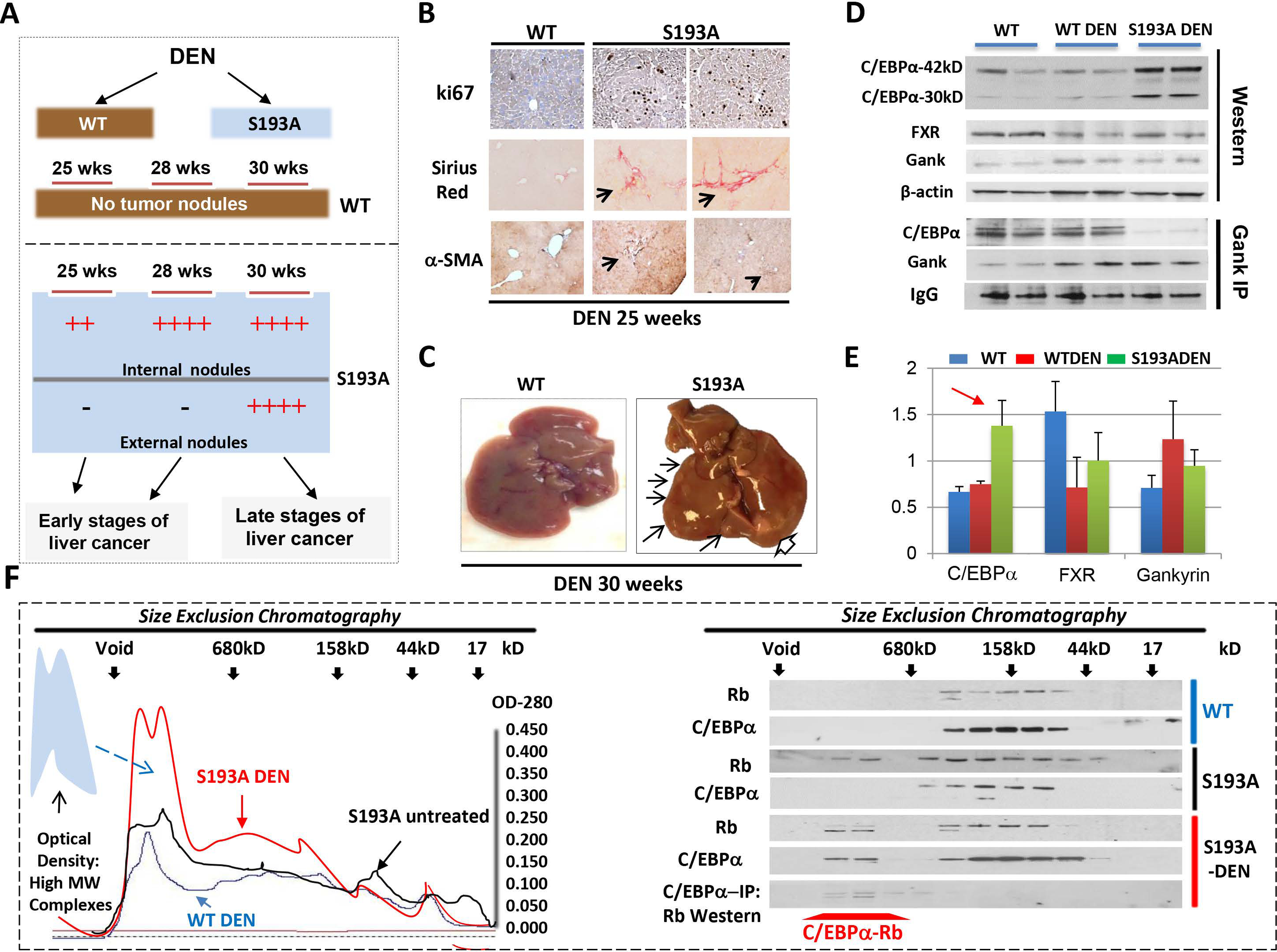
Figure 2. DEN-mediated liver cancer in C/EBPα-S193A mice has a molecular signature of aggressive HBL.

(A) A diagram shows results of the treatments of WT and C/EBPα-S193A mice with DEN. (B) WT and S193A livers of 25-wks DEN-treated mice were stained with ki67, Sirius Red and α-SMA. (C) Typical picture of external nodules in S193A mice treated with DEN for 30 weeks. Large nodule is shown by open arrow. (D) Nuclear extracts from livers of WT, WT DEN-treated and S193A DEN-treated mice were examined by Western blotting with Abs shown on the left. Gank-IP: Gank was immunoprecipitated from nuclear extracts of mice shown on the top. The IP were probed with antibodies to C/EBPα and Gank. IgG; heavy chains of IgGs. (E) Levels of proteins on “D” are calculated as ratios to β-actin. Red arrow shows the elevation of C/EBPα-S193A after DEN treatments. (F) Left: Optical Density analyses of nuclear extracts after SEC. The region of elevation of OD in the high MW sections of SEC is shown by arrow. Right: Proteins from fractions of SEC were analyzed by Western blotting with Abs to C/EBPα and Rb. The bottom panel shows results of C/EBPα-IP and Western with Rb for DEN-treated S193A mice.
