Abstract
Listeria monocytogenes is a foodborne pathogen. A source of infection can be artisanal cheeses. Identification of the Listeria species is important for the protection of public health and the food industry. This study aimed to examine artisanal cheeses for the presence of L. monocytogenes and the effectiveness of the MALDI-TOF MS method in the identification of the L. monocytogenes isolates. A total of 370 samples of artisanal cheeses were examined. L. monocytogenes was found in 23 cheese samples (6.2%). The reliability of L. monocytogenes identification achieved by MALDI-TOF MS was varied, and the vast majority of the isolates (27/32) were identified only to the secure genus, probable species level. This study showed that (i) the occurrence of L. monocytogenes in the artisanal cheeses was at a higher level than that in the other EU countries, (ii) the standard of species identification of L. monocytogenes isolates from artisanal cheeses achieved by MALDI-TOF MS was not satisfactory and (iii) the presence of L. monocytogenes in artisanal cheeses remains a problem with regard to the food safety criterion and a potential public health risk.
Keywords: Listeria monocytogenes, artisanal cheeses, MALDI-TOF MS, food safety
1. Introduction
Listeria monocytogenes is a real public health threat, as evidenced by the data of zoonosis and zoonotic agent monitoring in European Union (EU) countries. This pathogen is the etiological agent of listeriosis, a foodborne disease. Taking into account the number of reported human cases in the EU in 2018, listeriosis ranks fifth among 13 zoonoses and the highest number of hospitalisations and mortality rates have been noted for this zoonosis. In addition, in recent years (2009–2018), there has been an increase in the number of cases of listeriosis [1].
A source of infection may be artisanal cheeses contaminated with L. monocytogenes [2,3,4,5,6,7]. Artisanal cheeses belong to traditional dairy products [8,9,10,11,12], and they are constantly produced in many countries of the world. This type of foodstuff has cultural, social and economic importance [13].
Cheeses should meet the safety criterion concerning L. monocytogenes; a product that does not comply with this criterion is considered unsafe and cannot be offered for sale [14,15]. According to the literature, MALDI-TOF MS (matrix-assisted laser desorption ionisation–time of flight mass spectrometry) is considered a reliable, fast and cost-effective tool for the routine identification of Listeria [16,17]. This method consists of bacterial protein panel analysis in the mass range of 2–20 kDa, which mainly represents ribosomal proteins and basic metabolism proteins. These proteins create a bacteria-specific fingerprint, which, when compared with the protein profiles contained in a reference spectrum library, enables the determination of the taxonomic position of the microorganism. The MALDI-TOF MS analysis evaluates two parameters: the ion mass-to-charge ratio (m/z) and the relative ion intensity. The protein spectrum obtained in this procedure is processed to yield the protein code of the microorganism. A comparison of the obtained code against the reference codes contained in the library enables the identification of the microorganism at the genus, species, subspecies or strain level [18]. This technique enabled the identification of L. monocytogenes with 100% accuracy, regardless of its origin, whether from humans, animals, food or the environment [16,17]. On the other hand, other authors have shown that the MALDI-TOF MS method is not without limitations in identifying L. monocytogenes [19].
The objectives of this study were the examination of artisanal cheeses for the presence of L. monocytogenes and the evaluation of the usefulness of the MALDI-TOF MS method in the identification of L. monocytogenes isolates derived from these types of foodstuffs.
2. Results
2.1. Bacteriological Analysis and Identification by MALDI-TOF MS
A total of 370 samples of artisanal cheeses were analysed for the presence of L. monocytogenes. The pathogen was detected in 23/370 samples of the cheeses (6.2% of all samples tested). The products contaminated with L. monocytogenes came from 16 production sites. During the investigation period (2014–2018), the number of cheese dairies from which tested products contained L. monocytogenes was between two in 2014 and 2015 and eight in 2018; a rising trend was observed from 2016. A total of 32 isolates of L. monocytogenes were yielded: in three cases, three isolates from one batch of tested cheeses were obtained, and the relevant production facilities were G1, I and H; in another three cases, two isolates from one batch of tested cheeses were obtained, and the relevant production facilities were G2, F and I; in the remaining 17 cases, one isolate from one batch of tested cheeses was obtained (see Section 4.1. Materials and Table 1).
Table 1.
Identification of the L. monocytogenes isolates derived from the artisanal cheeses by MALDI-TOF MS.
| MALDI–TOF MS. | |||||
|---|---|---|---|---|---|
| Production facilities | Year of sampling |
Sample code |
Score values |
Species ID according to MALDI Biotyper 3.1 |
|
| G1 | 2014 | 1A | 2.041–2.272 | L. monocytogenes | DSM 20600T DSM |
| G1 | 2014 | 1B | 2.028–-2.055 | L. monocytogenes | DSM 20600T DSM |
| G1 | 2015 | 1C | 2.087–2.193 | L. monocytogenes | Mb 19348_1 CHB |
| D4 | 2014 | 16 | 1.803–2.036 | L. monocytogenes | Mb 19348_1 CHB |
| J | 2015 | 17 | 2.05–2.086 | L. monocytogenes | Mb 19348_1 CHB |
| I | 2015 | 2A | 1.915–2.141 | L. monocytogenes | DSM 20600T DSM |
| I | 2015 | 2B | 1.935–2.033 | L. monocytogenes | DSM 20600T DSM |
| I | 2015 | 2C | 2.033–2.152 | L. monocytogenes | Mb 19348_1 CHB |
| I | 2016 | 3 | 1.997–2.28 | L. monocytogenes | Mb 19348_1 CHB |
| D1 | 2016 | 4 | 2.239–2.29 | L. monocytogenes | DSM 20600T DSM |
| D2 | 2016 | 5 | 2.052–2.191 | L. monocytogenes | Mb 19348_1 CHB |
| A | 2016 | 18 | 2.249–2.268 | L. monocytogenes | Mb 19348_1 CHB |
| C | 2016 | 19 | 1.976–1.988 | L. monocytogenes | Mb 19348_1 CHB |
| G2 | 2016 | 6A | 2.101–2.137 | L. monocytogenes | DSM 20600T DSM |
| G2 | 2016 | 6B | 1.946–2.043 | L. monocytogenes | DSM 20600T DSM |
| F | 2017 | 7A | 2.042–2.122 | L. monocytogenes | DSM 20600T DSM |
| F | 2017 | 7B | 2.031–2.095 | L. monocytogenes | Mb 19348_1 CHB |
| J | 2017 | 21 | 2.052–2.153 | L. monocytogenes | Mb 19348_1 CHB |
| G2 | 2017 | 22 | 2.045–2.101 | L. monocytogenes | Mb 19348_1 CHB |
| C | 2017 | 23 | 1.905–2.02 | L. monocytogenes | CCUG 315227 CCUG |
| F | 2018 | 8 | 2.027–2.234 | L. monocytogenes | Mb 19348_1 CHB |
| G3 | 2018 | 9 | 2.127–2.164 | L. monocytogenes | Mb 19348_1 CHB |
| D3 | 2018 | 10 | 1.996–2.182 | L. monocytogenes | Mb 19348_1 CHB |
| K | 2018 | 12 | 1.901–1.929 | L. monocytogenes | Mb 19348_1 CHB |
| H | 2018 | 13A | 2.023–2.048 | L. monocytogenes | DSM 20600T DSM |
| H | 2018 | 13B | 1.903–2.112 | L. monocytogenes | Mb 19348_1 CHB |
| H | 2018 | 13C | 1.991–2.065 | L. monocytogenes | Mb 19348_1 CHB |
| I | 2018 | 14A | 2.306–2.368 | L. monocytogenes | Mb 19348_1 CHB |
| I | 2018 | 14B | 2.013–2.106 | L. monocytogenes | Mb 19348_1 CHB |
| I | 2018 | 15 | 1.812–2.048 | L. monocytogenes | Mb 19348_1 CHB |
| E | 2018 | 20 | 1.922–1.955 | L. monocytogenes | DSM 20600T DSM |
| B | 2018 | 11 | 1.96–1.977 | L. monocytogenes | DSM 20600T DSM |
Different capital letters (from A to K) indicate the production sites in different towns, while the same capital letters with different subscripts (D1–D4, G1–G3) show different production sites in one town. Sample code marked with a number and capital letter (1A–C; 2A–C; 6A and 6B; 7A and 7B; 13A–C; 14A and 14B) shown at some production sites (G1, I, G2, F, H, I, respectively) indicate the number of isolates out of 1 tested sample.
The score values for the tested isolates by MALDI-TOF MS indicated that one isolate (3.1%) was identified at the level of highly probable species identification (log(score) 2.306–2.368), 27 isolates (84.4%) were identified at the level of secure genus identification and probable species identification (log(score) ≥ 2.0–2.29) and four isolates (12.5%) were identified at the level of probable genus identification (log(score) 1.901–1.997) (Table 1; Tables S1 and S2).
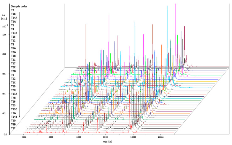
All of the mass spectra of the tested isolates showed a good resolution, with a variety of peaks with specific protein profiles. The isolates all had 20 peaks in common, with the masses at m/z (mean peak position ± standard deviation) 2755.864 ± 0.25, 3181.643 ± 0.30, 3251.036 ± 0.23, 3304.155 ± 0.34, 3320.206 ± 0.24, 3336.243 ± 0.25, 3702.389 ± 0.30, 4044.233 ± 0.43, 4324.443 ± 0.36, 4630.144 ± 0.52, 4876.781 ± 0.38, 4942.374 ± 0.40, 5118.111 ± 0.41, 6007.757 ± 0.47, 6362.696 ± 0.50, 6421.715 ± 2.41, 8086.682 ± 0.80, 9258.724 ± 0.69, 9751.647 ± 0.85 and 9882.999 ± 0.88. These peaks can be considered characteristic of L. monocytogenes. Some peaks were characteristic of only some isolates. In the mass spectra of T1A, T4 and T5 isolates, there was no 4926.213 ± 1.58 peak. This peak was characteristic of the T3, T18, T14A, T8, T6A, T2A, T1B, T21, T17, T2C, T2B, T9, T22, T1C, T19, T20, T13A, T16, T7B, T7A, T14B, T10, T23, T6B, T11, T13B, T15, T12 and T13C isolates. Also, except for three isolates (T13B, T15 and T12), all isolates had a peak m/z of 9810.927 ± 0.789 (Figure 1).
Figure 1.
Spectra of the L. monocytogenes isolates generated by MALDI-TOF MS. The intensities and masses of the ions are shown on the Y- and X-axes, respectively. Sample codes displayed on the Y-axis are in accordance with those in Table 1. The m/z value is the mass-to-charge ratio.
2.2. Dendrogram for the L. monocytogenes Isolates
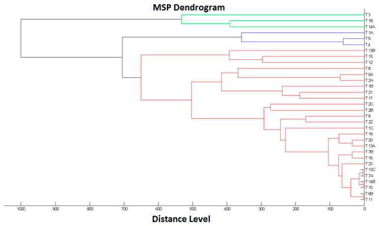
The dendrogram (Figure 2) of the tested isolates indicates two main clusters. Clusters one (shown in green) and two (shown in blue and red) contained 3 and 29 isolates, respectively. Additionally, in cluster two, the isolates were grouped into three internal branches (of 3, 3 and 23 isolates), the most proteomically diverse (red) being in one of these branches.
Figure 2.
MALDI-TOF MS dendrogram showing cluster analysis of mass spectral profiles from 32 L. monocytogenes isolates.
3. Discussion
Ready to eat (RTE) food contaminated with L. monocytogenes poses a risk to public health [1]. According to current food law, the safety criterion for the products tested was the absence of L. monocytogenes in 25 g of the sample; therefore, 23 samples in total, i.e., 6.2% of samples of the artisanal cheeses, did not meet the food safety criterion [14]. It emerges clearly from the data presented in Table 1 that the number of cheese dairies of which the products contained L. monocytogenes increased between 2016 and 2018. Among them, there were new productions (e.g., G3, D3, K, H, E and B), as well as those where the pathogen was already shown in the products in previous years (e.g., I, J, C and F) (Table 1). This indicates both the need for the constant monitoring of artisanal cheeses for L. monocytogenes and the need for corrective action at production sites [14]. It is worth noting that, in some cases, proteomically variant isolates of L. monocytogenes were identified in the same sample (Figure 2, Table 1), which may indicate various co-existent sources of contamination.
According to European Food Safety Authority (EFSA) data in the same years, i.e., 2014–2018, the frequency of L. monocytogenes occurrence in cheeses produced from raw or low-heat-treated milk ranged from 0.9% to 2.6% for soft and semi-soft cheeses and from 0.1% to 2.0% for hard cheeses [1,20]. Given that these data come from many European Union countries (depending on the type of cheese, data are from 9 to 22 countries), it should be concluded that the frequency of L. monocytogenes occurrence in the tested artisanal cheeses was higher than in other EU countries. The pathogen was more often found in traditional homemade cheeses from Turkey, such as cokelek and kumak cheese (both 30%), farm cheese (20%) [21] and white cheese (9.2%) [22], compared to the tested artisanal cheeses (6.2%). It is clear from the data presented that the frequency of L. monocytogenes occurrence in traditional cheeses varies, and the bacterium remains a problem concerning food safety. According to the literature, the risk of L. monocytogenes contamination of the final product may be due to the contamination of the raw material, milking equipment, production environment or farm workers [21,23,24,25,26,27].
In the present study, the identification of L. monocytogenes by MALDI-TOF MS was varied (highly probable species identification; secure genus identification, probable species identification; and probable genus identification). The vast majority of the L. monocytogenes isolates (27/32) were recognised to secure genus identification, probable species identification, and for the next group of isolates, as probable genus identification (4/32). Using the MALDI-TOF MS technique, it is possible to record the mass spectra reflecting the protein profiles of the analysed bacteria; however, a possibility of identification exists only for the protein profiles whose sequences are deposited in the database. Given that the analysed isolates were identified as L. monocytogenes by PCR, it should be concluded that the MALDI-TOF MS system database needs to be extended with spectra for strains within artisanal cheeses to increase the potential for thorough identification. Thouvenot et al. also indicated the importance of a continuously updated, high-quality reference library for the exact identification of Listeria [17]. Research by other authors has shown that the MALDI-TOF MS method is not without limitations in identifying L. monocytogenes. Pusztahelyi et al. reported that more than half of the tested bacterial strains (n = 18) were scored with values that gave secure identification only at the genus level (2.000–2.299), while only seven strains gave a highly probable species identification (>2.300). Furthermore, the same study showed the misidentification of L. monocytogenes at the species level [19]. In contrast, Thouvenot et al. reported the complete reliability of MALDI-TOF MS mass spectrometry to identify Listeria in human, animal, food and environmental microbiology, with 100% accuracy for identifying eight species, including L. monocytogenes [17]. The presented study results did not confirm the complete reliability of the MALDI-TOF MS method for the isolates derived from the artisanal cheeses. In view of the above, the MALDI-TOF MS method alone was not enough for a certain L. monocytogenes identification. As a consequence, this method was not a suitable for the routine diagnostics of L. monocytogenes in the foods.
In the mass spectra of the tested isolates, prominent peaks were between 2 and 11 kDa, with the highest-intensity peaks between 4 and 10 kDa (Figure 1). Studies by other authors showed that prominent peaks in the mass spectra of L. monocytogenes were noted in a similar range (2–10 kDa) [28].
Dendrogram analysis (Figure 2) indicated that closely related isolates, e.g., T11 and T6B, came from different administrative divisions and different cheese dairies. These were also isolated in different calendar years. Other isolates that were less related, such as T21 and T17, occurred only locally in the same administrative division and in the same cheese dairies and were also isolated in different calendar years. Therefore, the data indicated that the proteomic relationship of the L. monocytogenes isolates studied did not correlate with the cheese dairy, administrative division or year of isolation.
4. Materials and Methods
4.1. Materials
The research was conducted between 2014 and 2018. A total of 370 samples of artisanal cheeses were tested from cheese dairies located in Southern Poland. The cheeses were produced by traditional methods and according to long-standing recipes indigenous to the region. Advanced technological solutions were not used in the cheese production process, nor were production norms implemented (production diagram is included in Figure S1). The raw material for the production of artisanal cheeses was unpasteurised milk.
The sampling procedure was followed: a total of 5 samples of tested cheeses from each batch were taken in each of the cheese dairies [14]. The samples were taken at the producers before the cheeses were put on the market (for sale) under sterile conditions and then transferred to the laboratory at the cold-store temperature (0–4 °C).
4.2. Bacteriological Analysis
The presence of L. monocytogenes was determined according to the ISO standards [29,30]. Briefly, the mass of the sample was 25 g; selective multiplication of isolates was carried out on half-Fraser and Fraser broth; then, selective and differential media ALOA (agar Listeria according to Ottaviani and Agosti) and PALCAM (polymyxin acriflavine lithium chloride ceftazidime aesculin mannitol) media were used. For confirmation, 5 suspect colonies were selected from each plate of the selective and differential medium (ALOA and PALCAM), and from July 2017, from 1 to 5 suspected colonies, according to the revised procedure set out in the new version of ISO 11290-1. Material from suspect colonies was streaked on TSYEA (tryptone soya yeast extract agar), in a manner that allowed isolated colonies to develop and was then incubated at 37 °C for 24 h. The pure cultures thus obtained were tested for belonging to the Listeria spp. (Gram staining and catalase and bacterial motility tests) and followed by L. monocytogenes confirmatory tests (haemolysis test, CAMP test and the ability to decompose rhamnose and xylose). The strains thus selected were further confirmed using the MicrobactListeria 12L identification test. Each identification strip consisted of 12 tests. The following biochemical features were analysed: hydrolysis of aesculin, utilisation of specific 10 carbohydrates (mannitol, xylose, arabitol, ribose, rhamnose, trehalose, tagatose, glucose-1-phosphate and methyl-d-glucose and methyl-d-mannose), and a rapid haemolysis test was performed (micro-haemolysis). Material from confirmed pure cultures was emulsified in a suspending medium and mixed to obtain an inoculum to the MacFarland 0.5 standard. Subsequently, 4 drops of the bacterial suspension were transferred into each well. The inoculated strips were incubated at 35 ± 2 °C for 4 h. After incubation, the reactions were read visually and interpreted using the data tables provided with the test. Merck Millipore, Biomaxima and Thermo Fisher Scientific culture media were used in the bacteriological study. All isolates underwent identification using multiplex PCR. The procedure was carried out by an accredited laboratory commissioned by the Veterinary Inspectorate as part of official monitoring imposed by the applicable food law (detailed data on the molecular serotyping of L. monocytogenes by multiplex PCR are included in Tables S3–S6).
4.3. Identification of L. monocytogenes Isolates by MALDI-TOF MS
The identification of bacterial strains was preceded by the preliminary extraction of proteins with ethanol and formic acid. For this purpose, a single colony of bacterial culture was suspended in 150 µL of sterile deionised water, after which 450 µL of pure ethanol (Merck, Darmstadt, Germany) was added. Then, each sample was mixed thoroughly by vortexing. The resulting solution was then centrifuged for 5 min at 13,000 rpm. Next, the supernatant was discarded, 40 µL of 70% aqueous formic acid and 40 µL of acetonitrile (Merck, Darmstadt, Germany) were added to the precipitate and the sample was thoroughly mixed by vortexing again. After centrifugation at 13,000 rpm for 2 min, 1 µL of the obtained supernatant was applied to a metal plate and allowed to dry at room temperature. Then, 1 µL of matrix solution (cyano-4-hydroxycinnamic acid, Bruker, Bremen, Germany) was applied, and the sample was again left to dry at room temperature. The metal plate with the samples was subsequently placed in a MALDI chamber for analysis. A measurement of the spectrum and a comparative analysis with reference spectra of bacteria were performed using an UltrafleXtreme mass spectrometer at m/z range 2000–20,000 Da and MALDI Biotyper 3.1 software (Bruker Daltonik, Bremen, Germany). The results were shown as the top 10 identification matches, along with confidence scores ranging from 0.00 to 3.00. According to the criteria recommended by the manufacturer, a log (score) below 1.70 does not allow for reliable identification; a log (score) between 1.70 and 1.99 allows identification at the genus level; a log (score) between 2.00 and 2.29 indicates highly probable identification at the genus level and probable identification at the species level; and a log (score) between 2.30 and 3.00 indicates highly probable identification at the species level. Analysis of each sample was performed in triplicate, i.e., 3 spots for each sample. The identification result was considered reliable when at least the two best matches—log (score) 1.70–3.00—with the MALDI Biotyper database indicated the same species. For samples in which the top two matches indicated different species, we considered the first match—the log (score) was greater than the value for the second match.
4.4. Dendrogram Construction for L. monocytogenes Isolates
A dendrogram was created by matching main spectra peaks (MSPs) of the tested L. monocytogenes isolates. Each MSP was matched against all MSPs of the analysed set. The list of score values was used to calculate normalised distance values between strains, resulting in a matrix of matching scores. The visualisation of the relationship between the MSPs was displayed in a dendrogram using MALDI Biotyper 3.1 software (Bruker Daltonik, Bremen, Germany) [31].
5. Conclusions
There is no doubt that there is a need to monitor cheeses produced by traditional methods for L. monocytogenes. Studies have unequivocally shown that the presence of L. monocytogenes in artisanal cheeses is a current problem for food safety, and these cheeses pose a potential public health risk.
The standard of species identification of L. monocytogenes isolates from artisanal cheeses achieved by MALDI-TOF MS was unsatisfactory, indicating the need to extend the database for mass spectra of L. monocytogenes strains isolated from this type of foodstuff. The MALDI-TOF MS method alone was not enough for a reliable L. monocytogenes identification, and as a consequence, this method cannot be considered as a suitable tool for the routine diagnostics of L. monocytogenes from a food safety point of view.
Supplementary Materials
The following are available online at https://www.mdpi.com/article/10.3390/pathogens10060632/s1, Figure S1: The main stages in the production of artisanal cheeses; Table S1: Identification of the L. monocytogenes isolates by MALDI-TOF MS; Table S2: Meaning of score values; Table S3–S6: Molecular serotyping of L. monocytogenes by multiplex PCR.
Author Contributions
Conceptualization, R.P.-Ł.; methodology, R.P.-Ł., M.G. and M.Z.; validation, K.M. and D.W.; formal analysis, A.P.-K., A.P., K.M. and D.W.; data curation, W.P.; writing—original draft preparation, R.P.-Ł. and K.M.; writing—review and editing, R.P.-Ł., M.G., M.Z. and W.P.; visualization, K.M. and W.P.; funding acquisition, R.P.-Ł. and W.P. All authors have read and agreed to the published version of the manuscript.
Funding
The study was supported by the Ministry of Science and Higher Education project WKH/S/41/2020/WET of the University of Life Sciences in Lublin.
Institutional Review Board Statement
Not applicable.
Informed Consent Statement
Not applicable.
Data Availability Statement
Data is contained within the article or supplementary material.
Conflicts of Interest
The authors declare no conflict of interest.
Footnotes
Publisher’s Note: MDPI stays neutral with regard to jurisdictional claims in published maps and institutional affiliations.
References
- 1.EFSA and ECDC (European Food Safety Authority and European Centre for Disease Prevention and Control) The European Union one health 2018 zoonoses report. EFSA J. 2019;17:e05926. doi: 10.2903/j.efsa.2019.5926. [DOI] [PMC free article] [PubMed] [Google Scholar]
- 2.Bille J., Blanc D.S., Schmid H., Boubaker K., Baumgartner A., Siegrist H.H., Tritten M.L., Lienhard R., Berner D., Anderau R., et al. Outbreak of human listeriosis associated with tomme cheese in northwest Switzerland, 2005. Eurosurveillance. 2006;11:11–12. doi: 10.2807/esm.11.06.00633-en. [DOI] [PubMed] [Google Scholar]
- 3.Choi M.J., Jackson K.A., Medus C., Beal J., Rigdon C.E., Cloyd T.C., Forstner M.J., Ball J., Bosch S., Bottichio L., et al. Notes from the field: Multistate outbreak of listeriosis linked to soft-ripened cheese—United States, 2013. Morb. Mortal. Wkly. Rep. 2014;63:294–295. [PMC free article] [PubMed] [Google Scholar]
- 4.Jackson K.A., Biggerstaff M., Tobin-D’Angelo M., Sweat D., Klos R., Nosari J., Garrison O., Boothe E., Saathoff-Huber L., Hainstock L., et al. Multistate outbreak of Listeria monocytogenes associated with mexican-style cheese made from pasteurized milk among pregnant, hispanic women. J. Food Prot. 2011;74:949–953. doi: 10.4315/0362-028X.JFP-10-536. [DOI] [PubMed] [Google Scholar]
- 5.Jackson K.A., Gould L.H., Hunter J.C., Kucerova Z., Jackson B. Listeriosis outbreaks associated with soft cheeses, United States, 1998–20141. Emerg. Infect. Dis. 2018;24:1116–1118. doi: 10.3201/eid2406.171051. [DOI] [PMC free article] [PubMed] [Google Scholar]
- 6.Koch J., Dworak R., Prager R., Becker B., Brockmann S., Wicke A., Wichmann-Schauer H., Hof H., Werber D., Stark K. Large listeriosis outbreak linked to cheese made from pasteurized milk, Germany, 2006–2007. Foodborne Pathog. Dis. 2010;7:1581–1584. doi: 10.1089/fpd.2010.0631. [DOI] [PubMed] [Google Scholar]
- 7.McIntyre L., Wilcott L., Naus M. Listeriosis outbreaks in British Columbia, Canada, caused by soft ripened cheese contaminated from environmental sources. BioMed. Res. Int. 2015;2015:131623. doi: 10.1155/2015/131623. [DOI] [PMC free article] [PubMed] [Google Scholar]
- 8.Margalho L.P., Feliciano M.D., Silva C.E., Abreu J.S., Piran M.V.F., Sant’Ana A.S. Brazilian artisanal cheeses are rich and diverse sources of nonstarter lactic acid bacteria regarding technological, biopreservative, and safety properties—Insights through multivariate analysis. J. Dairy Sci. 2020;103:7908–7926. doi: 10.3168/jds.2020-18194. [DOI] [PubMed] [Google Scholar]
- 9.Aldalur A., Bustamante M.Á., Barron L.J.R. Characterization of curd grain size and shape by 2-dimensional image analysis during the cheesemaking process in artisanal sheep dairies. J. Dairy Sci. 2019;102:1083–1095. doi: 10.3168/jds.2018-15177. [DOI] [PubMed] [Google Scholar]
- 10.Johler S., Macori G., Bellio A., Acutis P., Gallina S., Decastelli L. Short communication: Characterization of staphylococcus aureus isolated along the raw milk cheese production process in artisan dairies in Italy. J. Dairy Sci. 2018;101:2915–2920. doi: 10.3168/jds.2017-13815. [DOI] [PubMed] [Google Scholar]
- 11.Meng Z., Zhang L., Xin L., Lin K., Yi H., Han X. Technological characterization of Lactobacillus in semihard artisanal goat cheeses from different Mediterranean areas for potential use as nonstarter lactic acid bacteria. J. Dairy Sci. 2018;101:2887–2896. doi: 10.3168/jds.2017-14003. [DOI] [PubMed] [Google Scholar]
- 12.Carrascosa C., Millán R., Saavedra P., Jaber J.R., Raposo A., Sanjuán E. Identification of the risk factors associated with cheese production to implement the hazard analysis and critical control points (HACCP) system on cheese farms. J. Dairy Sci. 2016;99:2606–2616. doi: 10.3168/jds.2015-10301. [DOI] [PubMed] [Google Scholar]
- 13.Mata G.M.S.C., Martins E., Machado S.G., Pinto M.S., De Carvalho A.F., Vanetti M.C.D. Performance of two alternative methods for Listeria detection throughout Serro Minas cheese ripening. Braz. J. Microbiol. 2016;47:749–756. doi: 10.1016/j.bjm.2016.04.006. [DOI] [PMC free article] [PubMed] [Google Scholar]
- 14.European Commission Commission Regulation (EC) No 2073/2005 of 15 November 2005 on Microbiological Criteria for Foodstuffs. Off. J. Eur. Union. 2005;338:1–26. [Google Scholar]
- 15.European Commission Regulation (EC) No 178/2002 of the European Parliament and of the Council of 28 January 2002 Laying down the General Principles and Requirements of Food Law, Establishing the European Food Safety Authority and Laying down Procedures in Matters of Food Safety. Off. J. Eur. Union. 2002 Jan 28;31:1–24. [Google Scholar]
- 16.Barbuddhe S.B., Maier T., Schwarz G., Kostrzewa M., Hof H., Domann E., Chakraborty T., Hain T. Rapid identification and typing of listeria species by matrix-assisted laser desorption ionization-time of flight mass spectrometry. Appl. Environ. Microbiol. 2008;74:5402–5407. doi: 10.1128/AEM.02689-07. [DOI] [PMC free article] [PubMed] [Google Scholar]
- 17.Thouvenot P., Vales G., Bracq-Dieye H., Tessaud-Rita N., Maury M.M., Moura A., Lecuit M., Leclercq A. MALDI-TOF mass spectrometry-based identification of Listeria species in surveillance: A prospective study. J. Microbiol. Methods. 2018;144:29–32. doi: 10.1016/j.mimet.2017.10.009. [DOI] [PubMed] [Google Scholar]
- 18.De Koster C.G., Brul S. MALDI-TOF MS identification and tracking of food spoilers and food-borne pathogens. Curr. Opin. Food Sci. 2016;10:76–84. doi: 10.1016/j.cofs.2016.11.004. [DOI] [Google Scholar]
- 19.Pusztahelyi T., Szabó J., Dombrádi Z., Kovács S., Pócsi I. Foodborne Listeria monocytogenes: A real challenge in quality control. Scientifica. 2016;2016:5768526. doi: 10.1155/2016/5768526. [DOI] [PMC free article] [PubMed] [Google Scholar]
- 20.European Food Safety Authority and European Centre for Disease Prevention and Control (EFSA and ECDC) The European Union summary report on trends and sources of zoonoses, zoonotic agents and food-borne outbreaks in 2017. EFSA J. 2018;16:e05500. doi: 10.2903/j.efsa.2018.5500. [DOI] [PMC free article] [PubMed] [Google Scholar]
- 21.Kevenk T.O., Gulel G.T. Prevalence, antimicrobial resistance and serotype distribution of Listeria monocytogenes isolated from raw milk and dairy products. J. Food Saf. 2016;36:11–18. doi: 10.1111/jfs.12208. [DOI] [Google Scholar]
- 22.Arslan S., Özdemir F. Prevalence and antimicrobial resistance of Listeria spp. in homemade white cheese. Food Control. 2008;19:360–363. doi: 10.1016/j.foodcont.2007.04.009. [DOI] [Google Scholar]
- 23.Barría C., Singer R.S., Bueno I., Estrada E., Rivera D., Ulloa S., Fernández J., Mardones F.O., Moreno-Switt A.I. Tracing Listeria monocytogenes contamination in artisanal cheese to the processing environments in cheese producers in southern chile. Food Microbiol. 2020;90:103499. doi: 10.1016/j.fm.2020.103499. [DOI] [PubMed] [Google Scholar]
- 24.Hunt K., Drummond N., Murphy M., Butler F., Buckley J., Jordan K. A case of bovine raw milk contamination with Listeria monocytogenes. Ir. Vet. J. 2012;65:13. doi: 10.1186/2046-0481-65-13. [DOI] [PMC free article] [PubMed] [Google Scholar]
- 25.Kroplewska M., Paluszak Z., Breza-Boruta B., Marosz A. The study on the survival skills of Listeria monocytogenes inhabit-ing a food processing plant. Res. Pap. Wrocław Univ. Econ. 2017;494:114–122. [Google Scholar]
- 26.Mansouri-Najand L., Kianpour M., Sami M., Jajarmi M. Prevalence of Listeria monocytogenes in raw milk in Kerman, Iran. Vet. Res. Forum Int. 2015;6:223–226. [PMC free article] [PubMed] [Google Scholar]
- 27.Tahoun A.B., Elez R.M.A., Abdelfatah E.N., Elsohaby I., El-Gedawy A.A., Elmoslemany A.M. Listeria monocytogenes in raw milk, milking equipment and dairy workers: Molecular characterization and antimicrobial resistance patterns. J. Glob. Antimicrob. Resist. 2017;10:264–270. doi: 10.1016/j.jgar.2017.07.008. [DOI] [PubMed] [Google Scholar]
- 28.Culak R.A., Fang M., Simon S.B., Dekio I., Rajakaruna L.K., Shah H.N. Changes in the matrix markedly enhance the resolution and accurate identification of human pathogens by MALDI-TOF MS. J. Anal. Bioanal. Tech. 2014;S2:2. doi: 10.4172/2155-9872.s2-002. [DOI] [Google Scholar]
- 29.PN-EN ISO 11290-1:1999. Microbiology of Food and Animal Feeding Stuffs—Horizontal Method for the Detection and Enumeration of Listeria monocytogenes—Part 1: Detection Method. PKN; Warszawa, Poland: 1999. [Google Scholar]
- 30.PN-EN ISO 11290-1: 2017-07. Microbiology of the Food Chain—Horizontal Method for the Detection and Enumeration of Listeria monocytogenes and of Listeria spp.—Part 1: Detection Method. PKN; Warszawa, Poland: 2017. [Google Scholar]
- 31.Sauer S., Freiwald A., Maier T., Kube M., Reinhardt R., Kostrzewa M., Geider K. Classification and identification of bacteria by mass spectrometry and computational analysis. PLoS ONE. 2008;3:e2843. doi: 10.1371/journal.pone.0002843. [DOI] [PMC free article] [PubMed] [Google Scholar]
Associated Data
This section collects any data citations, data availability statements, or supplementary materials included in this article.
Supplementary Materials
Data Availability Statement
Data is contained within the article or supplementary material.


