Abstract
Background
To survey variation in management of congenital nasolacrimal duct obstruction (CNLDO) by oculoplastic and paediatric ophthalmologists in the UK.
Methods
A 14-question online survey was sent to all members of the British Oculoplastic Surgery Society (BOPSS) and the British and Irish Paediatric Ophthalmology and Strabismus Association (BIPOSA) in February 2020. The aim was to establish preferred primary, secondary and tertiary interventions for CNLDO treatment, with emphasis on the use of nasoendoscopy and ductal intubation. Results were compared with a national survey from 2007 to observe trends in management.
Results
One hundred and three responses from single-speciality consultants were analysed. In total, 71.8% of CNLDO patients were assessed by paediatric ophthalmologists. Fluorescein dye disappearance test was the commonest investigation, and paediatric consultants were five times more likely to perform Jones test. No clinicians performed outpatient probing. Age of first intervention was most commonly 12 months, although more interventions are being conducted at younger ages than in 2007. Preferred primary procedure for both subspecialties was syringe and probe under general anaesthetic, with 43.9% of oculoplastic consultants using nasoendoscopy vs 12.9% of paediatric consultants. Most common re-do procedure for both subspecialties was nasoendoscopy-guided syringe and probe ± intubation. In contrast to 2007, dacryocystorhinostomy is now the commonest tertiary procedure, with endonasal approach twice as common as external.
Conclusion
Despite changes in approach since 2007, there is still considerable variation between oculoplastic and paediatric ophthalmologists regarding treatment preferences for CNLDO, particularly the use of nasoendoscopy. We propose a national audit of CNLDO treatment outcomes to potentially standardise treatment protocols.
Subject terms: Paediatrics, Diagnosis
Introduction
Congenital nasolacrimal duct obstruction (CNLDO) is the commonest cause of paediatric epiphora [1]. It can occur as a result of a distal obstruction in the nasolacrimal duct, which is most often due to an imperforate valve of Hasner; it can also result from bony abnormalities or stenosis of the inferior nasal meatus.
It is widely accepted that in the majority of infants, CNLDO spontaneously resolves within the first 12 months of life [2]. Based on this, a conservative approach is favoured as first-line management for this period. However, more recent data suggest that there is a plateau in the rate of spontaneous resolution as early as 9 months [3].
Conservative management includes monitoring of visual development and lacrimal sac massage, plus topical antibiotics as required. However, in case of non-resolution, further intervention involving invasive procedures under general anaesthesia is indicated. Whilst tear drainage can be assessed in clinic by use of the fluorescein dye disappearance test (FDDT), detailed investigation of the nasolacrimal system would require imaging in the form of dacryocystography. Surgical options include probing of the nasolacrimal duct, insertion of stents or dacryocystorhinostomy (DCR)—all of which can be performed with or without nasoendoscopy.
There is evidence to suggest that the timing of probing can affect the success rate [4]. Whilst it seems to be more successful at an early age, there have been cases when it has been effective at a later stage [5]. However, some authors advocate caution in delaying the initial probing as prolonged inflammation may reduce the likely success [6].
Thus, when considering the timing of intervention, one must balance the risk of general anaesthesia especially as there is a high chance of spontaneous resolution against the potential risks of delaying treatment.
Puvanachandra et al. [7] conducted a survey in 2007 which showed significant variability in the timing and order of the aforementioned surgical interventions. We were keen to see if, over a decade later, there has been any move towards a consensus approach in managing these infants. We also wanted to compare if there was any difference in management plans between paediatric ophthalmologists and oculoplastic surgeons.
Methods
A 14-question survey was designed using Google Forms (Google; California, United States). The aim was to survey oculoplastic and paediatric ophthalmology consultants across the UK. To reach as many consultants as possible from these subspecialties, two professional organisations were identified—British Oculoplastic Surgery Society (BOPSS) and British and Irish Paediatric Ophthalmology and Strabismus Association (BIPOSA). A link to the survey was sent out via their respective mailing lists to 168 members of BOPSS and 260 members of BIPOSA. Responses were collected between 31 January and 29 February 2020.
The survey asked whether patients were managed primarily by paediatrics or oculoplastics; which diagnostic tests were performed in the clinic setting; the age at which surgical intervention is considered; whether any stents or balloons were used; and whether the procedures were performed with nasoendoscopy. The authors also aimed to establish each respondent’s preference for primary and any subsequent procedures. Each survey question also had an option for free text response.
Results
The overall response rate from both societies was ~25% (108/428); 41 (38%) respondents were oculoplastic consultants, 62 (57%) were consultants with paediatric and strabismus interest and 4 (3.7%) were general ophthalmologists of non-consultant grade. One individual (<1%) had dual paediatric and oculoplastic consultant accreditation. For the purposes of this study, responses from single-specialty consultant ophthalmologists have been analysed (n = 103) and are described below.
The majority of survey respondents (71.8%) identified that CNLDO patients enter the hospital eye service through the paediatric ophthalmology clinic, compared to ~1 in 7 (14.6%) who are initially assessed by oculoplastic teams. In total, 7.8% of survey participants report that patients are referred to either subspecialty.
Once in the clinic, the majority of survey participants from both paediatrics (62.9%) and oculoplastics (61.0%) choose to perform the FDDT to aid initial diagnosis. Where the use of FDDT was equally represented in both subspecialties, there was a more notable difference in those choosing to exclude diagnostic tests and perform clinical examination only; 24.3% of paediatric and 36.6% of oculoplastic consultants prefer this unaided approach. In further contrast, paediatric consultants (11.7%) are five times more likely to utilise the Jones test than oculoplastic colleagues (2.4%).
As previously mentioned, there is no defined upper or lower age limit on the treatment of CNLDO. We asked an open-ended survey question to better understand the patient age range for which clinicians would consider primary intervention. Answers ranged from as young as 3 months to a maximum of 10 years old, with the most common response regarding lower age limit being 12 months of age. Table 1 shows the distribution of answers between the subspecialties.
Table 1.
Percentage distribution of answers between surveyed consultants categorised by subspecialty.
| % of surveyed sub-speciality ophthalmologists | ||
|---|---|---|
| Oculoplastics | Paediatrics | |
| Diagnostic tests | ||
| FDDT | 61.0% | 62.9% |
| Examination only | 36.6% | 24.2% |
| Jones I/II test | 2.4% | 11.7% |
| Lower age limit to consider primary intervention | ||
| <1 year | 12.2% | 19.4% |
| >1 year | 48.8% | 53.2% |
| >18 months | 19.5% | 17.7% |
| >2 years | 14.6% | 6.5% |
| Primary procedure | ||
| Simple S + P | 53.7% | 77.4% |
| S + P (endoscopy guided) | 43.9% | 12.9% |
| S + P + stent | 0.0% | 1.6% |
| S + P + stent (endoscopy guided) | 0.0% | 3.2% |
| Secondary procedure | ||
| Simple S + P | 17.1% | 6.5% |
| S + P (endoscopy guided) | 36.6% | 29.0% |
| S + P + stent | 9.8% | 14.5% |
| S + P + stent (endoscopy guided) | 26.8% | 14.5% |
| Balloon cathether dilatation (endoscopy guided) | 4.9% | 3.2% |
| Refer | 4.9% | 27.4% |
| Tertiary procedure | ||
| Simple S + P | 2.4% | 0.0% |
| S + P (endoscopy guided) | 0.0% | 1.6% |
| S + P + stent | 4.9% | 4.8% |
| S + P + stent (endoscopy guided) | 29.3% | 14.5% |
| DCR | 63.4% | 17.7% |
| Refer | 9.8% | 54.8% |
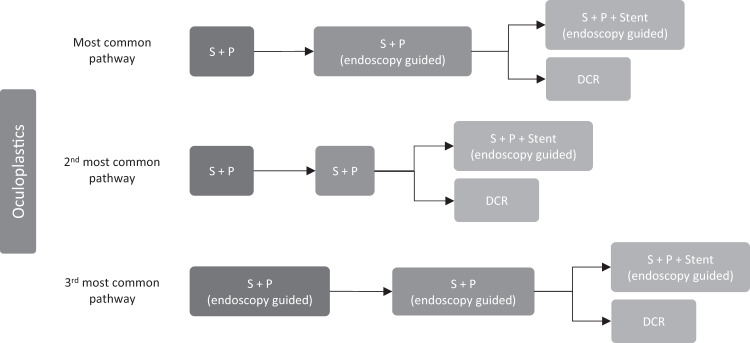
In total, 100% of survey participants agreed that all procedures should be conducted in theatre under general anaesthetic; however, there were notable differences in the preferred primary intervention between both subspecialties. Just over half (53.7%) of all oculoplastics consultants prefer simple syringe and probe procedures with no nasoendoscopy, compared to 43.9% of the same specialty who use nasoendoscopy at this stage. Of the 19 oculoplastic individuals who use nasoendoscopy, only 3 (15.8%) required ENT involvement. In contrast, three quarters of paediatric consultants (77.4%) choose unguided syringe and probe as primary intervention compared to eight individuals (12.9%) who utilise endoscopy assistance. Of these eight paediatric colleagues, only two (25%) sought ENT input. Perhaps surprisingly, just under 5% of paediatric consultants also consider stenting as part of the primary procedure. In summary, as shown in Figs. 1 and 2, syringe and probe with no nasoendoscopy is the most common choice of primary intervention for both subspecialties. Oculoplastic surgeons are more likely to use nasoendoscopy-guided procedures, and less likely to ask for ENT assistance, than their paediatric colleagues.
Fig. 1. Oculoplastic management pathway.
Most common management pathways between surveyed oculoplastic consultants for primary, secondary and tertiary interventions of CNLDO.
Fig. 2. Paediatric management pathway.
Most common management pathways between surveyed consultant paediatric ophthalmologists for primary, secondary and tertiary interventions of CNLDO.
The most common secondary procedures for oculoplastic surgeons are syringe and probe under nasoendoscopic guidance (36.6%), followed by nasoendoscopy-assisted lacrimal intubation (26.8%). In total, 17.1% of oculoplastic participants repeat a simple syringe and probe procedure as their second management step. Management preferences for paediatric subspecialty colleagues regarding re-do procedures are as follows: 29.0% choose syringe and probe with nasoendoscopy as their re-do procedure; the same percentage (29.0%) choose to stent with an equally observed split for with (14.5%) or without (14.5%) nasoendoscopy, and slightly fewer would refer onwards (27.4%), although it is not specified if that would be to a different subspecialty or unit. Less than 5% of each subspecialty perform balloon catheter dilatation, with or without endoscopy.
Lastly, we asked participants what their tertiary management step would be if the re-do procedure is unsuccessful. Of the oculoplastic surgeons who go on to perform DCR, 68% prefer endonasal DCR and 32% choose an external approach. Of the paediatric consultants who perform DCR procedures (15%), there was an equal divide between endonasal and external approach. Tertiary management by paediatric ophthalmologists is most commonly (54.8%) to refer to oculoplastic teams.
Discussion
We conducted a national survey to observe how children with CNLDO are managed by oculoplastic and paediatric subspecialties in the UK. We were also keen to note if there had been any changes in management preferences over the last decade in comparison to a similar survey in 2007 by Puvanachandra et al. [7]. This comparison is detailed in Table 2.
Table 2.
Comparison of our results to national survey of CNLDO management from 2007 (Puvanachandra et al.).
| % of surveyed ophthalmologists | ||
|---|---|---|
| 2007: Puvanachandra et al. | 2020: Current study | |
| (n = 100) | (n = 103) | |
| Investigation | ||
| Sub-speciality mostly managing CNLDO patients | Paediatrics (69%) | Paediatrics (71.8%) |
| Routinely performing FDDT | 49.0% | 62.1% |
| Lower age limit of 12 months for syringe and probe | 74.0% | 51.5% |
| Office probing | 0.0% | 0.0% |
| Primary procedure | ||
| Syringe and probe | 100.0% | 96.1% |
| % of all primary procedures using nasoendoscopy | 25.0% | 26.2% |
| % of nasoendoscopy cases requiring ENT involvement | 24.0% | 19.2% |
| Secondary procedure | ||
| Refer | 7.0% | 18.4% |
| Repeat syringe and probe | 64.5% | 42.7% |
| Lacrimal intubation | 35.5% | 32.0% |
| Balloon catheter dilatation | 0.0% | 3.9% |
| Tertiary procedure | ||
| Refer | 37.6% | 36.9% |
| Lacrimal intubation | 48.4% | 25.2% |
| Endonasal DCR | 28.0% | 58.0% |
| External DCR | 50.0% | 33.0% |
The majority of children with CNLDO today continue to enter hospital eye services through the paediatric ophthalmology clinic, similar to referral patterns in 2007. This referral pathway is particularly appropriate given the strengthening evidence of association between CNLDO and amblyopia in recent years. Ramkumar et al. [8] found that the prevalence of defined amblyopic risk factors in children with CNLDO to be 14.78%, and Mocanu et al. [9] report an odds ratio of 4.32% between CNLDO and amblyopia. Astigmatism of greater severity has also been noted in eyes with CNLDO compared to healthy contralateral eyes of the same patient [10], with hypermetropia being the most common astigmatic error [11]. For these reasons, the importance of a thorough refractive examination of children with CNLDO should be highlighted. Where possible, paediatric ophthalmologists are therefore best suited to initially assess children with CNLDO.
Regarding initial diagnosis, FDDT remains the most commonly performed investigation. In comparison to Puvanachandra et al., our survey reports increased use of FDDT since 2007, although reasons for this have not been identified.
Our survey shows outpatient clinic probing continues to remain highly uncommon in the UK, despite international paediatric ophthalmologists stating it is the second most common intervention in their practice [12]. Data from the Paediatric Eye Disease Investigator Group (PEDIG) in 2014 reports a success rate of office probing of 75% with nil complications encountered [13]. Success rates were lower in bilateral symptoms and children with multiple signs of CNLDO. The PEDIG technique involves stat topical anaesthesia and infant restraint with assistance, where Kothari et al. [12] describe a trialled technique of topical antibiotics pre- and post-probing with oral dextrose solution to supplement the anaesthetic effect of topical anaesthesia. Despite the reported high success rates, there are no randomised control trials to identify any clinical advantage between early outpatient probing and probing under anaesthetic. With this in mind, one cannot exclude the risks of general anaesthetic in young children, especially in children with genetic anomalies causing craniofacial malformations and cardiac compromise.
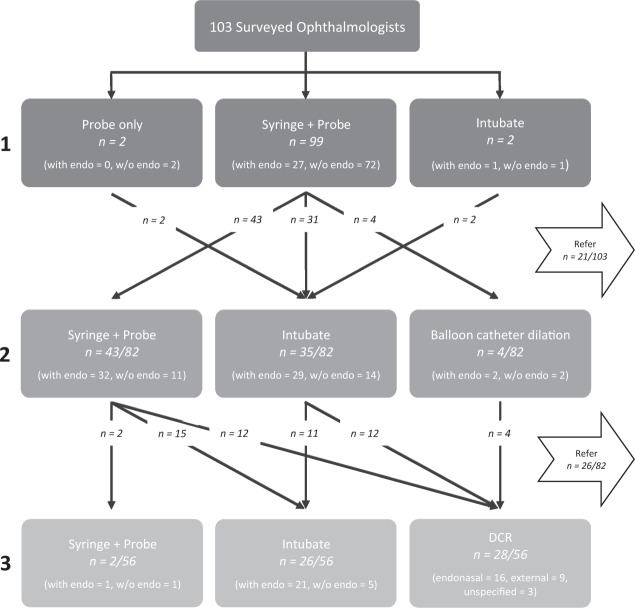
Almost unanimously from our survey, the primary intervention for CNLDO remains syringe and probe under general anaesthetic in theatre (96%). A minority perform probing only, claiming syringing would not change management, and two consultants report stenting as primary intervention to theoretically reduce need for recurrent procedures. As shown in Fig. 3, our survey reports 27% of all primary procedures are done with endoscopic assistance. Despite increasing evidence to support higher success rates of endoscopically assisted probing [14], this rate is similar to Puvanachandra et al. in 2007 (25%). This possibly reflects plateaued levels of endoscopically trained personnel and/or access to specialist equipment. Nevertheless, the advantages of nasoendoscopy are well recognised. The technique allows for better visualisation of obstruction and implementation of probing with the option to carry out additional treatments like inferior turbinate infracture, if required, under a single anaesthetic.
Fig. 3. Overview of survey results.
Flowchart showing the various management pathways of CNLDO as detailed by surveyed ophthalmologists.
Our own departmental audit data in 2019 of 142 eyes of CNLDO patients found an overall (with and without endoscopic guidance) initial probing success rate of 80.9%. Of the successful 115 initial procedures, 43% were done with endoscopic guidance and 57% without, with the caveat that the majority of CNLDO patients are seen through the paediatric clinic and thus primarily treated by paediatric ophthalmologists who do not routinely use nasoendoscopy. This trend is reflected in our national survey results, where oculoplastic consultants are nearly four times more likely (43.9%) than paediatric colleagues (12.9%) to use endoscopic guidance with primary probing. Of the 27 patients in our audit who required re-do procedures, two-thirds had undergone a primary procedure without nasoendoscopy compared to one-third with endoscopic guidance. Our departmental audit suggests a lower chance of needing re-do procedures if the primary procedure is done with endoscopic guidance.
The optimal age for interventional management of CNLDO remains debated. By asking an open-ended question in our survey regarding age limits, we were able to assess the true variance in practice. We found that the majority of surveyed ophthalmologists (51.5%) continue to benchmark 12 months as a lower age limit, although this is a lower percentage majority than in 2007 (74%). This suggests clinicians are considering a wider age range at which to intervene, as encouraged by Sathiamoorthi et al. [2] who found spontaneous resolution to plateau after 9 months of age and initial probing success to diminish after 15 months. However, many studies report no age-related correlation between success and interventions such as outpatient probing [13], primary and repeat probing [15] or endoscopically guided probing [16]. These contradicting results leave us far from a firm consensus regarding optimal age for intervention.
Regarding re-do procedures, our survey indicates nasoendoscopy-guided syringe and probe is the most favourable option with both paediatric ophthalmologists and oculoplastic colleagues; however the preference to refer (18.4%) has increased than when previously surveyed in 2007 (7%). This may be confounded by the larger proportion of paediatric than oculoplastic consultants replying to our survey, further emphasising the sub-speciality nature of ophthalmology. Overall rates for second-stage management with ductal intubation remain similar between the two national surveys in 2007 (35.5%) and 2020 (32%) (Table 2). In contrast, balloon catheter dilatation was more favourably represented in our survey (4%) than previously (0%). However, it is unclear whether this is due to truly increased use of the technique or simply representative of the participants surveyed.
When it comes to the third stage of management, the current survey would appear to suggest a change of preferences over time. Our survey highlights DCRs are nearly three times (35.9%) more commonly performed than in 2007 (13%). This is in parallel with a reduced rate of lacrimal intubation at the third step of management, from 48% in 2007 to 25.2%. This could indicate a shift in management trend from ductal stenting to DCR, as the latter provides more definitive treatment. Indeed, reported success rates of paediatric DCR are as high as 100% for certain sub-groups [17, 18] and 97% for lacrimal intubation [19].
It was interesting to observe a considerable shift in DCR technique since the previous survey. In 2007, external DCR was preferred by 50% of participants compared to 28% expressing preference for an endonasal approach. Our survey yields combined subspecialty figures of 33% for external DCR and 58% for endonasal, showing an increased preference for endonasal surgery.
We would like to acknowledge the limitations of our survey that have been considered whilst conducting this comparative study. The response rate to our survey (25%) could have been higher from both oculoplastic and paediatric subspecialties. By disseminating the survey through BOPSS and BIPOSA, some non-member clinicians may not have been able to participate, potentially explaining the small sample size (n = 103). For true comparison between the surveys in 2007 and 2020, the same participants would need to be questioned. Nevertheless, although this would allow direct comparison, it may not reflect true changes in management; individual clinicians may become comfortable with familiar interventions and be reluctant to change technique, whereby a new snapshot survey of different participants may reflect current preferences more accurately. Whilst one must consider the limiting factors of the survey, it is undoubtedly useful in showing trends across the country in the management of CNLDO.
As shown by this national survey, although there have been some changes in treatment preferences for CNLDO, considerable variation is still prevalent. Figure 3 summarises the stepwise variance described by individuals who responded to our survey. To better streamline CNLDO management, we propose consideration for a national audit of CNLDO intervention outcomes. Considering children with CNLDO can undergo repeated episodes of general anaesthetic to facilitate treatment, and the amblyopic association of the condition, there is value in gathering national data that has the potential to standardise treatment protocols.
Summary
What was known before
The majority of CNLDO patients in the UK are initially assessed by paediatric ophthalmologists.
Most common initial procedure by surveyed ophthalmologists in the UK was probe and syringe.
Most common re-do procedure was repeat probe and syringe, and most common tertiary procedure was ductal intubation.
What this study adds
Oculoplastic and paediatric ophthalmologists report different pathways in managing CNLDO, with oculoplastic surgeons more likely to use nasoendoscopy-guided procedures. The overall use of nasoendoscopy has increased compared to the 2007 national survey.
Most common initial procedure for both subspecialties remains syringe and probe under anaesthetic in theatre, with outpatient clinic probing being highly uncommon in the UK.
Paediatric DCR surgery is nearly three times more commonly performed compared to 2007, with the majority now being performed endonasally. This is in parallel to a reduction in ductal intubation.
Compliance with ethical standards
Conflict of interest
The authors declare that they have no conflict of interest.
Footnotes
Publisher’s note Springer Nature remains neutral with regard to jurisdictional claims in published maps and institutional affiliations.
References
- 1.Clarke WN. The child with epiphora. Paediatr Child Health. 1999;4:325–6. doi: 10.1093/pch/4.5.325. [DOI] [PMC free article] [PubMed] [Google Scholar]
- 2.MacEwen CJ, Young JDH. Epiphora in the first year of life. Eye. 1991;5:596–600. doi: 10.1038/eye.1991.103. [DOI] [PubMed] [Google Scholar]
- 3.Sathiamoorthi S, Frank RD, Mohney BG. Spontaneous resolution and timing of intervention in congenital nasolacrimal duct obstruction. JAMA Ophthalmol. 2018;136:1281–6. doi: 10.1001/jamaophthalmol.2018.3841. [DOI] [PMC free article] [PubMed] [Google Scholar]
- 4.Perveen S, Sufi AR, Rashid S, Khan A. Success rate of probing for congenital nasolacrimal duct obstruction at various ages. J Ophthalmic Vis Res. 2014;9:60–9. [PMC free article] [PubMed] [Google Scholar]
- 5.Kuschner BJ. The management of naso-lacrimal duct obstruction in children between 18 months and 4 years old. J AAPOS. 1998;2:57–60. doi: 10.1016/S1091-8531(98)90112-4. [DOI] [PubMed] [Google Scholar]
- 6.Robb RM. Success rate of nasolacrimal duct probing at time intervals after 1 year of age. Ophthalmology. 1998;105:1308–10. doi: 10.1016/S0161-6420(98)97038-5. [DOI] [PubMed] [Google Scholar]
- 7.Puvanachandra N, Trikha S, MacEwen C. A national survey of the management of congenital nasolacrimal duct obstruction in the United Kingdom. J Pedatr Ophthalmol. 2010;47:76–80. doi: 10.3928/01913913-20100308-04. [DOI] [PubMed] [Google Scholar]
- 8.Ramkumar VA, Agarkar S, Mukherjee B. Nasolacrimal duct obstruction: does it really increase the risk of amblyopia in children? Indian J Ophthalmol. 2016;64:496–9. doi: 10.4103/0301-4738.190101. [DOI] [PMC free article] [PubMed] [Google Scholar]
- 9.Mocanu V, Horhat R. Prevalence and risk factors of amblyopia among refractive errors in an Eastern European population. Medicina (Kaunas) 2018;54:6. doi: 10.3390/medicina54010006. [DOI] [PMC free article] [PubMed] [Google Scholar]
- 10.Eshraghi B, Akbari MR, Fard MA, Shahsanaei A, Assari R, Mirmohammadsadeghi A. The prevalence of amblyogenic factors in children with persistent congenital nasolacrimal duct obstruction. Graefes Arch Clin Exp Ophthalmol. 2014;252:1847–52. doi: 10.1007/s00417-014-2643-1. [DOI] [PubMed] [Google Scholar]
- 11.Saleem AA, Siddiqui SN, Wakeel U, Asif M. Anisometropia and refractive status in children with unilateral congenital nasolacrimal duct obstruction. Taiwan J Ophthalmol. 2018;8:31–5. doi: 10.4103/tjo.tjo_77_17. [DOI] [PMC free article] [PubMed] [Google Scholar]
- 12.Kothari M, Rathod V, Shah K, Shikhangi K, Singhania R. Congenital nasolacrimal duct obstruction: should we continue lacrimal massage till 1 year or perform an office probing at 6 months? A clinical decision analysis approach. Indian J Ophthalmol. 2017;65:167–9. doi: 10.4103/ijo.IJO_245_16. [DOI] [PMC free article] [PubMed] [Google Scholar]
- 13.Miller AM, Chandler DL, Repka MX, Hoover DL, Lee KA, Melia M, et al. Office probing for treatment of nasolacrimal duct obstruction in infants. J AAPOS. 2014;18:26–30. doi: 10.1016/j.jaapos.2013.10.016. [DOI] [PMC free article] [PubMed] [Google Scholar]
- 14.Galindo-Ferreiro A, Khandekar R, Akaishi PM, Cruz A, Gálvez-Ruiz A, Dolmetsch A, et al. Success rates of endoscopic-assisted probing compared to conventional probing in children 48 months or older. Semin Ophthalmol. 2018;33:435–42. doi: 10.1080/08820538.2017.1284872. [DOI] [PubMed] [Google Scholar]
- 15.Valcheva KP, Murgova SV, Krivoshiiska EK. Success rate of probing for congenital nasolacrimal duct obstruction in children. Folia Med (Plovdiv) 2019;61:97–103. doi: 10.2478/folmed-2018-0054. [DOI] [PubMed] [Google Scholar]
- 16.Gupta N, Neeraj C, Smriti B, Sima D. A comparison of the success rates of endoscopic-assisted probing in the treatment of membranous congenital nasolacrimal duct obstruction between younger and older children and its correlation with the thickness of the membrane at the Valve of Hasner. Orbit. 2018;37:257–61. doi: 10.1080/01676830.2017.1383483. [DOI] [PubMed] [Google Scholar]
- 17.Chan W, Wilcsek G, Ghabrial R, Goldberg RA, Dolman P, Selva D, et al. Pediatric endonasal dacryocystorhinostomy: a multicenter series of 116 cases. Orbit. 2017;36:311–6. doi: 10.1080/01676830.2017.1337168. [DOI] [PubMed] [Google Scholar]
- 18.Komínek P, Cervenka S, Matousek P, Pniak T, Zeleník K. Primary pediatric endonasal dacryocystorhinostomy-a review of 58 procedures. Int J Pediatr Otorhinolaryngol. 2010;74:661–4. doi: 10.1016/j.ijporl.2010.03.015. [DOI] [PubMed] [Google Scholar]
- 19.Komínek P, Cervenka S, Pniak T, Zeleník K, Tomášková H, Matoušek P. Monocanalicular versus bicanalicular intubation in the treatment of congenital nasolacrimal duct obstruction. Graefes Arch Clin Exp Ophthalmol. 2011;249:1729–33. doi: 10.1007/s00417-011-1700-2. [DOI] [PMC free article] [PubMed] [Google Scholar]


