Abstract
Background
Acute kidney injury (AKI) in Covid-19 patients admitted to the intensive care unit (ICU) is common, and its severity may be associated with unfavorable outcomes. Severe Covid-19 fulfills the diagnostic criteria for acute respiratory distress syndrome (ARDS); however, it is unclear whether there is any relationship between ventilatory management and AKI development in Covid-19 ICU patients.
Purpose
To describe the clinical course and outcomes of Covid-19 ICU patients, focusing on ventilatory management and factors associated with AKI development.
Methods
Single-center, retrospective observational study, which assessed AKI incidence in Covid-19 ICU patients divided by positive end expiratory pressure (PEEP) tertiles, with median levels of 9.6 (low), 12.0 (medium), and 14.7 cmH2O (high-PEEP).
Results
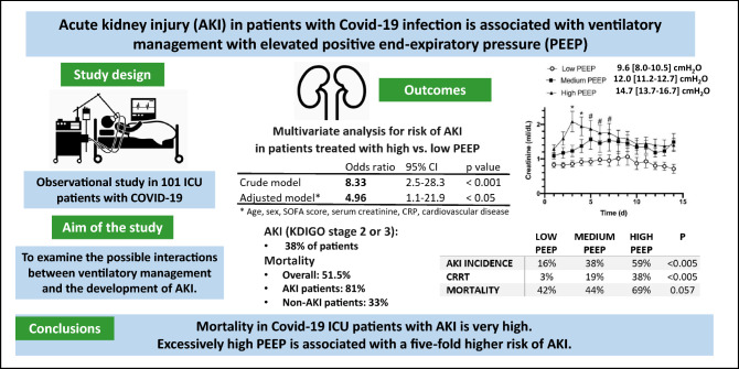
Overall mortality was 51.5%. AKI (KDIGO stage 2 or 3) occurred in 38% of 101 patients. Among the AKI patients, 19 (53%) required continuous renal replacement therapy (CRRT). In AKI patients, mortality was significantly higher versus non-AKI (81% vs. 33%, p < 0.0001). The incidence of AKI in low-, medium-, or high-PEEP patients were 16%, 38%, and 59%, respectively (p = 0.002). In a multivariate analysis, high-PEEP patients showed a higher risk of developing AKI than low-PEEP patients (OR = 4.96 [1.1–21.9] 95% CI p < 0.05). ICU mortality rate was higher in high-PEEP patients, compared to medium-PEEP or low-PEEP patients (69% vs. 44% and 42%, respectively; p = 0.057).
Conclusion
The use of high PEEP in Covid-19 ICU patients is associated with a fivefold higher risk of AKI, leading to higher mortality. The cause and effect relationship needs further analysis.
Graphic abstract
Supplementary Information
The online version contains supplementary material available at 10.1007/s40620-021-01100-3.
Keywords: Covid-19, ARDS, AKI, PEEP, Intensive care
Introduction
The severity of the disease caused by the novel ‘severe acute respiratory distress syndrome coronavirus 2’ (SARS-CoV-2) varies largely from asymptomatic cases to more severe presentations. Five to ten percent of hospitalized patients require admission to the intensive care unit (ICU) [1, 2], due to acute hypoxemic respiratory failure requiring mechanical ventilation.
Based on pre-existing evidence and guidelines on acute respiratory distress syndrome (ARDS), mechanical ventilation of Covid-19 patients with ARDS involves the use of low tidal volume (Vt), positive end-expiratory pressure (PEEP), low driving pressure and low plateau pressure [3]. However, various authors reported that Covid-19 ARDS often presents with preserved lung mechanics and well-aerated lungs on computed tomography (CT) scan [4]. To explain the severe hypoxemia conflicting with the respiratory system mechanics and CT findings, alternative mechanisms such as impairment of hypoxic pulmonary vasoconstriction and micro-thrombi formation in the pulmonary circulation have been proposed [5]. Based on these observations, specific ventilatory management, including low PEEP (8–10 cmH2O) and more liberal Vt (7–8 mL/kg), has been proposed [5, 6].
Acute kidney injury (AKI) has been reported in 20–30% of Covid-19 ICU patients [7, 8]. Multiple mechanisms have been proposed, including cytokine storm [9], direct virus-mediated renal damage [10], and pre-renal etiology due to aggressive diuretic use [11]. Previous studies found no clear correlation between ventilatory management and the risk of developing AKI in patients with ARDS [12]. Whether there is any relationship between ventilatory management and AKI development in Covid-19 ICU patients has not been established yet.
In this single-center retrospective study, we describe the clinical course and outcome of 101 Covid-19 ICU patients, focusing on ventilator management and factors associated with AKI development.
Methods
Study design and data collection
This single-center retrospective observational study included 101 consecutive adult patients admitted to the Sacco Hospital ICU in Milan from February 21st to April 28th, 2020. The Ethics Committee of L. Sacco Hospital approved the study, and informed consent was waived considering the observational, non-interventional nature of the research and the ICU clinical setting (Comitato Etico interaziendale Area 1, Milan, approval number 2020/ST/116).
Demographic and clinical characteristics of all patients were collected and recorded in a dedicated database. Daily laboratory data, including basic metabolic panel, complete blood count, liver function tests, coagulation profile, and arterial blood gas analysis, were recorded for the entire duration of the ICU stay. Ventilatory settings and adjunctive therapies such as pronation, neuromuscular blockade, and inhaled nitric oxide use were also recorded. The static (Cstat) and dynamic (Cdyn) respiratory system compliance were calculated according to standard formulae (see Supplement details). AKI was defined according to the 2012 Kidney Disease: Improving Global Outcomes (KDIGO) clinical practice guidelines [13]. The KDIGO guidelines stage AKI according to severity (stages 1–3). In this study, we considered only patients with stage 2 (serum creatinine 2.0–2.9 times of baseline values, urine output < 0.5 mL/kg/h for ≥ 12 h) and stage 3 (serum creatinine three times of baseline values, or ≥ 4.0 mg/dL (≥ 353.6 μmol/L) increase, or the initiation of renal replacement therapy (RRT); urine output < 0.3 mL/kg/h for ≥ 24 h or anuria ≥ 12 h).
Autopsy renal samples of two patients who died with Covid-19 and with severe AKI were analyzed. Renal tissues were fixed in formalin; the histological examination was performed on PAS-stained slides. In addition, immunohistochemistry for Sars-CoV-2 (nucleocapsid protein monoclonal antibody, Novus Biological) was performed according to the Ventana-Roche protocol.
Statistical analysis
All descriptive data are expressed as median [25th–75th inter quartile range (IQR)] unless specified otherwise. Categorical data were compared via χ2 test or Fisher’s exact test, as appropriate. Between-group differences of continuous variables were analyzed with Mann–Whitney U test or non-parametric one-way ANOVA, as appropriate. A two-way ANOVA was used for between-group comparison over time. We used logistic regression models to assess the association between AKI within 7 days after admission and the average PEEP level applied during the first 7 days after admission. We modeled the probability of having AKI-Injury or failure against the likelihood of AKI-Risk and no-AKI. We performed multivariate analyses, adjusting for confounders associated with both exposure (PEEP) and outcome (AKI). We compared models with different baseline characteristics using the Akaike information criterion. We derived odds ratios, 95% CIs, and p values (Supplemental Material). Statistical analysis was performed using SAS 9.2 and GraphPad 8.0. Statistical significance was defined as a p value of less than 0.05.
Results
Study population
One hundred and one patients were included in the study. The majority of the patients were male (77%), and the median age was 61 [53–68] years. Cardiovascular disease was the most common comorbidity, present in 51% of the study population. Sixty-one patients were transferred from outside hospitals, and 25 had already been admitted to an ICU for a median ICU stay before the transfer of 2 [0–16] days. The overall mortality rate was 51.5%. Patients who died had a higher incidence of cardiovascular disease and cancer at baseline and were more likely to be male. Inflammatory markers on admission, including C-reactive protein (CRP), lactate dehydrogenase (LDH), and D-dimer, were elevated, but there was no significant difference between survivors and non-survivors (Table 1).
Table 1.
Demographic and clinical characteristics on admission of the study population
| Overall population (n = 101) | Survivors (n = 49) | Non-survivors (n = 52) | p | |
|---|---|---|---|---|
| Baseline features | ||||
| Age (years) |
101/101 61 [53–68] |
49/49 57 [46–64] |
52/52 63 [60–70] |
0.096 |
| Male sex (n-%) |
101/101 78 (77) |
49/49 30 (61) |
52/52 48 (92) |
0.0002 |
| BMI |
95/101 28 [25–31] |
47/49 28 [26–32] |
48/52 28 [25–31] |
0.760 |
| Smoking, n (%) |
100/101 5 (5%) |
48/49 2 (4) |
52/52 3 (6) |
0.202 |
| Cardiovascular disease, n (%) |
100/101 51 (51) |
48/49 19 (40) |
52/52 32 (62) |
0.028 |
| Chronic lung disease, n (%) |
100/101 7 (7) |
48/49 3 (6) |
52/52 4 (8) |
0.999 |
| Immunodepression, n (%) |
100/101 4 (4) |
48/49 1 (2) |
52/52 3 (6) |
0.618 |
| Diabetes, n (%) |
100/101 14 (14) |
48/49 7 (15) |
52/52 7 (13) |
0.872 |
| Cancer, n (%) |
100/101 6 (6) |
48/49 0 (0) |
52/52 6 (12) |
0.027 |
| SOFA score |
101/101 9 [7–11] |
49/49 8 [4–11] |
52/52 9 [8–11] |
0.346 |
| Ventilatory and laboratory data | ||||
| Vt (mL/kg IBW) |
81/101 7.6 [7.0–8.2] |
37/49 7.8 [7.4–8.9] |
44/52 7.4 [6.9–8.1] |
0.226 |
| PEEP (cmH2O) |
94/101 13 [12–16] |
46/49 13 [10–15] |
48/52 14 [12–18] |
0.101 |
| Peak pressure (cmH2O) |
79/101 32 [29–35] |
35/49 31 [28–34] |
44/52 33 [30–35] |
0.024 |
| Arterial pH |
96/101 7.35 [7.29–7.42] |
47/49 7.39 [7.31–7.45] |
49/52 7.32 [7.27–7.37] |
0.009 |
| PaCO2 (mmHg) |
96/101 46 [40–53] |
47/49 45 [38–50] |
49/52 46 [42–56] |
0.360 |
| FiO2 (%) |
95/101 0.8 [0.6–0.9] |
46/49 0.7 [0.6–0.9] |
49/52 0.8 [0.7–0.9] |
0.170 |
| PaO2 (mmHg) |
96/101 86 [72–106] |
47/49 80 [67–115] |
49/52 87 [74–101] |
0.310 |
| PaO2:FiO2 |
95/101 113 [92–152] |
46/49 113 [92–176] |
49/52 113 [93–142] |
0.921 |
| Lactate (mmol/L) |
92/101 1.3 [1.0–1.5] |
46/49 1.1 [0.9–1.4] |
46/52 1.3 [1.2–1.7] |
0.013 |
| WBC (*109/L) |
82/101 8715 [6020–11900] |
37/49 8390 [5760–11900] |
45/52 8750 [6390–11890] |
0.825 |
| Neutrophils (%WBC) |
75/101 88 [80–91] |
34/49 86 [81–91] |
41/52 90 [79–91] |
0.201 |
| Lymphocytes (%WBC) |
73/101 6 [4–12] |
34/49 8 [6–15] |
41/52 6 [4–11] |
0.016 |
| Platelets (*109/L) |
82/101 227 [172–297] |
37/49 232 [163–289] |
45/52 200 [176–297] |
0.508 |
| D-Dimer (µg/L) |
49/101 2042 [1169–5202] |
27/49 1944 [1015–7000] |
22/52 2069 [1169–5202] |
0.486 |
| LDH (U/L) |
58/101 531 [447–664] |
28/49 523 [416–643] |
30/52 561 [449–691] |
0.602 |
| CRP (mg/L) |
74/101 171 [88–288] |
35/49 137 [67–254] |
39/52 231 [109–307] |
0.106 |
| PCT within 48 h (µg/L) |
83/101 0.6 [0.2–1,7] |
44/49 0.3 [0.1–1.0] |
39/52 0.9 [0.2–2.0] |
0.142 |
| Creatinine (mg/dL) |
89/101 0.9 [0.7–1.2] |
42/49 0.9 [0.7–1.1] |
47/52 1.0 [0.8–1.3] |
0.369 |
| BUN (mg/dL) |
48/101 50 [30–64] |
24/49 48 [30–58] |
24/52 50 [31–80] |
0.999 |
| Outcomes | ||||
| AKI-Risk, n (%) | 14/96 (15) | 8/47 (17) | 6/49 (12) | |
| AKI-Injury, n (%) | 8 (8) | 1 (2) | 7 (14) | |
| AKI-Failure, n (%) | 28 (29) | 6 (13) | 22 (45) | |
| AKI-Injury + Failure, n (%) | 36 (38) | 7 (15) | 29 (59) | < .0001 |
| CRRT, n (%) | 19 (20) | 5 (11) | 14 (29) | 0.028 |
| ICU LOS (days) | 15 [9–21] | 14 [9–25] | 15 [8–20] | 0.822 |
| Mechanical ventilation (days) |
90/101 13 [8–19] |
38/49 11 [8–19] |
52/52 14 [6–21] |
0.671 |
| ICU mortality, n (%) | 52/101 (51) | 0 (0) | 52 (100) | NA |
Data represent number of patients and percentage (%) or median [IQR]. For each parameter, if missing values are present, the number of available data is expressed. p values represent comparisons between survivors and non-survivors, Mann–Whitney U test, χ2 test, or Fisher’s exact test, as appropriate
BMI body mass index, SOFA sequential organ failure assessment, Vt tidal volume, PEEP positive end expiratory pressure, PaCO2 arterial carbon dioxide partial pressure, PaO2 arterial oxygen partial pressure, FiO2 fraction of inspired oxygen, WBC White blood cell count, LDH lactic dehydrogenase, CRP C-reactive protein, PCT procalcitonin, AKI acute kidney injury, CRRT continuous renal replacement therapy, ICU LOS ICU length of stay
AKI: incidence, mortality, and histopathologic features
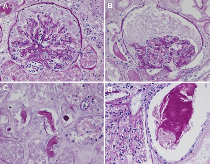
Among 96 patients in whom creatinine data were available, 36 (38%) developed AKI (KDIGO AKI stage 2 or 3) within 28 days of ICU stay (Table 1). Among the 36 patients who developed AKI, 19 (53%) required continuous renal replacement therapy (CRRT). Patients who developed AKI (AKI-group) had a higher SOFA score (10 [8–12] vs. 8 [5–11], p = 0.049) and higher serum creatinine at admission (1.0 [0.9–1.9] vs. 0.8 [0.7–1.1] mg/dL, p = 0.018) compared with patients who did not develop AKI (no-AKI-group). ICU mortality in AKI group was significantly higher, compared with the no-AKI group: 81% vs. 33%, p < 0.0001. No difference in the number of patients transferred from another ICU was found in the two groups (Table 2). The histopathological findings observed in the post-mortem kidneys of two Covid-19 ICU patients with AKI (Fig. 1) were glomeruli with mild or moderate tuft collapse but without hypertrophy or hyperplasia of the overlying visceral epithelium; other features were interstitial expansion by edema and tubules with protein casts. No inflammation or vascular lesions were found. Immunohistochemistry for SARS-CoV-2 antigens gave negative results.
Table 2.
Clinical features and outcomes in patients who did or did not develop AKI
| AKI (n = 36) | No-AKI (n = 60) | p | |
|---|---|---|---|
| Baseline features | |||
| Age (years) |
36/36 62 [58–69] |
60/60 61 [48–68] |
0.413 |
| Male sex (n-%) | 32 (89) | 43 (72) | 0.048 |
| BMI |
35/36 29 [26–33] |
57/60 28 [25–31] |
0.388 |
| Smoke, n (%) | 3(8) | 2 (3) | 0.304 |
| Cardiovascular disease, n (%) | 23 (64) | 26 (43) | 0.051 |
| Chronic lung disease, n (%) | 3 (8) | 3 (5) | 0.669 |
| Immunodepression, n (%) | 1 (3) | 3 (5) | 1.000 |
| Diabetes, n (%) | 7 (19) | 5 (8) | 0.125 |
| Cancer, n (%) | 2 (6) | 4 (7) | 0.999 |
| SOFA score |
36/36 10 [8–12] |
60/60 8 [5–11] |
0.049 |
| Patients transferred from another ICU, n (%) | 25 (69) | 31 (52) | 0.087 |
| Laboratory data | |||
| PaO2:FiO2 |
36/36 108 [78–128] |
59/60 118 [93–173] |
0.109 |
| Creatinine admission (mg/dL) |
33/36 1.0 [0.9–1.9] |
56/60 0.8 [0.7–1.1] |
0.018 |
| BUN (mg/dL) |
16/36 60 [38–83] |
32/60 43 [30–56] |
0.226 |
| CRP (mg/L) |
33/36 238 [129–294] |
40/60 141 [68–255] |
0.027 |
| D-Dimer (µg/L) |
17/36 1417 [1105–4075] |
32/60 2045 [1394–10353] |
0.846 |
| Lactate (mmol/L) |
32/36 1.3 [1.1–1.6] |
58/60 1.2 [0.9–1.4] |
0.390 |
| WBC (*109/L) |
34/36 8565 [6390–12350] |
48/60 9225 [5970–11895] |
0.656 |
| Neutrophils (%WBC) |
32/36 89.3 [83.1–91.2] |
43/60 86.7 [77.0–91.6] |
0.574 |
| Lymphocytes (%WBC) |
31/36 6.0 [4.1–12.0] |
42/60 6.9 [4.5–14.6] |
0.122 |
| Ventilatory and hemodynamic data | |||
| PEEP at admission ICU (cmH2O) |
35/36 14.0 [12.0-1m8.0] |
59/60 12.5 [10.0–14.5] |
0.019 |
| Mean PEEP (cmH2O) |
36/36 14.5 [12.4–17.0] |
60/60 12.1 [10.4–14.3] |
0.003 |
| Cdyn (mL/cmH2O) |
34/36 31.4 [26.4–37.9] |
44/60 31.3 [26.4–35.3] |
0.754 |
| Neuromuscular blockade, n (%) | 35 (97) | 48 (80) | 0.028 |
| Neuromuscular blockade duration (days) |
36/36 9.0 [5.5–12.0] |
60/60 5.5 [1–10.5] |
0.036 |
| Prone positioning ICU, n (%) | 27 (75) | 33 (55) | 0.050 |
| Prone positioning duration (days) | 3 [1–5] | 1 [0–3] | 0.015 |
| Nitric oxide use, n (%) | 3 (8) | 8 (13) | 0.528 |
| Diuretic use, n (%) | 34 (94) | 55 (92) | 0.612 |
| Diuretic duration (days) |
36/36 7 [3–13] |
60/60 8 [3–13] |
0.542 |
| Cumulative urine output (L/28 days) |
36/36 19 [4–33] |
59/60 27 [12–43] |
0.109 |
| Fluid balance (L/28 days) |
34/34 1.3[(-5.3)-(+ 4.5)] |
58/60 -0.5 [(-5.3)-(+ 2.6)] |
0.091 |
| Outcomes | |||
| ICU LOS (days) |
36/36 15 [10–19] |
60/60 13 [7–21] |
0.208 |
| Mechanical ventilation within 48 h, n (%) | 36 (100) | 51 (85) | 0.024 |
| Mechanical ventilation (days) |
36/36 14 [8–18] |
50/60 11 [7–21] |
0.385 |
| ICU mortality, n (%) | 29 (81) | 20 (33) | < 0.0001 |
Data represent number of patients and percentage (%) or median [IQR]. For each parameter, if missing values are present, the number of available data is expressed. p values were calculated by non-parametric One-way ANOVA, χ2 test, or Fisher’s exact test, as appropriate
BMI body mass index, SOFA sequential organ failure assessment, WBC white blood cell count, Mean PEEP positive end expiratory pressure, for each patient, the average of the first 7 days after ICU admission was calculated, Cdyn dynamic respiratory system compliance, PaO2 arterial oxygen partial pressure, FiO2 fraction of inspired oxygen, BUN blood urea nitrogen, CRP C-Reactive protein, ICU LOS intensive care unit length of stay
Fig. 1.
Representative post-mortem histological features in kidneys of patients with AKI-associated severe Covid-19. A, B Glomerular tuft collapse to the vascular pole, without hyperplasia or hypertrophy of podocytes. C, D Interstitial edema and tubular casts, without inflammation. Most of the tubules are lytic because of autopsy samples. Thus, tubular epithelial cells are not evaluable. A–D PAS staining, OM × 20
Inflammatory markers and AKI
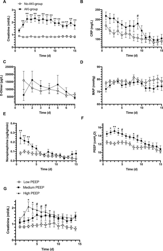
To investigate the possible relationship between the development of AKI and inflammation, we analyzed inflammatory markers and coagulation function in patients who developed AKI and those who did not. D-dimer levels on admission were similar between the two groups. However, upon admission, CRP was higher in patients who developed AKI compared to those who did not (238 [129–294] vs. 141 [68–255] mg/L, p = 0.027, Table 2), and tended to remain higher during the first 9 days after ICU admission (Fig. 2B).
Fig. 2.
Laboratory data, PEEP and hemodynamics in patients who did or did not develop AKI. Trend in A creatinine, B C-reactive protein, C D-Dimer, D mean arterial pressure (MAP), E norepinephrine dose, F PEEP; G creatinine levels over time in patients treated with low (n = 31), medium (n = 32) or high (n = 32) PEEP. Two-way ANOVA. **p < 0.001, *p < 0.01, #p < 0.05. All data represent mean ± SEM
Hemodynamics and AKI
We examined the mean arterial pressure, vasopressor requirement, diuretic use, and fluid balance in patients divided by AKI group to evaluate the relationship between hemodynamics, fluid management, and renal function. During the first 15 days of ICU stay, there was no difference in mean arterial pressure in patients who developed AKI compared to those who did not (Fig. 2D). However, patients in the AKI group were treated with significantly higher doses of norepinephrine (Fig. 2E). Nevertheless, diuretic use (94% vs. 92%) and duration (7 [3–13] vs. 8 [3–13] days), cumulative urine output, and the total cumulative fluid balance over the 28 days of observation were similar in the AKI and no-AKI-groups (Table 2).
Respiratory data and ventilatory management
Upon admission, the PaO2:FiO2 was similar in patients who developed AKI and patients who did not. The dynamic compliance of the respiratory system was similar in the two groups (31.4 [26.4–37.9] vs. 31.3 [26.4–35.3] mL/cmH2O, p = 0.754, Table 2). On admission and during the first week of ICU stay, patients who developed AKI were ventilated with higher PEEP (Fig. 2F). More frequently patients in the AKI-group underwent prone positioning (75% vs. 55%, p = 0.05) and for a longer duration (3 [1–5] vs. 1 [0–3] days, p = 0.015). Neuromuscular blockade was used for a longer duration in the AKI-group than in the no-AKI-group (9 [6–12] vs. 6 [1–11] days, p = 0.036), while no difference in the use of inhaled nitric oxide was found in the two groups (Table 2).
Effect of PEEP level on kidney function and outcome
To further investigate any possible relationship between PEEP and the risk of developing AKI, we divided the entire population into tertiles of the average PEEP level applied during the first 7 days after admission. Patients in the low-PEEP group were treated with an average PEEP of 9.6 [8.0–10.5] cmH2O (n = 31), in the medium-PEEP group with PEEP 12.0 [11.2–12.7] cmH2O (n = 32), and in the high-PEEP group with PEEP 14.7 [13.7–16.7] cmH2O (n = 32) (Table 3). The PaO2:FiO2 on admission was similar in the three groups. However, the FiO2 was significantly higher in patients treated with high PEEP. Patients treated with high PEEP had higher SOFA scores on admission compared with patients with low PEEP (11 [8–12] vs. 8 [5–11], p = 0.057, Table 3).
Table 3.
Clinical features and outcomes of the study population divided into tertiles of PEEP level applied during the first 7 days of ICU stay
| Low PEEP | Medium PEEP | High PEEP | p | |
|---|---|---|---|---|
| N = 31 | N = 32 | N = 32 | ||
| Baseline features | ||||
| Age (years) | 60 [53–68] | 61 [51–69] | 61 [53–68] | 0.478 |
| Male sex (n-%) | 20 (65) | 26 (81) | 29 (91) | 0.037 |
| SOFA score |
31/31 8 [5–11] |
32/32 9 [7–11] |
32/32 11 [8–12] |
0.057 |
| Laboratory data | ||||
| PaO2:FiO2 |
31/31 124 [96–156] |
32/32 111[92–148] |
32/32 109 [79–142] |
0.495 |
| FiO2 (%) |
31/31 0.7 [0.6–0.8] |
32/32 0.8 [0.6–0.8] |
32/32 0.8 [0.7–1.0] |
0.028 |
| Creatinine (mg/dL) |
29/31 0.8 [0.6–0.9] |
27/32 1.0 [0.7–1.3] |
32/32 1.0 [0.9–1.5] |
0.012 |
| CRP (mg/L) |
23/31 90 [45–172] |
24/32 237 [143–325] |
26/32 215 [109–282] |
0.007 |
| WBC |
26/31 10,895 [5100–14040] |
28/32 8420 [5970–10935] |
28/32 8280 [6515–11345] |
0.641 |
| Ventilatory and diuretic data | ||||
| PEEP (cmH2O) |
31/31 9.6 [8.0–10.5] |
32/32 12.0 [11.2–12.7] |
32/32 14.7 [13.7–16.7] |
< 0.0001 |
| Prone positioning, n (%) | 5 (16) | 8 (25) | 10 (31) | 0.372 |
| Cdyn (mL/cmH2O) |
23/31 26 [22–34] |
26/32 31 [27–35] |
29/32 32 [29–39] |
0.057 |
| Diuretic use, n (%) | 29 (94) | 30 (94) | 29 (91) | 0.999 |
| Diuretic therapy duration (days) |
31/31 6 [3–11] |
32/32 11 [5–13] |
32/32 7 [3–14] |
0.486 |
| Outcomes | ||||
| AKI-Risk, n (%) | 3 (10) | 7 (22) | 4 (13) | |
| AKI-Injury, n (%) | 1 (3) | 4 (13) | 3 (9) | |
| AKI-Failure, n (%) | 4 (13) | 8 (25) | 16 (50) | |
| AKI-Injury + Failure, n (%) | 5 (16) | 12 (38) | 19 (59) | 0.002 |
| CRRT, n (%) | 1 (3) | 6 (19) | 12 (38) | 0.003 |
| ICU mortality, n (%) | 13 (42) | 14 (44) | 22 (69) | 0.057 |
| Hospital mortality, n (%) | 14 (48) | 15 (52) | 22 (71) | 0.157 |
Data represent number of patients and percentage (%) or median [IQR]. p values were calculated by non-parametric ONE-way ANOVA, χ2 test, or Fisher’s exact test, as appropriate. Low-PEEP group: 9.6 [8.0–10.5] cmH2O; medium-PEEP group: 12.0 [11.2–12.7] cmH2O; high-PEEP group 14.7 [13.7–16.7] cmH2O
PEEP positive end expiratory pressure, SOFA sequential organ failure assessment, PaO2 arterial oxygen partial pressure, FiO2 fraction of inspired oxygen, AKI acute kidney injury, CRRT continuous renal replacement therapy
Patients treated with high PEEP had higher creatinine at baseline (Table 3) and creatinine further increased over time, whereas it remained significantly lower in patients treated with low PEEP (Fig. 2G). The incidence of AKI (injury + failure) in low-, medium-, and high-PEEP groups were 16%, 38%, and 59%, respectively (p = 0.002), while the need for CRRT occurred in 3%, 19%, and 38%, respectively (p = 0.003) (Table 3).
To assess whether PEEP could be an independent factor affecting AKI incidence, we performed a multivariate analysis in patients intubated within 48 h from ICU admission. Without any adjustment, the odds ratio of developing AKI in patients with high PEEP was significantly higher than in patients with low PEEP (OR = 8.3 [2.5–28.3] 95% CI p = 0.0007). After adjusting for confounders associated with both the development of AKI (Table 2) and the level of PEEP applied (Table 3) (including sex, SOFA score, serum creatinine, CRP at the time of ICU entry, and the presence of cardiovascular disease), the risk of developing AKI remained significantly higher in patients with high PEEP compared with those with low PEEP (OR = 4.96 [1.1–21.9] 95% CI p = 0.034) (Table 4).
Table 4.
Multivariate analysis for risk of AKI in patients treated with low or high PEEP
| Crude model | |||||
|---|---|---|---|---|---|
| Outcome | Comparisons | OR | 95% CI | p value | |
| Acute kidney | Medium vs low | 3.75 | 0.98 | 14.39 | 0.0541 |
| injury or failure | High vs. low | 8.33 | 2.45 | 28.29 | 0.0007 |
| Adjusted model | |||||
|---|---|---|---|---|---|
| Outcome | Comparisons | OR | 95% CI | p value | |
| Acute kidney injury or failure | Medium vs. low | 1.91 | 0.35 | 10.48 | 0.454 |
| High vs low | 4.96 | 1.13 | 21.91 | 0.034 | |
| Confounders | |||||
| Male | 0.64 | 0.13 | 3.12 | 0.580 | |
| SOFA score | 1.09 | 0.91 | 1.32 | 0.347 | |
| Creatinine | 2.61 | 0.61 | 11.23 | 0.198 | |
| CRP | 1.00 | 1.00 | 1.01 | 0.744 | |
| Cardiovascular disease | 2.80 | 0.88 | 8.89 | 0.081 | |
Logistic regression models to assess the association between AKI within 7 days after admission and average PEEP level applied during the first 7 days after admission modeling the probability of having AKI-Injury or failure against the likelihood of AKI-Risk and no-AKI. Multivariate analyses, adjusting for baseline characteristics known a priori to be potential confounders and factors associated with both exposure to PEEP and outcome. Odds ratio (OR), 95% CIs, and p values. Low-PEEP group: 9.6 [8.0–10.5] cmH2O; medium-PEEP group: 12.0 [11.2–12.7] cmH2O; high-PEEP group 14.7 [13.7–16.7] cmH2O
The ICU mortality in patients with high PEEP was higher than in patients with low or medium PEEP, although not statistically significant (69% vs. 44% and 42% respectively, p = 0.057) (Table 3).
Discussion
This study describes the demographic and clinical characteristics of 101 ICU patients with Covid-19 hypoxemic respiratory failure, mainly focusing on the clinical course, laboratory findings, and ventilatory management related to AKI development. The mortality rate in our patient population was 51.5%, with a greater risk of death in male patients and patients with pre-existing cardiovascular disease or cancer. Many patients received prone positioning and neuromuscular blockade, with a prolonged ICU course and extended duration of mechanical ventilation. Patients treated with high PEEP levels had a higher incidence of AKI and requirement of CRRT (Supplementary Fig. 1).
The 51.5% mortality rate observed in our study is consistent with previous reports [7, 14–16]. Whether patients with Covid-19 lung disease present with a clinical picture resembling typical ARDS or rather have a distinct disease has been the subject of debate [17, 18]. Despite the presence of hypoxemia and bilateral infiltrates on chest X-rays, the respiratory system compliance in the early phases of the disease is often preserved, with CT scan images frequently showing maintained lung aeration and low weight [19].
Among hospitalized Covid-19 patients, AKI is a severe and common complication. In this study, we observed a 38% incidence of AKI (KDIGO stage 2 and 3) [7, 8]. AKI was associated with a 2.5-fold increase in ICU mortality. A meta-analysis of 39 studies (25,566 patients) including ICU and non-ICU patients showed a pooled incidence of AKI of 15.4% with significant heterogeneity. However, the AKI incidence in patients with severe Covid-19 was 53% [8]. Prior studies in patients with all-cause ARDS showed an incidence of AKI in the 30–45% range [20, 21], while 17.7% of patients with H1N1 ARDS developed AKI [22]. In a recent study on AKI risk factors in patients with ARDS, no association was found between ventilatory parameters and the presence of AKI. In contrast, ‘ARDS-related’ risk factors associated with AKI were sepsis, non-cardiogenic shock, transfusion-related acute lung injury (TRALI), and pancreatitis [12]. These pathologies were rare in our population, and other factors may have accounted for the high incidence of AKI.
Our histological observations do not support direct renal damage caused by the virus. A direct effect could be mediated by SARS-CoV-2 interaction with the ACE2 receptor, which is the cell-entry receptor for SARS-CoV-2, widely expressed in the renal epithelium [10]. SARS-CoV-2 nucleocapsid protein has been demonstrated in the kidneys of deceased patients with Covid-19 [23]. Werion et al. described specific manifestations of proximal tubule dysfunction, including low molecular weight proteinuria, aminoaciduria, and defective handling of uric acid and phosphate, associated with ultrastructural signs of tubular injury [24]. In post-mortem analysis of kidney tissue, Braun et al. [25] found SARS-CoV-2 RNA in 60% of 63 patients (72% of those with AKI), suggesting that viral renal tropism is associated with disease severity and AKI development. However, other studies failed to demonstrate the presence of the virus at the ultrastructural level in the kidneys in most patients. Schurink et al. [26] showed that sporadic SARS-CoV-2-positive cells were present in the tubular epithelium of the kidney in only 9% of patients at an early stage of the disease (8 days).
Other mechanisms may include kidney damage secondary to cytokine storm [9]. In addition, the strong inflammatory response mounted by some patients in response to the SARS-CoV-2 infection and the activation of the coagulation cascade and platelets may contribute to renal injury via inflammatory cells invasion and micro-thrombi formation. In our cohort, CRP was significantly higher in patients who developed AKI. They were treated with higher doses of vasopressors, which may have been related to the higher degree of inflammation and the hemodynamic effects of positive pressure ventilation (PPV) with higher PEEP levels.
The use of diuretics and PPV to correct hypoxemia may cause hypovolemia, venous stasis, and renal hypoperfusion, contributing to AKI development via hemodynamic mechanisms. In our study, diuretics were used in over 90% of the patients; however, no difference in diuretic use, duration, or fluid balance was found in patients who developed AKI compared with those who did not.
It has been previously demonstrated that PPV can affect renal function by reducing renal perfusion and glomerular filtration [27]. During PPV, the pulmonary resistance rises during inspiration, causing an impaired emptying of the right ventricle. Also, the increased intrathoracic pressure and the displacement of the diaphragm towards the abdominal organs and vessels causes a reduction of venous return. In turn, these changes produce a decrease in cardiac output [28].
Previous studies on ARDS patients (who typically had low respiratory system compliance) did not find any relationship between PEEP levels and AKI [12]. Thus, in patients with Covid-19 presenting with lung compliance higher than the typical ARDS patient, the effect of PEEP on renal perfusion and glomerular filtration may be more pronounced, and the use of high PEEP may contribute to the development of renal dysfunction. This hypothesis is supported by the finding of a more pronounced impairment in renal blood flow in mechanically ventilated patients with SARS-CoV-2 ARDS, compared with patients with “classical” ARDS [29].
Our study found that patients who developed AKI were treated with higher PEEP levels than those who did not. Also, they more frequently underwent prone positioning and paralysis, had higher inflammatory markers, and were treated with higher doses of vasopressors. All these factors were likely related to a higher degree of inflammation and a more severe disease presentation in patients who developed AKI.
To investigate whether PEEP could be an independent factor associated with the development of AKI, we divided the population by the average PEEP level applied during the first 7 days of ICU stay, and we found that patients treated with higher PEEP more frequently developed AKI compared to patients who were treated with lower PEEP levels. To account for differences in disease severity and baseline characteristics, we performed a multivariate analysis correcting for confounding factors associated with both the exposure to PEEP and outcome (AKI injury or failure). These included sexes, SOFA score, serum creatinine, CRP at the time of ICU entry, and the presence of cardiovascular disease. We found that in patients exposed to high levels of PEEP, the odds of developing AKI were five times greater than the odds for those exposed to low levels of PEEP; moreover, for those exposed to medium levels of PEEP, the odds of developing AKI were two times greater than the odds for those exposed to low levels of PEEP.
These results suggest that in this patient population, a specific PEEP level should be set with caution, as the beneficial effect of higher PEEP on oxygenation may be counteracted by the harmful effects of overdistension and hemodynamic impairment, with negative consequences on renal function and outcome.
This study has some limitations. First, it is a single-center retrospective study, and any observed effect of various treatments on outcomes should be acknowledged cautiously. Second, patients were included in this study at the beginning of the pandemic, when steroids, monoclonal antibodies, anti-viral, and other adjunctive therapies were not available, and there was no evidence of their benefit. Third, given the rapidly changing guidelines at the beginning of the pandemic, differences in treatment over time could reflect changes in clinical management not accounted for in the analysis. Fourth, histology findings are presented for only two representative patients, limiting the generalizability of the results. Finally, the association between the use of high PEEP and the risk of developing AKI is only descriptive in nature since comparisons were not powered for hypothesis testing.
In conclusion, our findings suggest that the use of high-PEEP ventilation in Covid-19 ICU patients with hypoxemic respiratory failure may be linked to a higher risk of developing AKI, which in turn further increases mortality in patients who are already at increased risk of an unfavorable outcome.
Supplementary Information
Below is the link to the electronic supplementary material.
Author contributions
Concept and design: DO and LZ. Acquisition, analysis, and interpretation of data: all authors. Drafting of the manuscript: DO, LZ, LT, AA, MG, MN. Critical revision of the manuscript for important intellectual content: all authors. Statistical analysis: LT, DO, AZ. Supervision: RC, EC.
Funding
Open access funding provided by Università degli Studi di Milano within the CRUI-CARE Agreement. Intramural source only.
Data availability
The database is available on request.
Declarations
Conflict of interest
All authors have nothing to disclose.
Ethical approval
The Ethics Committee of L. Sacco Hospital approved the study on 26/10/2020. Informed consent was waived considering the observational, non-interventional nature of the research and the ICU clinical setting (Comitato Etico interaziendale Area 1, Milan, approval number 2020/ST/116).
Consent to participate
Not applicable.
Consent for publication
Not applicable.
Footnotes
Publisher's Note
Springer Nature remains neutral with regard to jurisdictional claims in published maps and institutional affiliations.
Davide Ottolina and Luca Zazzeron have contributed equally.
References
- 1.Grasselli G, Pesenti A, Cecconi M. Critical care utilization for the Covid-19 outbreak in Lombardy, Italy: early experience and forecast during an emergency response. JAMA. 2020;323:1545–1546. doi: 10.1001/jama.2020.4031. [DOI] [PubMed] [Google Scholar]
- 2.Guan WJ, Ni ZY, Hu Y, Liang WH, Ou CQ, He JX, Liu L, Shan H, Lei CL, Hui DSC, Du B, Li LJ, Zeng G, Yuen KY, Chen RC, Tang CL, Wang T, Chen PY, Xiang J, Li SY, Wang JL, Liang ZJ, Peng YX, Wei L, Liu Y, Hu YH, Peng P, Wang JM, Liu JY, Chen Z, Li G, Zheng ZJ, Qiu SQ, Luo J, Ye CJ, Zhu SY, Zhong NS, China Medical Treatment Expert Group for Covid-19 Clinical characteristics of coronavirus disease 2019 in China. N Engl J Med. 2020;382:1708–1720. doi: 10.1056/NEJMoa2002032. [DOI] [PMC free article] [PubMed] [Google Scholar]
- 3.Alhazzani W, Moller MH, Arabi YM, Loeb M, Gong MN, Fan E, Oczkowski S, Levy MM, Derde L, Dzierba A, Du B, Aboodi M, Wunsch H, Cecconi M, Koh Y, Chertow DS, Maitland K, Alshamsi F, Belley-Cote E, Greco M, Laundy M, Morgan JS, Kesecioglu J, McGeer A, Mermel L, Mammen MJ, Alexander PE, Arrington A, Centofanti JE, Citerio G, Baw B, Memish ZA, Hammond N, Hayden FG, Evans L, Rhodes A. Surviving sepsis campaign: guidelines on the management of critically ill adults with coronavirus disease 2019 (Covid-19) Intensive Care Med. 2020;46:854–887. doi: 10.1007/s00134-020-06022-5. [DOI] [PMC free article] [PubMed] [Google Scholar]
- 4.Gattinoni L, Coppola S, Cressoni M, Busana M, Rossi S, Chiumello D. Covid-19 does not lead to a “typical” acute respiratory distress syndrome. Am J Respir Crit Care Med. 2020;201:1299–1300. doi: 10.1164/rccm.202003-0817LE. [DOI] [PMC free article] [PubMed] [Google Scholar]
- 5.Marini JJ, Gattinoni L. Management of Covid-19 respiratory distress. JAMA. 2020;323:2329–2330. doi: 10.1001/jama.2020.6825. [DOI] [PubMed] [Google Scholar]
- 6.Roesthuis L, van den Berg M, van der Hoeven H. Advanced respiratory monitoring in Covid-19 patients: use less PEEP! Crit Care. 2020;24:230. doi: 10.1186/s13054-020-02953-z. [DOI] [PMC free article] [PubMed] [Google Scholar]
- 7.Yang X, Yu Y, Xu J, Shu H, Xia J, Liu H, Wu Y, Zhang L, Yu Z, Fang M, Yu T, Wang Y, Pan S, Zou X, Yuan S, Shang Y. Clinical course and outcomes of critically ill patients with SARS-CoV-2 pneumonia in Wuhan, China: a single-centered, retrospective, observational study. Lancet Respir Med. 2020;8:475–481. doi: 10.1016/S2213-2600(20)30079-5. [DOI] [PMC free article] [PubMed] [Google Scholar]
- 8.Fabrizi F, Alfieri CM, Cerutti R, Lunghi G, Messa P. Covid-19 and acute kidney injury: a systematic review and meta-analysis. Pathogens. 2020;9:1052. doi: 10.3390/pathogens9121052. [DOI] [PMC free article] [PubMed] [Google Scholar]
- 9.Ye Q, Wang B, Mao J. The pathogenesis and treatment of the ‘cytokine storm’ in Covid-19. J Infect. 2020;80:607–613. doi: 10.1016/j.jinf.2020.03.037. [DOI] [PMC free article] [PubMed] [Google Scholar]
- 10.Hamming I, Timens W, Bulthuis ML, Lely AT, Navis G, van Goor H. Tissue distribution of ACE2 protein, the functional receptor for SARS coronavirus. A first step in understanding SARS pathogenesis. J Pathol. 2004;203:631–637. doi: 10.1002/path.1570. [DOI] [PMC free article] [PubMed] [Google Scholar]
- 11.Nadim MK, Forni LG, Mehta RL, Connor MJ, Jr, Liu KD, Ostermann M, Rimmelé T, Zarbock A, Bell S, Bihorac A, Cantaluppi V, Hoste E, Husain-Syed F, Germain MJ, Goldstein SL, Gupta S, Joannidis M, Kashani K, Koyner JL, Legrand M, Lumlertgul N, Mohan S, Pannu N, Peng Z, Perez-Fernandez XL, Pickkers P, Prowle J, Reis T, Srisawat N, Tolwani A, Vijayan A, Villa G, Yang L, Ronco C, Kellum JA. Covid-19-associated acute kidney injury: consensus report of the 25th Acute Disease Quality Initiative (ADQI) Workgroup. Nat Rev Nephrol. 2020;16:747–764. doi: 10.1038/s41581-020-00356-5. [DOI] [PMC free article] [PubMed] [Google Scholar]
- 12.Rezoagli E, McNicholas B, Pham T, Bellani G, Laffey JG. Lung-kidney cross-talk in the critically ill: insights from the Lung Safe study. Intensive Care Med. 2020;46:1072–1073. doi: 10.1007/s00134-020-05962-2. [DOI] [PMC free article] [PubMed] [Google Scholar]
- 13.Kidney Disease: Improving Global Outcomes (KDIGO) Acute Kidney Injury Work Group KDIGO clinical practice guideline for acute kidney injury. Kidney Int Suppl. 2012;2:1–138. doi: 10.1038/kisup.2012.1. [DOI] [Google Scholar]
- 14.Grasselli G, Zangrillo A, Zanella A, Antonelli M, Cabrini L, Castelli A, Cereda D, Coluccello A, Foti G, Fumagalli R, Iotti G, Latronico N, Lorini L, Merler S, Natalini G, Piatti A, Ranieri MV, Scandroglio AM, Storti E, Cecconi M, Pesenti A, Covid-19 Lombardy ICU Network Baseline characteristics and outcomes of 1591 patients infected with SARS-CoV-2 admitted to ICUs of the Lombardy region, Italy. JAMA. 2020;323:1574–1581. doi: 10.1001/jama.2020.5394. [DOI] [PMC free article] [PubMed] [Google Scholar]
- 15.Bhatraju PK, Ghassemieh BJ, Nichols M, Kim R, Jerome KR, Nalla AK, Greninger AL, Pipavath S, Wurfel MM, Evans L, Kritek PA, West TE, Luks A, Gerbino A, Dale CR, Goldman JD, O'Mahony S, Mikacenic C. Covid-19 in critically ill patients in the Seattle region - case series. N Engl J Med. 2020;382:2012–2022. doi: 10.1056/NEJMoa2004500. [DOI] [PMC free article] [PubMed] [Google Scholar]
- 16.Bellani G, Laffey JG, Pham T, Fan E, Brochard L, Esteban A, Gattinoni L, van Haren F, Larsson A, McAuley DF, Ranieri M, Rubenfeld G, Thompson BT, Wrigge H, Slutsky AS, Pesenti A, LUNG SAFE Investigators. ESICM Trials Group Epidemiology, patterns of care, and mortality for patients with acute respiratory distress syndrome in intensive care units in 50 countries. JAMA. 2016;315:788–800. doi: 10.1001/jama.2016.0291. [DOI] [PubMed] [Google Scholar]
- 17.Ziehr DR, Alladina J, Petri CR, Maley JH, Moskowitz A, Medoff BD, Hibbert KA, Thompson BT, Hardin CC. Respiratory pathophysiology of mechanically ventilated patients with Covid-19: a cohort study. Am J Respir Crit Care Med. 2020;201:1560–1564. doi: 10.1164/rccm.202004-1163LE. [DOI] [PMC free article] [PubMed] [Google Scholar]
- 18.Gattinoni L, Chiumello D, Rossi S. Covid-19 pneumonia: ARDS or not? Crit Care. 2020;24:154. doi: 10.1186/s13054-020-02880-z. [DOI] [PMC free article] [PubMed] [Google Scholar]
- 19.Gattinoni L, Chiumello D, Caironi P, Busana M, Romitti F, Brazzi L, Camporota L. Covid-19 pneumonia: different respiratory treatments for different phenotypes? Intensive Care Med. 2020;46:1099–1102. doi: 10.1007/s00134-020-06033-2. [DOI] [PMC free article] [PubMed] [Google Scholar]
- 20.McNicholas BA, Rezoagli E, Pham T, Madotto F, Guiard E, Fanelli V, Bellani G, Griffin MD, Ranieri M, Laffey JG, ESICM Trials Group and the Large observational study to UNderstand the Global impact of Severe Acute respiratory FailurE (LUNG SAFE) Investigators Impact of early acute kidney injury on management and outcome in patients with acute respiratory distress syndrome: a secondary analysis of a multicenter observational study. Crit Care Med. 2019;47:1216–1225. doi: 10.1097/CCM.0000000000003832. [DOI] [PubMed] [Google Scholar]
- 21.Darmon M, Clec'h C, Adrie C, Argaud L, Allaouchiche B, Azoulay E, Bouadma L, Garrouste-Orgeas M, Haouache H, Schwebel C, Goldgran-Toledano D, Khallel H, Dumenil AS, Jamali S, Souweine B, Zeni F, Cohen Y, Timsit JF. Acute respiratory distress syndrome and risk of AKI among critically ill patients. Clin J Am Soc Nephrol. 2014;9:1347–1353. doi: 10.2215/CJN.08300813. [DOI] [PMC free article] [PubMed] [Google Scholar]
- 22.Martin-Loeches I, Papiol E, Rodriguez A, Diaz E, Zaragoza R, Granada RM, Socias L, Bonastre J, Valverdú M, Pozo JC, Luque P, Juliá-Narvaéz JA, Cordero L, Albaya A, Serón D, Rello J, H1N1 SEMICYUC Working Group Acute kidney injury in critical ill patients affected by influenza A (H1N1) virus infection. Crit Care. 2011;15:R66. doi: 10.1186/cc10046. [DOI] [PMC free article] [PubMed] [Google Scholar]
- 23.Puelles VG, Lütgehetmann M, Lindenmeyer MT, Sperhake JP, Wong MN, Allweiss L, Chilla S, Heinemann A, Wanner N, Liu S, Braun F, Lu S, Pfefferle S, Schröder AS, Edler C, Gross O, Glatzel M, Wichmann D, Wiech T, Kluge S, Pueschel K, Aepfelbacher M, Huber TB. Multiorgan and renal tropism of SARS-CoV-2. N Engl J Med. 2020;383:590–592. doi: 10.1056/NEJMc2011400. [DOI] [PMC free article] [PubMed] [Google Scholar]
- 24.Werion A, Belkhir L, Perrot M, Schmit G, Aydin S, Chen Z, Penaloza A, De Greef J, Yildiz H, Pothen L, Yombi JC, Dewulf J, Scohy A, Gérard L, Wittebole X, Laterre PF, Miller SE, Devuyst O, Jadoul M, Morelle J, Cliniques universitaires Saint-Luc (CUSL) Covid-19 Research Group SARS-CoV-2 causes a specific dysfunction of the kidney proximal tubule. Kidney Int. 2020;98:1296–1307. doi: 10.1016/j.kint.2020.07.019. [DOI] [PMC free article] [PubMed] [Google Scholar]
- 25.Braun F, Lütgehetmann M, Pfefferle S, Wong MN, Carsten A, Lindenmeyer MT, Nörz D, Heinrich F, Meißner K, Wichmann D, Kluge S, Gross O, Pueschel K, Schröder AS, Edler C, Aepfelbacher M, Puelles VG, Huber TB. SARS-CoV-2 renal tropism associates with acute kidney injury. Lancet. 2020;396:597–598. doi: 10.1016/S0140-6736(20)31759-1. [DOI] [PMC free article] [PubMed] [Google Scholar]
- 26.Schurink B, Roos E, Radonic T, Barbe E, Bouman CSC, de Boer HH, de Bree GJ, Bulle EB, Aronica EM, Florquin S, Fronczek J, Heunks LMA, de Jong MD, Guo L, du Long R, Lutter R, Molenaar PCG, Neefjes-Borst EA, Niessen HWM, van Noesel CJM, Roelofs JJTH, Snijder EJ, Soer EC, Verheij J, Vlaar APJ, Vos W, van der Wel NN, van der Wal AC, van der Valk P, Bugiani M. Viral presence and immunopathology in patients with lethal Covid-19: a prospective autopsy cohort study. Lancet Microbe. 2020;1:e290–e299. doi: 10.1016/S2666-5247(20)30144-0. [DOI] [PMC free article] [PubMed] [Google Scholar]
- 27.Marquez JM, Douglas ME, Downs JB, Wu WH, Mantini EL, Kuck EJ, Calderwood HW. Renal function and cardiovascular responses during positive airway pressure. Anesthesiology. 1979;50:393–398. doi: 10.1097/00000542-197905000-00004. [DOI] [PubMed] [Google Scholar]
- 28.Nanas S, Magder S. Adaptations of the peripheral circulation to PEEP. Am Rev Respir Dis. 1992;146:688–693. doi: 10.1164/ajrccm/146.3.688. [DOI] [PubMed] [Google Scholar]
- 29.Fogagnolo A, Grasso S, Dres M, Gesualdo L, Murgolo F, Morelli E, Ottaviani I, Marangoni E, Volta CA, Spadaro S. Focus on renal blood flow in mechanically ventilated patients with SARS-CoV-2: a prospective pilot study. J Clin Monit Comput. 2021;1:1–7. doi: 10.1007/s10877-020-00633-5. [DOI] [PMC free article] [PubMed] [Google Scholar]
Associated Data
This section collects any data citations, data availability statements, or supplementary materials included in this article.
Supplementary Materials
Data Availability Statement
The database is available on request.


