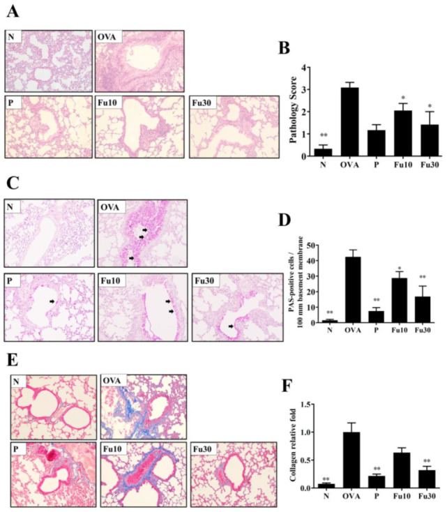
Figure 7.

The effects of fucoxanthin (Fu) on asthmatic lung tissue. Histological sections of lung tissues from normal (N) and OVA-stimulated (OVA) mice with or without Fu (10 or 30 μM) treatment. (A) Fu reduced eosinophil infiltration. HE stain, 200× magnification. (B) Inflammation was scored by a pathological evaluation of inflammatory cell infiltration in lung sections. (C) PAS-stained lung sections show goblet cell hyperplasia. Goblet cells are indicated by arrows. 200× magnification. (D) Results were expressed as the number of PAS-positive cells per 100 μm of the basement membrane. (E) Lung sections were stained with Masson’s trichrome stain to detect collagen expression. 200× magnification. (F) Quantitative analysis of collagen in lung sections. Three independent experiments were analyzed. Data are presented as mean ± SEM. * p < 0.05, ** p < 0.01 compared to the OVA control group. 10 mg/kg and 30 mg/kg fucoxanthin were named as Fu10 and Fu30, respectively. 5 mg/kg prednisolone was named as P.
