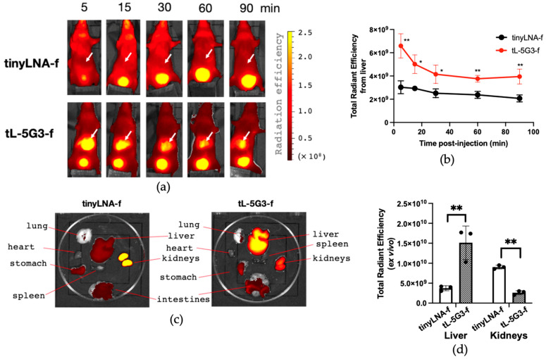
Figure 5.
In vivo and ex vivo visualization of the effect of GalNAc-conjugation on biodistribution of tiny LNA. (a) Representative fluorescence images of mice (Balb/cSlc-nu/nu, male, 6-week-old, n = 3) at different time points after administration of each Alexa647-labeled oligonucleotide (300 pmol) via tail vein (See Supplementary Figure S4 for all images). Arrows point to the liver. (b) Semi-quantitative fluorescence analysis of the liver of each mouse using IVIS imager (Total Radiation efficiency = (photons/sec)/(µW/cm2)). (c) Ex vivo images of representative tissues from mice after 90 min of administration (See Supplementary Figure S5 for all images). (d) Semi-quantitative fluorescent analysis for the tissue accumulation of each tiny LNA in the liver and kidneys using IVIS. Each fluorescence image was overlayed with a corresponding photograph. For (b,d), a multiple unpaired t-test was utilized. ** p < 0.01 and * p < 0.05.

