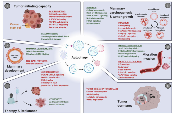
Figure 5.
The role of autophagy in cancer initiation, progression, and treatment. (A) Apart from its ambiguous role in normal mammary development and differentiation (D&D), autophagy influences virtually each step of cancer progression in a context-dependent manner. (B) Under normal conditions, high autophagic activity is predominantly cytoprotective and hence prevents cancer initiation. At the same time, autophagy facilitates the maintenance of CD44+/ALDH+ BCSC population. (C) During carcinogenesis, autophagy is frequently hijacked to support tumor expansion by promoting activation of the Ras pathway and the NRF2 oncogene, particularly in TNBC. However, inhibitory effects are often seen, which are here exemplified by the contribution of autophagy to block HER2- and WNT1-driven mammary tumorigenesis. (D) It is also evident that high autophagy activity may limit EMT traits, since several EMT-transcription factors are degraded via autophagy, including SNAIL and TWIST. In contrast, the inhibition of anoikis in malignant cells and accelerated cell migration (due to turnover of focal adhesions) were also attributed to enforced autophagy. (E) Furthermore, high levels of autophagy maintain the dormancy of the BCSCs, which is a main source of disease relapse, protecting them from metabolic stress. (F) Commonly induced autophagic activity upon anti-cancer therapy leads to an excessive autophagy potentiating drug cytotoxicity via the AMPK/mTOR/ULK pathway. In contrast, increased autophagy activates ERK and p53 signaling to limit the effect of chemotherapeutics, facilitating drug resistance. Green: tumor-suppressive roles, red: oncogenic roles. Created with BioRender.com, accessed on 3 May 2021.

