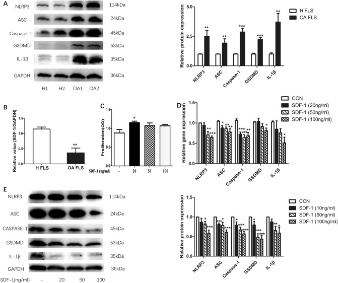
Fig. 1.
SDF-1 suppressed NLRP3 inflammasome and pyroptosis of OA FLS. NLRP3, ASC, caspase-1, GSDMD and IL-1β protein expression in healthy FLS and OA FLS were estimated by western blotting (a, n = 9). Data are expressed as means ± standard error of the mean (SEM). In addition, SDF-1 gene expressions in healthy FLS and OA FLS were detected by RT-PCR (b, n = 5). After treatment with different concentrations of SDF-1 (20, 50, and 100 ng/ml) for 24 h (n = 6), CCK-8 was added to OA FLS and incubated for 1 h. Absorbance (OD, optical density value) at 450 nm was detected by a BioTek microplate reader (c) (OD, optical density value). SDF-1 downregulated the gene (d) and protein (e) expression levels of NLRP3 inflammasome, GSDMD, and IL-1β, which represented pyroptosis (p < 0.05, n = 6–8). *p < 0.05; **p < 0.01; ***p < 0.001 versus control; CON, control: OA FLS treated only with the medium

