Abstract
Neurological manifestations, such as encephalitis, meningitis, ischemic, and hemorrhagic strokes, are reported with increasing frequency in patients affected by Coronavirus disease 2019 (COVID-19). In children, acute ischemic stroke is usually multifactorial: viral infection is an important precipitating factor for stroke. We present a case of a child with serological evidence of SARS-CoV-2 infection whose onset was a massive right cerebral artery ischemia that led to a malignant cerebral infarction. The patient underwent a life-saving decompressive hemicraniectomy, with good functional recovery, except for residual hemiplegia. During rehabilitation, the patient also developed a lower extremity peripheral nerve neuropathy, likely related to a long-Covid syndrome.
Keywords: Acute ischemic stroke, Decompressive craniectomy, Peripheral nerve neuropathy, Long-Covid syndrome
Introduction
Neurological manifestations in adult patients affected by severe acute respiratory syndrome coronavirus-2 (SARS-CoV-2), such as encephalitis, meningitis, ischemic, and hemorrhagic strokes, are reported with an increasing frequency in the scientific literature [1–4], whereas scarce shreds of evidence are portrayed for the pediatrics. SARS-CoV-2 disease promotes a hypercoagulable state through a cytokine storm using angiotensin-converting enzyme 2 (ACE2) as receptor binding [5, 6]. A few papers reported the onset of focal arteriopathies and ischemia in children affected by SARS-COV-2 [7, 8]. In these studies, the patients had a clear manifestation of the disease. We portray, instead, a case of a child with just serological evidence of SARS-CoV-2 infection whose onset was a massive right cerebral artery ischemia that led to a malignant cerebral infarction. During rehabilitation, the patient also developed a lower extremity peripheral nerve neuropathy, likely related to a long-Covid syndrome.
Case presentation
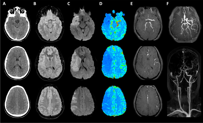
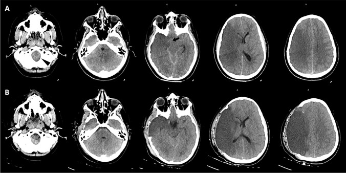
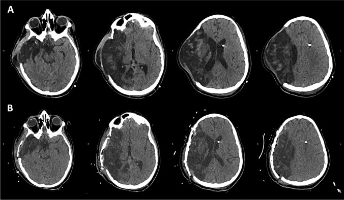
A previously healthy 11-year-old boy was admitted into our hospital with a new onset of left-sided hemiplegia, dysarthria, and lateral nystagmus. Upon arrival to the emergency care, the first SARS-CoV-2 viral nucleic acid nasopharyngeal swab resulted negative for the patient but positive for both parents. Two weeks before, he presented mild common cold symptoms characterized by cough and sore throat for a couple of days. Computed tomography (CT) and the magnetic resonance (MR) of the head showed a large right middle cerebral artery (RMCA) ischemia (Fig. 1) in early stage, as confirmed with the distinct DWI/FLAIR mismatch. Being in the right time window (< 4 h), he was treated with bridging systemic thrombolysis followed by endovascular thrombectomy. The patient remained intubated and sedated. Twelve hours later, he developed anisocoria; then, the pupils became dilated and fixed. A CT scan demonstrated malignant cerebral edema with a 1-cm midline shift to the left and concurrent right uncal herniation (Fig. 2). He underwent urgent decompressive right-sided hemicraniectomy (DHC) (Fig. 2). A large (12 × 15 cm) fronto-temporal-parietal bone flap (extended until the floor of the middle cranial fossa) was elevated. The dura was opened in a cruciate fashion. The brain was swollen and pale, with congested cortical veins. A synthetic not-resorbable patch was used to cover the dural defect. At the end of the procedure, an intraparenchymal probe for intracranial pressure (ICP) monitoring was placed in the right frontal lobe. Initially, intracranial pressure was normal, but on postoperative day (POD) 2, it persistently raised until pathological values (> 20 mmHg). A left frontal external ventricular drain (EVD) was placed. ICP immediately normalized. Afterwards, it was acknowledged that the mother had an altered coagulative axis with Factor V (Leiden)‘s absence, Factor II, and MTHFR mutations. The patient was screened as well, and the coagulability panel revealed high levels of fibrinogen (785 mg/dL), D-Dimer (3188 ng/mL), homocysteine (31.3 mcmol/L); he also had the same maternal Factor II mutation (Table 1). At the same time, he was tested for a complete virology panel that revealed high levels of SARS-COV-2 IgM. Anti-coagulant and anti-platelet therapy based on heparin and acetylsalicylic acid was suggested by hematological consultation. However, because of the presence of EVD, the use of anti-platelets was deferred until EVD removal. On POD 15, a gradual weaning process led to the patient awakening without ventilator support; he was transferred to the neurosurgical ward. The neurologic exam at the arrival was GCS 10 (E = 4; V = 2; M = 4) with left-sided hemiplegia. Surveillance head CT scans showed a natural course of progressive malacic phenomena in the right hemisphere (Fig. 3). Therefore, as soon as the neuro-clinical conditions became stationary, an uneventful autologous bone cranioplasty was performed, along with EVD removal (Fig. 3). The patient did not need further treatment for hydrocephalus. In the subsequent days, the patient experienced a significant neurologic improvement. With the recovery of consciousness and the ability to talk, he revealed a severe lower extremity peripheral nerve neuropathy characterized by pain and dysesthesia, described as a constant burning sensation in the bottom of the feet and sensitivity to light touch on the dorsum of the non-paretic foot. He was later transferred to the rehabilitation center. The neurologic exam at discharge was GCS 14 with the persistence of left-sided severe hemiparesis.
Fig. 1.
Early CT scan and fast MR study at admission. First CT scan (A) showed only slight hypoattenuation and obscuration of the head of the caudate nucleus as well as a loss of gray/white matter definition in the lateral margins of the right insula, findings suggestive of “early signs” of ischemic stroke; slight hyperattenuating band on the right middle cerebral artery (MCA) was also observed, due to intravascular occluding thrombus. Similarly, MR FLAIR images (B) were nearly normal, with only subtle hyperintensity along with the cortical insular ribbon. Instead, diffusion-weighted [b 1000 s/mm2] images (C) clearly showed acute stroke–induced cytotoxic edema in the right MCA territory; the discrepancy between DWI and FLAIR images was suggestive of an early stage of stroke. The MR perfusion CBF map (D) demonstrated a large wedge-shaped area of significantly reduced perfusion in the same region, also corresponding to a lack of vascular representation on MRA TOF3D MIP reconstructions (E). MR-angiography (F) performed with TOF3D, and contrast-enhanced techniques confirmed the occlusion of M1 segment of the right MCA
Fig. 2.
Brain CT monitoring. Post-endovascular treatment CT scan (A), performed after the onset of anisocoria, demonstrated extensive swelling in the right hemisphere, right transtentorial uncal herniation (black arrow), effacement of the CSF spaces in the posterior fossa (asterisk), incipient tonsillar herniation (arrowhead) and leftward midline shift due to malignant cerebral edema. These compressive findings were improved on CT images (B) obtained after DHC
Table 1.
Coagulation screening profile
| Basic profile | |||||||||
|---|---|---|---|---|---|---|---|---|---|
| PT | APTT | INR | Antithrombin | Fibrinogen | D-Dimer | ||||
| 10.5 | 26 |
0.93 (0.8–1–2) |
111% (n.v. 80–120) |
785 mg/DL* (n.v. 150–400) |
188 ng/mL* (n.v. 0–500) |
||||
| Advanced first level | |||||||||
| Protein C | Protein S | Homocysteine | Anti Xa | ||||||
|
95 (n.v. 70–140) |
104.5 (n.v. 74–146) |
31.3 mcmol/L* (n.v. 4–11) |
0.39 U/mL (n.v. 0.2–0.4) |
||||||
| Advanced second level | |||||||||
|
Factor II 20210G/20210A |
Factor V 1691G/1691A |
Lupus anticoagulant (dRVVT) |
Antiphospholipid antibody syndrome (APS) Antibodies |
||||||
| Heterozygote mutation | Absent |
1.13 (Ratio 0–1.2) |
Anti-cardiolipin IgG |
Anti-cardiolipin IgM |
Anti-Neta2Gp1 IgG |
Anti-Neta2Gp1 IgM |
|||
|
0.8 (n.v. 0–10) |
0.7 (n.v. 0–7) |
0.7 (n.v. 0–5) |
1.2 (n.v. 0–5) |
||||||
*bold values are the ones out of range
Fig. 3.
Late brain CT exams. Surveillance CT scan (A) showed progressive malacic evolution of MCA infarction, with brain expanded through the skull defect. Last CT exam at discharge (B) demonstrated a good result of bone cranioplasty
Discussion
Acute ischemic stroke (AIS) in the pediatric population has an incidence of 1.2–2.1 per 100,000 children per year and it is triggered, in most of the cases, by a multifactorial etiology [9]. It is well-known that risk factors associated with childhood AIS are different to adults. Among the correlated conditions, non-atherosclerotic arteriopathies represent a leading factor. Furthermore, cerebral vasculitis in the context of systemic disease (including Kawasaki disease) or infections (particularly herpes group viruses, like Varicella zoster virus) are considered important precipitants for stroke [10]. Our patient experienced a massive right cerebral artery ischemia, an alleged finding in the adult population with SARS-CoV-2 infection, but barely reported for children [11, 12]. A recent review endeavored to explain the complex neurologic complications framework related to SARS-CoV-2 infection. Due to the similarities with SARS-CoV, the authors speculate a possible neuroinvasive and neurotropic behavior, although the virus’s mechanism of invading and reaching the human central nervous system (CNS) is to be defined [13]. Hematogenous dissemination and neuronal retrograde dissemination are the two foremost pathogenetic mechanisms described. In our opinion, the patient’s unknown pro-coagulate pattern, synergistically boosted by its Factor II mutation, provided a fertile substrate for SARS-CoV-2 to lead the thrombotic event escalated into the malignant infarction that required an emergency decompressive craniectomy, as previously described for adult patients [14–16]. According to Vernuccio et al., a procoagulant pattern encountered in some patients, along with the progressive endothelial thrombo-inflammatory syndrome caused by SARS-CoV-2, may enhance the multisystemic microvascular thrombotic disease [17]. In accordance to our findings, a recent survey documented that SARS-CoV-2 may be a contributing factor for stroke in children with underlying risk factors, but its contribution, even in these cases, remains not fully understood [18]. Moreover, this assumption is reinforced by the subsequent development of a severe lower extremity peripheral nerve neuropathy characterized by pain and dysesthesia. Considering the involvement of the contralateral side to the malignant infarction, it is more likely to be correlated with a long post-Covid expression [19] and, to the best of our knowledge, this peripherical manifestation has never been reported before in a pediatric patient.
The prevalent and current hypothesis is a SARS-CoV-2 immune-mediated damage to the nerves induced after a latent period following the infectious illness. In addition, it can also be ascribable to the virus capacity of infecting leukocytes [20]. Once activated by the infection, these leukocytes can cross the blood–brain barrier to enter the CNS. This process has been entitled as “Trojan horse mechanism” and may explain the pro-inflammatory and the aberrant neuroinflammatory loop caused by SARS-CoV-2, which can lead to peripheral neuropathies and neuronal injuries.
Conclusions
With the massive spreading of the SARS-COV-2 pandemic, the time where this disease was mainly associated with respiratory failure seems out-of-date. Children are not excluded by the worst scenario triggered by previous and relatively asymptomatic SARS-CoV-2 infection. Moreover, this case report raises awareness on the importance of further studies of the cerebrovascular thromboembolic events linked to SARS-CoV-2 infection.
Declarations
Ethics approval
All procedures performed in the studies involving human participants were in accordance with the ethical standards of the institutional and/or national research committee and with the 1964 Helsinki Declaration and its later amendments or comparable ethical standard.
Conflict of interest
The authors declare that they have no conflict of interest.
Footnotes
Publisher's Note
Springer Nature remains neutral with regard to jurisdictional claims in published maps and institutional affiliations.
References
- 1.Koralnik IJ, Tyler KL. COVID-19: A global threat to the nervous system. Ann Neurol. 2020;88:1–11. doi: 10.1002/ana.25807. [DOI] [PMC free article] [PubMed] [Google Scholar]
- 2.Liu JM, Tan BH, Wu S, Gui Y, Suo JL, Li YC. Evidence of central nervous system infection and neuroinvasive routes, as well as neurological involvement, in the lethality of SARS-CoV-2 infection. J Med Virol. 2021;93:1304–1313. doi: 10.1002/jmv.26570. [DOI] [PMC free article] [PubMed] [Google Scholar]
- 3.Zhou Z, Kang H, Li S, Zhao X. Understanding the neurotropic characteristics of SARS-CoV-2: from neurological manifestations of COVID-19 to potential neurotropic mechanisms. J Neurol. 2020;267:2179–2184. doi: 10.1007/s00415-020-09929-7. [DOI] [PMC free article] [PubMed] [Google Scholar]
- 4.Zubair AS, McAlpine LS, Gardin T, Farhadian S, Kuruvilla DE, Spudich S. Neuropathogenesis and neurologic manifestations of the coronaviruses in the age of coronavirus disease 2019: a review. JAMA Neurol. 2020;77:1018–1027. doi: 10.1001/jamaneurol.2020.2065. [DOI] [PMC free article] [PubMed] [Google Scholar]
- 5.Ranucci M, Ballotta A, Di Dedda U, Bayshnikova E, Dei Poli M, Resta M, Falco M, Albano G, Menicanti L. The procoagulant pattern of patients with COVID-19 acute respiratory distress syndrome. J Thromb Haemost. 2020;18:1747–1751. doi: 10.1111/jth.14854. [DOI] [PMC free article] [PubMed] [Google Scholar]
- 6.Uhlén M, Fagerberg L, Hallström BM, Lindskog C, Oksvold P, Mardinoglu A, Sivertsson Å, Kampf C, Sjöstedt E, Asplund A, Olsson I, Edlund K, Lundberg E, Navani S, Szigyarto CA, Odeberg J, Djureinovic D, Takanen JO, Hober S, Alm T, Edqvist PH, Berling H, Tegel H, Mulder J, Rockberg J, Nilsson P, Schwenk JM, Hamsten M, von Feilitzen K, Forsberg M, Persson L, Johansson F, Zwahlen M, von Heijne G, Nielsen J, Pontén F (2015) Proteomics. tissue-based map of the human proteome. Science 347:1260419 [DOI] [PubMed]
- 7.Tiwari L, Shekhar S, Bansal A, Kumar S. COVID-19 associated arterial ischaemic stroke and multisystem inflammatory syndrome in children: a case report. Lancet Child Adolesc Health. 2021;5:88–90. doi: 10.1016/S2352-4642(20)30314-X. [DOI] [PMC free article] [PubMed] [Google Scholar]
- 8.Appavu B, Deng D, Dowling MM, Garg S, Mangum T, Boerwinkle V, Abruzzo T (2021) Arteritis and large vessel occlusive strokes in children after COVID-19 infection. Pediatrics 147 [DOI] [PubMed]
- 9.Mallick AA, Ganesan V, Kirkham FJ, Fallon P, Hedderly T, McShane T, Parker AP, Wassmer E, Wraige E, Amin S, Edwards HB, Tilling K, O’Callaghan FJ. Childhood arterial ischaemic stroke incidence, presenting features, and risk factors: a prospective population-based study. Lancet Neurol. 2014;13:35–43. doi: 10.1016/S1474-4422(13)70290-4. [DOI] [PubMed] [Google Scholar]
- 10.Mackay MT, Steinlin M. Recent developments and new frontiers in childhood arterial ischemic stroke. Int J Stroke. 2019;14:32–43. doi: 10.1177/1747493018790064. [DOI] [PubMed] [Google Scholar]
- 11.Tan YK, Goh C, Leow AST, Tambyah PA, Ang A, Yap ES, Tu TM, Sharma VK, Yeo LLL, Chan BPL, Tan BYQ. COVID-19 and ischemic stroke: a systematic review and meta-summary of the literature. J Thromb Thrombolysis. 2020;50:587–595. doi: 10.1007/s11239-020-02228-y. [DOI] [PMC free article] [PubMed] [Google Scholar]
- 12.Kihira S, Morgenstern PF, Raynes H, Naidich TP, Belani P. Fatal cerebral infarct in a child with COVID-19. Pediatr Radiol. 2020;50:1479–1480. doi: 10.1007/s00247-020-04779-x. [DOI] [PMC free article] [PubMed] [Google Scholar]
- 13.Pezzini A, Padovani A. Lifting the mask on neurological manifestations of COVID-19. Nat Rev Neurol. 2020;16:636–644. doi: 10.1038/s41582-020-0398-3. [DOI] [PMC free article] [PubMed] [Google Scholar]
- 14.Roy D, Hollingworth M, Kumaria A (2020) A case of malignant cerebral infarction associated with COVID-19 infection. Br J Neurosurg 1–4 [DOI] [PubMed]
- 15.Pisano TJ, Hakkinen I, Rybinnik I (2020) Large vessel occlusion secondary to COVID-19 hypercoagulability in a young patient: a case report and literature review. J Stroke Cerebrovasc Dis 29:105307 [DOI] [PMC free article] [PubMed]
- 16.Patel SD, Kollar R, Troy P, Song X, Khaled M, Parra A, Pervez M (2020) Malignant cerebral ischemia in a COVID-19 infected patient: case review and histopathological findings. J Stroke Cerebrovasc Dis 29:105231 [DOI] [PMC free article] [PubMed]
- 17.Vernuccio F, Lombardo FP, Cannella R, Panzuto F, Giambelluca D, Arzanauskaite M, Midiri M, Cabassa P. Thromboembolic complications of COVID-19: the combined effect of a pro-coagulant pattern and an endothelial thrombo-inflammatory syndrome. Clin Radiol. 2020;75:804–810. doi: 10.1016/j.crad.2020.07.019. [DOI] [PMC free article] [PubMed] [Google Scholar]
- 18.Beslow LA, Linds AB, Fox CK, Kossorotoff M, Zuñiga Zambrano YC, Hernández-Chávez M, Hassanein SMA, Byrne S, Lim M, Maduaka N, Zafeiriou D, Dowling MM, Felling RJ, Rafay MF, Lehman LL, Noetzel MJ, Bernard TJ, Dlamini N, Group IPSS (2021) Pediatric ischemic stroke: an infrequent complication of SARS-CoV-2. Ann Neurol 89:657–665 [DOI] [PubMed]
- 19.Soliman SB, Klochko CL, Dhillon MK, Vandermissen NR, van Holsbeeck MT. Peripheral polyneuropathy associated with COVID-19 in two patients: a musculoskeletal ultrasound case report. J Med Ultrasound. 2020;28:249–252. doi: 10.4103/JMU.JMU_150_20. [DOI] [PMC free article] [PubMed] [Google Scholar]
- 20.Desforges M, Le Coupanec A, Brison E, Meessen-Pinard M, Talbot PJ. Neuroinvasive and neurotropic human respiratory coronaviruses: potential neurovirulent agents in humans. Adv Exp Med Biol. 2014;807:75–96. doi: 10.1007/978-81-322-1777-0_6. [DOI] [PMC free article] [PubMed] [Google Scholar]


