The hallmark pneumonia of SARS-CoV-2 infection (coronavirus disease 2019, COVID-19) is often accompanied by important extra-pulmonary manifestations. Liver dysfunction occurs in up to 45% of patients and manifests predominantly as moderate transaminitis. Although the hepatic expression of angiotensin-converting-enzyme-2 (ACE2) receptor is largely restricted to cholangiocytes, reports of cholestatic injury have been rare [1].
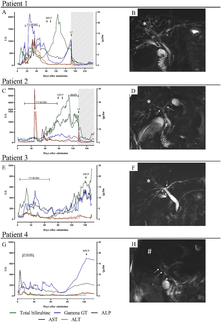
In the first 12 weeks of the pandemic, 3/114 COVID-19 patients admitted to our tertiary intensive care unit (ICU) developed a rapidly progressive cholestatic liver injury that persisted after the acute respiratory distress syndrome (ARDS) had resolved, and evolved to a condition reminiscent of secondary sclerosing cholangitis in critically ill patients (SSC-CIP), a rare but often fatal complication in patients receiving prolonged critical care [2]. During the same time period, a fourth patient with this condition was referred to our center (Fig. 1).
Fig. 1.
Temporal evolution of liver tests, treatments & MRCP images. For each individual patient (rows), the temporal evolution of liver enzymes, along with time points when critical diagnostic or treatment events occurred (left). Levels of gamma-glutamyltransferase (Gamma GT), alkaline phosphatase (ALP), aspartate transaminase (AST) and alanine transaminase (ALT) are projected on the left vertical axis and expressed as U/L, total bilirubin is projected on the right vertical axis and is expressed as mg/dl. VV-ECMO veno-venous extracorporeal membrane oxygenation, ERCP endoscopic retrograde cholangiopancreatography, MARS molecular absorbent recirculating system, LT liver transplantation. Cholestasis was defined as alkaline phosphatase > 1.5 times the upper limit. Also shown are a representative MRCP image for each patient (right) illustrating: in patient 1, diffuse beading of the intrahepatic biliary system (*); in patient 2 and 3, diffuse beading of the intrahepatic biliary ducts (*) and focal strictures on the left and right hepatic ducts (arrows); in patient 4, focal strictures on the right hepatic duct (arrows) and diminished arborisation of the intrahepatic biliary tree (#). All these findings are consistent with the diagnosis of ‘Secondary Sclerosing Cholangitis in Critically Ill Patients’ (SSC-CIP)
The patients were male, aged 48–68, and required prolonged mechanical ventilation, renal support, and veno-venous extracorporeal membrane oxygenation (VV-ECMO, supplementary Table 1). Magnetic resonance cholangiopancreatography (MRCP) showed focal strictures in intrahepatic bile ducts with intraluminal sludge and casts, the radiological hallmark of SSC-CIP. Liver biopsies showed findings consistent with biliary obstruction, typical for SSC (supplementary Fig. 2). Patients 1 and 2 ultimately required liver transplantation because of refractory cholangitis with irreversible biliary damage: patient 1 is currently doing well but patient 2 died of post-transplant pneumonia and septic shock. Patient 3 experienced a milder form of SSC-CIP and is currently doing well, while patient 4 died as a result of a lethal hepatic haemorrhage.
With an estimated prevalence of 1/2000 (0.05%) ICU admissions, SSC-CIP was remarkably frequent with 3/114 ICU patients (2.6%) over 3 months and represented 3/74 (4.1%) of mechanically ventilated and 3/13 (23.1%) of VV-ECMO-treated patients [3]. COVID-19-specific disease and treatment factors may have precipitated biliary ischemia and cholangiopathy, including varying degrees of hemodynamic instability, high positive end-expiratory pressures reducing hepatosplanchnic blood flow, drug-induced bile duct injury by sedatives such as ketamine, parenteral nutrition, and the exaggerated pro-inflammatory cytokine storm that interferes with the biliary epithelium’s physiological defense against hydrophobic bile salts [2, 4].
Importantly, SARS-CoV-2 RNA and nucleo-capsid protein have been detected in the cholangiocytes and bile of patients with fatal COVID-19 pneumonia, suggesting that a direct cytopathic effect may occur [5]. Moreover, endothelialitis resulting in hypercoagulability and microthrombi deposition in the peribiliary vascular plexus may aggravate ischemia of the biliary epithelium.
This report aims to raise awareness about the risk for COVID-19 patients to develop severe cholestatic liver dysfunction reminiscent of SSC-CIP. As COVID-19 becomes better understood, more patients may recover from ARDS and require prolonged critical care with its associated risks. Our data—although from a small cohort—indicate a spectrum of severity, ranging from asymptomatic bile duct abnormalities to cholangiosepsis. Infamous for its bleak prognosis, early diagnosis with MRCP is critical. Whether mild forms confer a risk for secondary biliary cirrhosis is unknown. The outcome of liver transplantation for COVID-19-cholangiopathy remains to be determined, but a timely multidisciplinary evaluation is warranted. A direct causal role of SARS-CoV-2 in COVID-19-associated SSC-CIP is the subject of ongoing investigations.
Supplementary Information
Below is the link to the electronic supplementary material.
Acknowledgements
Collaborators Leuven Liver Transplant program members are: Joost Wauters (Department of Microbiology, Immunology and Transplantation, Laboratory for Clinical Infectious and Inflammatory Disorders, KU Leuven, Leuven, Belgium; Department of General Internal Medicine, Medical Intensive Care Unit, University Hospitals Leuven, Leuven, Belgium), Alexander Wilmer (Department of Microbiology, Immunology and Transplantation, Laboratory for Clinical Infectious and Inflammatory Disorders, KU Leuven, Leuven, Belgium; Department of General Internal Medicine, Medical Intensive Care Unit, University Hospitals Leuven, Leuven, Belgium), Nicholas Gilbo (Department of Microbiology, Immunology and Transplantation, Laboratory of Abdominal Transplantation, KU Leuven, Leuven, Belgium; Department of Abdominal Transplant Surgery and Coordination, University Hospitals Leuven, Leuven, Belgium), Ina Jochmans (Department of Microbiology, Immunology and Transplantation, Laboratory of Abdominal Transplantation, KU Leuven, Leuven, Belgium; Department of Abdominal Transplant Surgery and Coordination, University Hospitals Leuven, Leuven, Belgium), Jacques Pirenne (Department of Microbiology, Immunology and Transplantation, Laboratory of Abdominal Transplantation, KU Leuven, Leuven, Belgium; Department of Abdominal Transplant Surgery and Coordination, University Hospitals Leuven, Leuven, Belgium), Mauricio Sainz Barriga (Department of Microbiology, Immunology and Transplantation, Laboratory of Abdominal Transplantation, KU Leuven, Leuven, Belgium; Department of Abdominal Transplant Surgery and Coordination, University Hospitals Leuven, Leuven, Belgium), Yves Debaveye (Department of Cellular and Molecular Medicine, Laboratory of Intensive Care Medicine, KU Leuven, Leuven, Belgium; Department of Intensive Care Medicine, Intensive Care Unit, Leuven, University Hospitals Leuven, Leuven, Belgium), Jan Gunst (Department of Cellular and Molecular Medicine, Laboratory of Intensive Care Medicine, KU Leuven, Leuven, Belgium; Department of Intensive Care Medicine, Intensive Care Unit, Leuven, University Hospitals Leuven, Leuven, Belgium), Geert Meyfroidt (Department of Cellular and Molecular Medicine, Laboratory of Intensive Care Medicine, KU Leuven, Leuven, Belgium; Department of Intensive Care Medicine, Intensive Care Unit, Leuven, University Hospitals Leuven, Leuven, Belgium), Wim Laleman (Department of Chronic Diseases and Metabolism, Hepatology, KU Leuven, Leuven, Belgium; Department of Gastroenterology and Hepatology, University Hospitals Leuven, Leuven, Belgium), Frederik Nevens (Department of Chronic Diseases and Metabolism, Hepatology, KU Leuven, Leuven, Belgium; Department of Gastroenterology and Hepatology, University Hospitals Leuven, Leuven, Belgium), Hannah Van Malenstein (Department of Chronic Diseases and Metabolism, Hepatology, KU Leuven, Leuven, Belgium; Department of Gastroenterology and Hepatology, University Hospitals Leuven, Leuven, Belgium), Jef Verbeek (Department of Chronic Diseases and Metabolism, Hepatology, KU Leuven, Leuven, Belgium; Department of Gastroenterology and Hepatology, University Hospitals Leuven, Leuven, Belgium), Chris Verslype (Department of Chronic Diseases and Metabolism, Hepatology, KU Leuven, Leuven, Belgium; Department of Gastroenterology and Hepatology, University Hospitals Leuven, Leuven, Belgium), Tania Roskams (Department of Imaging and Pathology, Translational Cell & Tissue Research, KU Leuven, Leuven, Belgium; Department of Imaging and Pathology, University Hospitals Leuven, Leuven, Belgium), Vincent Vandecaveye (Department of Imaging and Pathology, Translational MRI, KU Leuven, Leuven, Belgium; Department of Radiology, University Hospitals Leuven, Leuven, Belgium), Tim Balthazar (Department of Cardiovascular Diseases, Cardiology, KU Leuven, Leuven, Belgium; Department of Cardiovascular Diseases, Leuven, University Hospitals Leuven, Leuven, Belgium), Christophe Vandenbriele (Department of Cardiovascular Diseases, Cardiology, KU Leuven, Leuven, Belgium; Department of Cardiovascular Diseases, Leuven, University Hospitals Leuven, Leuven, Belgium), Greet Hermans (Department of Cellular and Molecular Medicine, Laboratory of Intensive Care Medicine, KU Leuven, Leuven, Belgium; Department of General Internal Medicine, Medical Intensive Care Unit, Leuven, University Hospitals Leuven, Leuven, Belgium), Peter Rogiers (ZNA Middelheim, Intensive Care Unit, Antwerp, Belgium), Marleen Verhaegen (Anesthesiology and Algology, KU Leuven, Leuven, Belgium; Department of Anesthesiology, University Hospitals Leuven, Leuven, Belgium)
Funding
Not applicable.
Availability of data and material
Not applicable.
Code availability
Not applicable.
Declarations
Conflicts of interest
The authors declare that they have no conflict of interest.
Ethics approval
Not applicable.
Consent to participate
Not applicable.
Consent for publication
Not applicable.
Footnotes
The members of the Collaborators Leuven Liver Transplant program are listed in acknowledgements.
Publisher's Note
Springer Nature remains neutral with regard to jurisdictional claims in published maps and institutional affiliations.
Philippe Meersseman and Joris Blondeel contributed equally.
Contributor Information
Philippe Meersseman, Email: philippe.meersseman@uzleuven.be.
Joris Blondeel, Email: joris.blondeel@uzleuven.be.
Greet De Vlieger, Email: greet.devlieger@uzleuven.be.
Schalk van der Merwe, Email: schalk.vandermerwe@uzleuven.be.
Diethard Monbaliu, Email: diethard.monbaliu@uzleuven.be.
Collaborators Leuven Liver Transplant program:
Joost Wauters, Alexander Wilmer, Nicholas Gilbo, Ina Jochmans, Jacques Pirenne, Mauricio Sainz Barriga, Yves Debaveye, Jan Gunst, Geert Meyfroidt, Wim Laleman, Frederik Nevens, Hannah Van Malenstein, Jef Verbeek, Chris Verslype, Tania Roskams, Vincent Vandecaveye, Tim Balthazar, Christophe Vandenbriele, Greet Hermans, Peter Rogiers, and Marleen Verhaegen
References
- 1.Gupta A, Madhavan MV, Sehgal K, et al. Extrapulmonary manifestations of COVID-19. Nat Med. 2020;26(7):1017–1032. doi: 10.1038/s41591-020-0968-3. [DOI] [PMC free article] [PubMed] [Google Scholar]
- 2.Martins P, Verdelho MM. Secondary sclerosing cholangitis in critically ill patients: an underdiagnosed entity. GE Port J Gastroenterol. 2020;27(2):103–114. doi: 10.1159/000501405. [DOI] [PMC free article] [PubMed] [Google Scholar]
- 3.Van Aerde N, Van den Berghe G, Wilmer A, et al. Intensive care unit acquired muscle weakness in COVID-19 patients. Intensive Care Med. 2020 doi: 10.1007/s00134-020-06244-7. [DOI] [PMC free article] [PubMed] [Google Scholar]
- 4.Gudnason HO, Björnsson ES. Secondary sclerosing cholangitis in critically ill patients: current perspectives. Clin Exp Gastroenterol. 2017;10:105–111. doi: 10.2147/CEG.S115518. [DOI] [PMC free article] [PubMed] [Google Scholar]
- 5.Kaltschmidt B, Fitzek ADE, Schaedler J, et al. Hepatic vasculopathy and regenerative responses of the liver in fatal cases of COVID-19. Clin Gastroenterol Hepatol. 2021 doi: 10.1016/j.cgh.2021.01.044. [DOI] [PMC free article] [PubMed] [Google Scholar]
Associated Data
This section collects any data citations, data availability statements, or supplementary materials included in this article.
Supplementary Materials
Data Availability Statement
Not applicable.
Not applicable.


