Abstract
Background
Catabolism of serine via serine hydroxymethyltransferase2 (SHMT2) through the mitochondrial one-carbon unit pathway is important in tumorigenesis. Therefore, SHMT2 may play a role in thyroid cancer.
Methods
Thyroid tissue samples and The Cancer Genome Atlas (TCGA) database were used to evaluate SHMT2 expression in thyroid tissues and the association with clinical outcomes.
Results
SHMT2 protein expression was evaluated in thyroid tissues consisting of 52 benign nodules, 129 papillary thyroid carcinomas (PTC) and matched normal samples, and 20 anaplastic thyroid carcinomas (ATC). ATCs presented the highest (95.0%) positivity of SMHT2 protein expression. PTCs showed the second highest (73.6%) positivity of SHMT2 expression, which was significantly higher than that of benign nodules (19.2%, P = 0.016) and normal thyroid tissues (0%, P < 0.001). Analysis of TCGA data showed that SHMT2 messenger RNA (mRNA) expression was significantly higher in tumors than in normal tissues (P < 0.001). When we classified thyroid cancer into high and low groups according to SHMT2 mRNA expression levels, the thyroid differentiation score for the high SHMT2 group was significantly lower than that of the low SHMT2 group (P < 0.001). There was also a significant correlation between SHMT2 mRNA expression and the stemness index (r = 0.41, P < 0.001). The high SHMT2 group had more advanced TNM stages and shorter progression-free survival rates than the low SHMT2 group (P < 0.01 and P = 0.007, respectively).
Conclusion
SHMT2 expression is higher in thyroid cancers than normal or benign tissues and is associated with de-differentiation and poor clinical outcomes. Thus, SHMT2 might be useful as a diagnostic and prognostic marker for thyroid cancer.
Keywords: thyroid cancer, SHMT2, diagnosis, prognosis
Introduction
The ability of cancer cells to change their metabolism pattern from oxidative phosphorylation to glycolysis is a well-known hallmark of cancer and is called the Warburg effect (1). This metabolic reprogramming is essential for cancer cells to maintain rapid cell proliferation, tumor progression, and resistance to cell death (2, 3). However, metabolic reprogramming in thyroid cancers is not well known.
In a previous study, we uncovered that phosphoglycerate dehydrogenase (PHGDH), an important enzyme in serine biosynthesis, is associated with proliferation, tumorigenesis, and stemness of thyroid cancer cells (4). As PHGDH expression resulted in aggressive thyroid cancers, it might be a potential therapeutic target (4). Additionally, we focused on serine hydroxymethyltransferase (SHMT), which metabolizes the biosynthesized serine (5). The mitochondrial isoform of SHMT2 is the first rate-limiting enzyme of the mitochondrial serine and one-carbon (1C) unit pathway and catalyzes serine to glycine. Glycine is essential for the production of glutathione, heme, and 5,10-methylentetrahydrofolate, which is a 1C unit carrier, indispensable for several anabolic pathways including de novo nucleotide biosynthesis (6, 7). Catabolism of the serine through the mitochondrial 1C unit pathway is critical for maintaining cellular redox control under stress conditions and biosynthesis that promotes tumorigenesis (8, 9, 10, 11). Furthermore, SHMT2 has shown prognostic and therapeutic value for many cancers, such as hepatic carcinoma, breast cancer, prostate cancer, and glioma (12, 13, 14, 15). However, due to a lack of investigation, it remains unclear whether SHMT2 has a similar prognostic role in thyroid cancer.
In this study, we first evaluated SHMT2 protein expression in various thyroid tissues, including normal, benign, and thyroid cancer tissues. Next, we tried to explore the association between SHMT2 messenger RNA (mRNA) expression and thyroid cancer differentiation and prognosis using The Cancer Genome Atlas (TCGA) database.
Methods
Thyroid tissue specimens and construction of tissue microarray blocks
Archival formalin-fixed, paraffin-embedded (FFPE) tissue from surgically removed thyroid samples taken between 1997 and 2013 at the Asan Medical Center in Korea were selected for use. One hundred and twenty-nine fresh frozen papillary thyroid carcinomas (PTC) with matched normal thyroid samples, 20 anaplastic thyroid carcinomas (ATC), 35 follicular adenomas (FA), and 17 nodular hyperplasia (NH) samples were used for immunohistochemistry (IHC) analysis and SHMT2 protein expression. An experienced pathologist reviewed the histopathology and immunohistology of the thyroid cancers and benign nodule specimens. The FFPE tissue samples were arrayed by using a tissue-arraying instrument (MTAII; Beecher Instruments, Silver Spring, Sun Prairie, WI), as previously described (16). This study protocol was approved by the institutional review board of the Asan Medical Center (IBR No: 2013-0539).
IHC analysis and SMHT2 protein expression
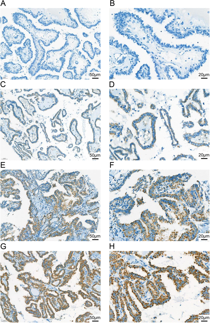
The degree of SHMT2 protein expression was evaluated by IHC staining with anti-SHMT2 (Invitrogen) antibody. IHC staining was performed on the tissue microarray section, using a BenchMark XT automated immunostaining device (Ventana Medical Systems, Tucson, AZ) with the OptiView DAB IHC Detection Kit (Ventana Medical Systems) according to the manufacturer’s instructions, as previously described (4, 16). It is well known that SHMT2 is found in mitochondria, therefore, the protein expression of SHMT2 assessed by IHC was mainly shown in the cytoplasm. The cytoplasmic staining intensity of SHMT2 was graded semi-quantitatively by an experienced pathologist according to the proportion of positively stained cells as follows: 0, negative; 1 (<10% positive), 2 (10–30% positive), and 3 (30–50%) (Fig. 1). The samples with intensity scores higher than 2 positive points were classified as having positive SMHT2 protein expression.
Figure 2.
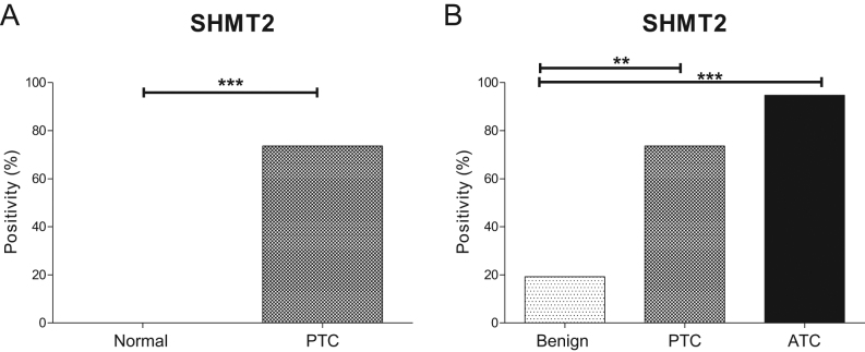
Protein expression of the serine hydroxymethyltransferase2 (SHMT2) in thyroid tissue. (A) Comparison of protein expression of SHMT2 between PTC (n = 129) and matched normal thyroid tissue. (B) Comparison of protein expression of SHMT2 between benign thyroid nodules (n = 52), PTCs (n = 129), and ATCs (n = 20). Asterisks (P < 0.05 (**), P < 0.001 (***)) indicate significant differences from the statistical analyses. SHMT2, serine hydroxymethyltransferase2; PTC, papillary thyroid carcinoma; ATC, anaplastic thyroid carcinoma.
Analysis of TCGA data
Transcriptomes and matched clinicopathological data from thyroid cancers deposited in The Cancer Genome Atlas (TCGA-THCA) were used to explore the biological and clinical significance of the SHMT2 gene. To do this, we divided the dataset into two groups according to SHMT2 expression: low (lower quartile) and high (upper quartile). We then compared thyroid differentiation scores (TDS) (17) and stemness indexes (SI) (18) that were generated by a machine-learning algorithm. To identify gene signatures underlying the biological characteristics, we performed gene set enrichment analysis (GSEA, http://software.broadinstitute.org/gsea/index.jsp) according to the SHMT2 expression groups. Among the ranked gene sets from the GSEA, those with a nominal P-value < 0.05 and a false discovery rate (FDR) q-value < 0.10 were considered statistically significant. For clinical outcomes, tumor-node-metastasis (TNM) stages, according to the 6thAmerican Joint Committee on Cancer staging system, and disease-free survival (DFS) were evaluated.
Statistical analysis
We used R (version 3.5.1, R Foundation for Statistical Computing, Vienna, Austria; https://www.r-project.org/) for statistical analysis. Continuous variables were presented as median and interquartile ranges (IQR), and categorical variables were presented as a number (percentage). Comparison of continuous variables was performed by using the Student’s t-test, and comparison of categorical variables was performed by using the Chi-square test. Pearson’s correlation coefficient (r) was calculated to evaluate the correlation between SHMT2 and the stemness index. GraphPad Prism version 5.01 (GraphPad Software, Inc. was used to draw graphs. Survival curves were plotted using the Kaplan–Meier method, and the log-rank test was used to determine significance. All P-values were two-sided, and a P-value < 0.05 was considered statistically significant.
Results
SHMT2 protein expression in thyroid tissues
We analyzed the protein expression of SHMT2 in thyroid tissue, which consisted of 52 benign thyroid nodules, 129 PTCs with matched normal thyroid tissue and 20 ATCs. Table 1 presents the baseline characteristics of patients included for SHMT2 protein expression analysis in this study. Among 201 patients, 159 patients (79.1%) were female. The median age of the patients was 49.7 years (IQR 40.0–59.1), and patients with ATC were older than those with benign nodules or PTC. In terms of thyroid surgery, 38 patients (73.1%) with benign nodules underwent lobectomy, while a majority of patients with PTC (93.0%) and ATC (100%) underwent total thyroidectomy. The median primary tumor size was 2.7 cm (IQR 2.0–4.0), with the median tumor size of ATC (4.5, IQR 3.8–5.8) being the largest.
Table 1.
Baseline characteristics of patients.
| Total patients (n = 201) | Benign nodule (n = 52) | PTC (n = 129) | ATC (n = 20) | |
|---|---|---|---|---|
| Sex | ||||
| Male | 42 (20.9%) | 14 (26.9%) | 23 (17.8%) | 5 (25.0%) |
| Female | 159 (79.1%) | 38 (73.1%) | 106 (82.2%) | 15 (75.0%) |
| Age (years) | 49.7 (40.0–59.1) | 45.0 (36.6–57.0) | 48.7 (40.0–56.6) | 68.3 (66.5–73.9) |
| Age ≥ 55 | 71 (35.3%) | 19 (36.5%) | 35 (27.1%) | 17 (85.0%) |
| Thyroid surgery | ||||
| Lobectomy | 47 (23.4%) | 38 (73.1%) | 9 (7.0%) | 0 (0.0%) |
| Total thyroidectomy | 154 (76.6%) | 14 (26.9%) | 120 (93.0%) | 20 (100%) |
| Tumor size (cm) | 2.7 (2.0–4.0) | 3.5 (2.8–4.5) | 2.2 (1.6–3.0) | 4.5 (3.8–5.8) |
| Tumor size > 4 cm | 41 (20.4%) | 16 (30.8%) | 15 (11.6%) | 10 (50.0%) |
Continuous variables are presented as median and interquartile range (IQR) and categorical variables are presented as numbers (percentage).
ATC, anaplastic thyroid carcinoma; PTC, papillary thyroid carcinoma.
Of 129 PTCs, 95 (73.6%) had positive SHMT2 protein expression, while all normal thyroid tissue showed negative SMHT2 protein expression (0%, P < 0.001, Fig. 2A). The SHMT2 positive protein expression in ATCs (95.0%) and PTCs (73.6%) were significantly higher than that of benign nodules (19.2%; P < 0.001 and P = 0.006, respectively, Fig. 2B). The rate of SHMT2 expression was not significantly different between PTCs and ATCs (P = 0.07). When we compared the clinicopathological characteristics of PTCs between positive and negative SHMT2 expression, there were also no significant differences except that patient age was older in the positive SHMT expression group (Supplementary Table 1, see section on supplementary materials given at the end of this article). There was no difference in pathologic subtypes (NH or FA), age, sex, and tumor size according to SHMT2 expression in benign nodules (data not shown).
SHMT2 is associated with de-differentiation and stemness of thyroid cancer
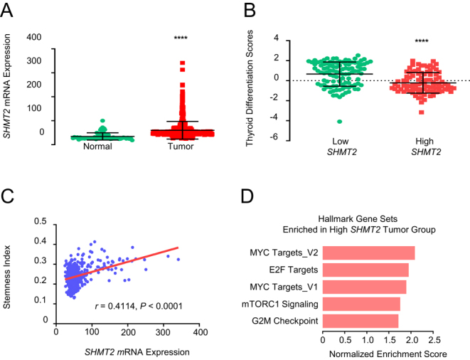
To validate the clinical importance of the SHMT2 in human thyroid cancer, we performed a comprehensive analysis using transcriptomes and matched clinicopathological data from the TCGA-THCA dataset. We first found that mRNA expression of SHMT2 was significantly higher in tumor tissue than in normal tissue, suggesting SHMT2 as an oncogene in thyroid cancer (Fig. 3 P < 0.001). To understand the biological characteristics of thyroid cancer according to SHMT2 expression, we divided the dataset into low and high SHMT2 tumor groups and then compared TDS (Fig. 3B) and SI (Fig. 3C) between the groups. High SHMT2 tumors exhibit lower TDS than low SHMT2 tumors (Fig. 3B, P < 0.0001), indicating oncogenic de-differentiation of these tumors, and SHMT2 expression consistently had a strong positive correlation with SI (Fig. 3C, r = 0.41, P < 0.0001), suggesting that high SHMT2 tumors possess more stem cell-like features after de-differentiation.
Figure 3.

Analysis of serine hydroxymethyltransferase2 (SHMT2) mRNA expression in The Cancer Genome Atlas (TCGA) data. (A) Comparison of mRNA expression of SHMT2 between normal thyroid (n = 59) and tumor tissue (n = 505). (B) Comparison of thyroid differentiation scores between low (n = 103) and high (n = 95) SHMT2 tumor groups. (C) Correlation between SHMT2 expression and stemness index using Pearson coefficient (n = 500). (D) Enriched hallmark gene sets (P < 0.05 and P < 0.10) in the high SHMT2 tumor group compared to the low SHMT2 group. Asterisks (P < 0.001 (****)) indicate significant differences from the statistical analyses. THCA, thyroid carcinoma.
Hallmark gene sets associated with SHMT2 expression
We also performed GSEA to identify gene signatures underlying the biological features and clinical outcomes (Fig. 3D). Interestingly, well-known oncogenic signaling pathways, such as MYC, E2F, and mTORC1, were highly enriched in the high SHMT2 tumor group, supporting the role of SHMT2 for thyroid cancer progression.
SHMT2 is associated with poor clinical outcomes of thyroid cancer
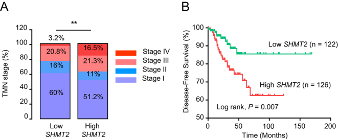
To identify the association between SHMT2 mRNA expression and clinical outcome of thyroid cancer, TNM stage and DFS were compared between low and high SHMT2 groups. The high SHMT2 tumor group exhibited more advanced tumor stages (Fig. 4A, P < 0.01). For example, in the high SHMT2 group, the proportion of TNM stage IV was 16.5%, whereas in the low SHMT2 group, the proportion of stage IV was only 3.2%. Furthermore, the high SHMT2 group showed significantly lower DFS than the low SHMT2 group (Fig. 4B, P = 0.007).
Figure 4.

Clinical outcomes according to serine hydroxymethyltransferase2 (SHMT2) mRNA expression from The Cancer Genome Atlas (TCGA) data. (A) Comparison of TNM stages between low and high SHMT2 tumor groups. (B) Comparison of disease-free survival between low and high SHMT2 tumor groups. Asterisks (P < 0.01 (**)) indicate significant differences. THCA, thyroid carcinoma.
Discussion
Although differentiated thyroid carcinomas generally exhibit excellent outcomes, recurrence in some patients and tumor progression lead to more aggressive phenotypes and loss of iodine uptake ability in approximately 10% of cases (19, 20). The clinical outcomes of poorly differentiated thyroid carcinomas and ATCs are worse, and these types of thyroid cancers are also refractory to radioactive iodine (21, 22). Early diagnosis and prognostication of aggressive thyroid cancer are important for appropriate management, but a better and unambiguous marker is needed. In this study, we showed that SHMT2 is expressed in thyroid cancers but not in normal thyroid tissue, associated with the degree of thyroid differentiation and stemness and related to the clinical outcomes of thyroid cancer.
We have shown that thyroid cancers, both PTC and ATC, had significantly high expression of SHMT2 in this study. High mRNA expression of SHMT2 in thyroid cancer was also related to worse clinical outcomes, evidenced by advanced initial TNM stages and higher disease recurrence. Taken together, these results suggest that SHMT2 might be useful as a diagnostic and prognostic marker for thyroid cancer. This is consistent with other studies conducted in other malignant tumors (10, 12, 13, 14, 15, 23, 24, 25). In glioma, high SHMT2 expression was seen more frequently in advanced grades of disease and showed significantly worse outcomes (14, 26). In breast cancer, high SHMT2 protein expression was also significantly correlated with poor overall survival (12). Similar results were also seen in renal cell cancer, prostate cancer, and gastrointestinal cancers, including hepatocellular carcinoma, cholangiocarcinoma, and gastric cancer (13, 15, 23, 24, 25).
Functional experimental studies have been conducted in other cancers and suggest the potential of SHMT2 as a therapeutic target. Wu et al. showed that knockdown of SHMT2 suppressed proliferation and invasion of glioma cells in vitro (14). In addition, Li et al. recently demonstrated that the mitochondrial serine and 1C unit pathway are upregulated in breast cancer subclones having increased metastatic potential (10). They showed that inhibition of SHMT2 suppressed proliferation of these metastatic subclones and impaired growth of lung metastatic subclones in mice. The possibility of SHMT2 as a novel therapeutic target in thyroid cancer has not been elucidated and further research is needed.
In the current study, thyroid cancers with high SHMT2 expression also had a high frequency of mutations in hallmark gene sets. It is well known that SHMT2 is a direct c-Myc target gene for cell survival during hypoxia (11, 27). This may explain why MYC target-gene mutations are enriched in the high SHMT2 tumor group in this study. In addition, mTOR, which is a central regulator of cell growth and proliferation that responds to diverse microenvironments, including cellular stresses (28, 29), is also frequently seen in high SHMT2 expression thyroid cancers. These results suggest the important role of SHMT2 as an oncogene in thyroid cancer progression and are consistent with clinical outcome data.
The present study has several limitations. First, SHMT2 protein expression was performed in a relatively small number of thyroid tissues from a single center, resulting in selection bias. Because of this limitation, we have assessed the association between SHMT2 and clinical outcomes of patients with PTC using TCGA data instead. Secondly, the intensity of SMHT2 protein expression was graded semi-quantitatively by an experienced pathologist, and there are concerns about bias in grading. Thirdly, in vitro and in vivo experiments were not conducted to validate the results. Despite these limitations, this study is the first to evaluate the association between SHMT2 expression and the prognosis of thyroid cancer and suggest a possible role for SHMT2 as a diagnostic and prognostic marker, facilitating accurate diagnosis and prognostication of thyroid cancer.
In conclusion, SHMT2 expression is associated with thyroid cancer de-differentiation and poor clinical outcomes. Therefore, SHMT2 might be useful as a diagnostic and prognostic marker for thyroid cancer.
Supplementary Material
Declaration of interest
The authors declare that there is no conflict of interest that could be perceived as prejudicing the impartiality of the research reported.
Funding
This study was supported by a grant (No. 2020IL0005) from the Asan Institute for Life Sciences, Asan Medical Center, Seoul, Korea and by a National Research Foundation of Korea (NRF) grant funded by the Korea government (MIST; No. 2020R1F1A1049312).
Author contribution statement
M J J and Y M L conceived and designed the manuscript. W K L, M Y, S C and M J J performed the experiments. MJ, WGK, MJJ and YML acquired the data. M J and A J analyzed and interpreted the data. M J drafted the article. All authors have accepted responsibility for the entire content of this manuscript and approved its submission.
Figure 1.
Representative images for immunohistochemistry (IHC) staining of serine hydroxymethyltransferase2 (SHMT2) in papillary thyroid carcinoma (PTC) tissue. Tissue without (0, (A) and (B)) or weak (1+, (C) and (D)) SHMT2 protein expression were classified as negative, and tissues with moderate (2+, (E) and (F)) or strong (3+, (G) and (H)) SHMT2 protein expression were classified as positive. (A), (C), (E), and (G) were low magnification images (×200) and (B), (D), (F), and (H) were high magnification images (×400).
Acknowledgements
The authors would like to thank Asan Institute for Life Sciences.
References
- 1.Warburg O.On the origin of cancer cells. Science 1956. 123 309–314. ( 10.1126/science.123.3191.309) [DOI] [PubMed] [Google Scholar]
- 2.Hanahan D, Weinberg RA. Hallmarks of cancer: the next generation. Cell 2011. 144 646–674. ( 10.1016/j.cell.2011.02.013) [DOI] [PubMed] [Google Scholar]
- 3.Coelho RG, Fortunato RS, Carvalho DP.Metabolic reprogramming in thyroid carcinoma. Frontiers in Oncology 2018. 8 82. ( 10.3389/fonc.2018.00082) [DOI] [PMC free article] [PubMed] [Google Scholar]
- 4.Jeon MJ, You MH, Han JM, Sim S, Yoo HJ, Lee WK, Kim TY, Song DE, Shong YK, Kim WG, et al. High phosphoglycerate dehydrogenase expression induces stemness and aggressiveness in thyroid cancer. Thyroid 2020. 30 1625–1638. ( 10.1089/thy.2020.0105) [DOI] [PMC free article] [PubMed] [Google Scholar]
- 5.Ubonprasert S, Jaroensuk J, Pornthanakasem W, Kamonsutthipaijit N, Wongpituk P, Mee-Udorn P, Rungrotmongkol T, Ketchart O, Chitnumsub P, Leartsakulpanich U, et al. A flap motif in human serine hydroxymethyltransferase is important for structural stabilization, ligand binding, and control of product release. Journal of Biological Chemistry 2019. 294 10490–10502. ( 10.1074/jbc.RA119.007454) [DOI] [PMC free article] [PubMed] [Google Scholar]
- 6.Newman AC, Maddocks ODK. One-carbon metabolism in cancer. British Journal of Cancer 2017. 116 1499–1504. ( 10.1038/bjc.2017.118) [DOI] [PMC free article] [PubMed] [Google Scholar]
- 7.Lucas S, Chen G, Aras S, Wang J.Serine catabolism is essential to maintain mitochondrial respiration in mammalian cells. Life Science Alliance 2018. 1 e201800036. ( 10.26508/lsa.201800036) [DOI] [PMC free article] [PubMed] [Google Scholar]
- 8.DeBerardinis RJ.Serine metabolism: some tumors take the road less traveled. Cell Metabolism 2011. 14 285–286. ( 10.1016/j.cmet.2011.08.004) [DOI] [PMC free article] [PubMed] [Google Scholar]
- 9.Amelio I, Cutruzzolá F, Antonov A, Agostini M, Melino G.Serine and glycine metabolism in cancer. Trends in Biochemical Sciences 2014. 39 191–198. ( 10.1016/j.tibs.2014.02.004) [DOI] [PMC free article] [PubMed] [Google Scholar]
- 10.Li AM, Ducker GS, Li Y, Seoane JA, Xiao Y, Melemenidis S, Zhou Y, Liu L, Vanharanta S, Graves EE, et al. Metabolic profiling reveals a dependency of human metastatic breast cancer on mitochondrial serine and one-carbon unit metabolism. Molecular Cancer Research 2020. 18 599–611. ( 10.1158/1541-7786.MCR-19-0606) [DOI] [PMC free article] [PubMed] [Google Scholar]
- 11.Ye J, Fan J, Venneti S, Wan YW, Pawel BR, Zhang J, Finley LW, Lu C, Lindsten T, Cross JR, et al. Serine catabolism regulates mitochondrial redox control during hypoxia. Cancer Discovery 2014. 4 1406–1417. ( 10.1158/2159-8290.CD-14-0250) [DOI] [PMC free article] [PubMed] [Google Scholar]
- 12.Bernhardt S, Bayerlová M, Vetter M, Wachter A, Mitra D, Hanf V, Lantzsch T, Uleer C, Peschel S, John J, et al. Proteomic profiling of breast cancer metabolism identifies SHMT2 and ASCT2 as prognostic factors. Breast Cancer Research 2017. 19 112. ( 10.1186/s13058-017-0905-7) [DOI] [PMC free article] [PubMed] [Google Scholar]
- 13.Woo CC, Chen WC, Teo XQ, Radda GK, Lee PT.Downregulating serine hydroxymethyltransferase 2 (SHMT2) suppresses tumorigenesis in human hepatocellular carcinoma. Oncotarget 2016. 7 53005–53017. ( 10.18632/oncotarget.10415) [DOI] [PMC free article] [PubMed] [Google Scholar]
- 14.Wu M, Wanggou S, Li X, Liu Q, Xie Y.Overexpression of mitochondrial serine hydroxyl-methyltransferase 2 is associated with poor prognosis and promotes cell proliferation and invasion in gliomas. OncoTargets and Therapy 2017. 10 3781–3788. ( 10.2147/OTT.S130409) [DOI] [PMC free article] [PubMed] [Google Scholar]
- 15.Marrocco I, Altieri F, Rubini E, Paglia G, Chichiarelli S, Giamogante F, Macone A, Perugia G, Magliocca FM, Gurtner A, et al. Shmt2: a Stat3 signaling new player in prostate cancer energy metabolism. Cells 2019. 8 1048. ( 10.3390/cells8091048) [DOI] [PMC free article] [PubMed] [Google Scholar]
- 16.Sung TY, Kim M, Kim TY, Kim WG, Park Y, Song DE, Park SY, Kwon H, Choi YM, Jang EK, et al. Negative expression of CPSF2 predicts a poorer clinical outcome in patients with papillary thyroid carcinoma. Thyroid 2015. 25 1020–1025. ( 10.1089/thy.2015.0079) [DOI] [PubMed] [Google Scholar]
- 17.Cancer Genome Atlas Research Network. Integrated genomic characterization of papillary thyroid carcinoma. Cell 2014. 159 676–690. ( 10.1016/j.cell.2014.09.050) [DOI] [PMC free article] [PubMed] [Google Scholar]
- 18.Malta TM, Sokolov A, Gentles AJ, Burzykowski T, Poisson L, Weinstein JN, Kamińska B, Huelsken J, Omberg L, Gevaert O, et al. Machine learning identifies stemness features associated with oncogenic dedifferentiation. Cell 2018. 173 338–354.e15. ( 10.1016/j.cell.2018.03.034) [DOI] [PMC free article] [PubMed] [Google Scholar]
- 19.Pacini F, Cetani F, Miccoli P, Mancusi F, Ceccarelli C, Lippi F, Martino E, Pinchera A.Outcome of 309 patients with metastatic differentiated thyroid carcinoma treated with radioiodine. World Journal of Surgery 1994. 18 600–604. ( 10.1007/BF00353775) [DOI] [PubMed] [Google Scholar]
- 20.Coelho SM, Vaisman M, Carvalho DP.Tumour re-differentiation effect of retinoic acid: a novel therapeutic approach for advanced thyroid cancer. Current Pharmaceutical Design 2005. 11 2525–2531. ( 10.2174/1381612054367490) [DOI] [PubMed] [Google Scholar]
- 21.Ibrahimpasic T, Ghossein R, Carlson DL, Nixon I, Palmer FL, Shaha AR, Patel SG, Tuttle RM, Shah JP, Ganly I.Outcomes in patients with poorly differentiated thyroid carcinoma. Journal of Clinical Endocrinology and Metabolism 2014. 99 1245–1252. ( 10.1210/jc.2013-3842) [DOI] [PubMed] [Google Scholar]
- 22.Nagaiah G, Hossain A, Mooney CJ, Parmentier J, Remick SC.Anaplastic thyroid cancer: a review of epidemiology, pathogenesis, and treatment. Journal of Oncology 2011. 2011 542358. ( 10.1155/2011/542358) [DOI] [PMC free article] [PubMed] [Google Scholar]
- 23.Liu Y, Yin C, Deng MM, Wang Q, He XQ, Li MT, Li CP, Wu H.High expression of SHMT2 is correlated with tumor progression and predicts poor prognosis in gastrointestinal tumors. European Review for Medical and Pharmacological Sciences 2019. 23 9379–9392. ( 10.26355/eurrev_201911_19431) [DOI] [PubMed] [Google Scholar]
- 24.Wang H, Chong T, Li BY, Chen XS, Zhen WB.Evaluating the clinical significance of SHMT2 and its co-expressed gene in human kidney cancer. Biological Research 2020. 53 46. ( 10.1186/s40659-020-00314-2) [DOI] [PMC free article] [PubMed] [Google Scholar]
- 25.Ning S, Ma S, Saleh AQ, Guo L, Zhao Z, Chen Y.SHMT2 overexpression predicts poor prognosis in intrahepatic cholangiocarcinoma. Gastroenterology Research and Practice 2018. 2018 4369253. ( 10.1155/2018/4369253) [DOI] [PMC free article] [PubMed] [Google Scholar]
- 26.Wang B, Wang W, Zhu Z, Zhang X, Tang F, Wang D, Liu X, Yan X, Zhuang H.Mitochondrial serine hydroxymethyltransferase 2 is a potential diagnostic and prognostic biomarker for human glioma. Clinical Neurology and Neurosurgery 2017. 154 28–33. ( 10.1016/j.clineuro.2017.01.005) [DOI] [PubMed] [Google Scholar]
- 27.Nikiforov MA, Chandriani S, O'Connell B, Petrenko O, Kotenko I, Beavis A, Sedivy JM, Cole MD.A functional screen for Myc-responsive genes reveals serine hydroxymethyltransferase, a major source of the one-carbon unit for cell metabolism. Molecular and Cellular Biology 2002. 22 5793–5800. ( 10.1128/mcb.22.16.5793-5800.2002) [DOI] [PMC free article] [PubMed] [Google Scholar]
- 28.Ben-Sahra I, Manning BD. mTORC1 signaling and the metabolic control of cell growth. Current Opinion in Cell Biology 2017. 45 72–82. ( 10.1016/j.ceb.2017.02.012) [DOI] [PMC free article] [PubMed] [Google Scholar]
- 29.He L, Gomes AP, Wang X, Yoon SO, Lee G, Nagiec MJ, Cho S, Chavez A, Islam T, Yu Y, et al. mTORC1 promotes metabolic reprogramming by the suppression of GSK3-dependent Foxk1 phosphorylation. Molecular Cell 2018. 70 949–960.e4. ( 10.1016/j.molcel.2018.04.024) [DOI] [PMC free article] [PubMed] [Google Scholar]
Associated Data
This section collects any data citations, data availability statements, or supplementary materials included in this article.


 This work is licensed under a
