

Abstract
Background
Chordoma is a rare primary bone tumour with a high propensity for local recurrence. Surgical resection is the mainstay of treatment, but complete resection is often morbid due to tumour location. Similarly, the dose of radiotherapy (RT) that surrounding healthy organs can tolerate is frequently below that required to provide effective tumour control. Therefore, clinicians have investigated different radiation delivery techniques, often in combination with surgery, aimed to improve the therapeutic ratio.
Objectives
To assess the effects and toxicity of proton and photon adjuvant radiotherapy (RT) in people with biopsy‐confirmed chordoma.
Search methods
We searched CENTRAL (2021, Issue 4); MEDLINE Ovid (1946 to April 2021); Embase Ovid (1980 to April 2021) and online registers of clinical trials, and abstracts of scientific meetings up until April 2021.
Selection criteria
We included adults with pathologically confirmed primary chordoma, who were irradiated with curative intent, with protons or photons in the form of fractionated RT, SRS (stereotactic radiosurgery), SBRT (stereotactic body radiotherapy), or IMRT (intensity modulated radiation therapy). We limited analysis to studies that included outcomes of participants treated with both protons and photons.
Data collection and analysis
The primary outcomes were local control, mortality, recurrence, and treatment‐related toxicity. We followed current standard Cochrane methodological procedures for data extraction, management, and analysis. We used the ROBINS‐I tool to assess risk of bias, and GRADE to assess the certainty of the evidence.
Main results
We included six observational studies with 187 adult participants. We judged all studies to be at high risk of bias. Four studies were included in meta‐analysis.
We are uncertain if proton compared to photon therapy worsens or has no effect on local control (hazard ratio (HR) 5.34, 95% confidence interval (CI) 0.66 to 43.43; 2 observational studies, 39 participants; very low‐certainty evidence).
Median survival time ranged between 45.5 months and 66 months. We are uncertain if proton compared to photon therapy reduces or has no effect on mortality (HR 0.44, 95% CI 0.13 to 1.57; 4 observational studies, 65 participants; very low‐certainty evidence).
Median recurrence‐free survival ranged between 3 and 10 years. We are uncertain whether proton compared to photon therapy reduces or has no effect on recurrence (HR 0.34, 95% CI 0.10 to 1.17; 4 observational studies, 94 participants; very low‐certainty evidence).
One study assessed treatment‐related toxicity and reported that four participants on proton therapy developed radiation‐induced necrosis in the temporal bone, radiation‐induced damage to the brainstem, and chronic mastoiditis; one participant on photon therapy developed hearing loss, worsening of the seventh cranial nerve paresis, and ulcerative keratitis (risk ratio (RR) 1.28, 95% CI 0.17 to 9.86; 1 observational study, 33 participants; very low‐certainty evidence). There is no evidence that protons led to reduced toxicity.
There is very low‐certainty evidence to show an advantage for proton therapy in comparison to photon therapy with respect to local control, mortality, recurrence, and treatment related toxicity.
Authors' conclusions
There is a lack of published evidence to confirm a clinical difference in effect with either proton or photon therapy for the treatment of chordoma. As radiation techniques evolve, multi‐institutional data should be collected prospectively and published, to help identify persons that would most benefit from the available radiation treatment techniques.
Plain language summary
Proton radiation therapy versus conventional radiation treatment (i.e. photon radiation) for the treatment of chordoma
What is the issue? Chordoma is a rare tumour that can grow at any location in the spine. Because some of the locations are difficult to reach, some of the tumour may be left behind, even after aggressive surgery. When this happens, radiation to the postoperative area can help reduce the chance of the tumour coming back. Given the closeness to the spinal cord and other important organs, special radiation techniques are required to maximise tumour control, while minimising harm to normal tissues in the body.
Why is this review important? At the time of this review, there is a lack of evidence to confirm a benefit with proton therapy compared to conventional photon radiation treatment. However, it is important to note that this review found a number of delivery techniques for both proton and photon therapy. As evidence comparing the latest techniques for proton and photon therapy emerges, we will update this review.
The aim of the review: The aim of this Cochrane Review was to find out whether proton therapy is more effective than photon therapy for improving local control, and reducing mortality, recurrence, and treatment‐related toxicity. We collected and analysed all relevant studies to answer this question.
What are the main findings? We included six studies that enrolled 187 adults with chordoma, four of which we included in a meta‐analysis. We are uncertain whether proton radiation therapy compared to conventional photon radiation treatment improves local control, because our confidence in the evidence was very low (2 observational studies, 39 participants). We are uncertain whether proton therapy compared to photon therapy reduces mortality (4 observational studies, 65 participants), recurrence (4 observational studies, 94 participants), or treatment‐related toxicity (1 observational study, 33 participants).
Quality of the Evidence: We found very low‐certainty evidence for all outcomes due to high risk of bias in all the included observational studies, very small sample sizes, and varying ranges in the duration of participants' follow‐up period between studies.
Authors' conclusions: From the currently available evidence, we do not know if proton therapy, used for adults with chordoma, is associated with clinically appreciable benefits and acceptable harms.
The existing research on this question is solely of non‐randomised design, and is underpowered, with substantial imprecision. Any estimates of effect based on the existing insufficient evidence is very uncertain and is likely to change with future research.
Summary of findings
Summary of findings 1. Protons compared to photons for the treatment of chordoma.
| Protons compared to photons for the treatment of chordoma | ||||||
| Patient or population: adults with chordoma Setting: hospitals Intervention: protons Comparison: photons | ||||||
| Outcomes | Anticipated absolute effects* (95% CI) | Relative effect (95% CI) | № of participants (studies) | Certainty of the evidence (GRADE) | Comments | |
| Risk with photons | Risk with protons | |||||
|
Local control (follow‐up: median ranged between 45.5 and 66 months) |
400 per 1000 | 935 per 1000 (286 to 1000) | HR 5.34 (0.66 to 43.43) | 39 (27 analysed, 2 non‐randomised studies) | ⊕⊝⊝⊝ Very lowa,b,c | We are uncertain if proton compared to photon therapy effects local control. |
|
Mortality (follow‐up: median ranged between 45.5 and 66 months) |
600 per 1000 | 332 per 1000 (112 to 763) | HR 0.44 (0.13 to 1.57) | 65 (39 analysed, 4 non‐randomised studies) | ⊕⊝⊝⊝ Very lowa,d,e,f,g | We reported mortality instead of overall survival for methodological reasons. We are uncertain if proton compared to photon therapy affects mortality. |
|
Recurrence (follow up: median ranged between 3 and 10 years) |
600 per 1000 | 268 per 1000 (88 to 658) | HR 0.34 (0.10 to 1.17) | 94 (81 participants, 4 non‐randomised studies) | ⊕⊝⊝⊝ Very lowd,f,h | We reported recurrence instead of recurrence‐free survival for methodological reasons. We are uncertain if proton compared to photon therapy effects recurrence. |
| Treatment‐related toxicity | 125 per 1000 | 160 per 1000 (21 to 1000) | RR 1.28 (0.17 to 9.86) | 33 (1 observational study) | ⊕⊝⊝⊝ Very lowg,i | We are uncertain if proton compared to photon therapy effects treatment‐related toxicity. |
| *The risk in the intervention group (and its 95% confidence interval) is based on the assumed risk in the comparison group and the relative effect of the intervention (and its 95% CI). The assumed risk in the photon group for local control, mortality, and recurrence outcomes are based on the event rate in Park 2006. CI: confidence interval; RR: risk ratio; HR: hazard ratio | ||||||
| GRADE Working Group grades of evidence High certainty. We are very confident that the true effect lies close to that of the estimate of the effect. Moderate certainty. We are moderately confident in the effect estimate; the true effect is likely to be close to the estimate of the effect, but there is a possibility that it is substantially different. Low certainty. Our confidence in the effect estimate is limited; the true effect may be substantially different from the estimate of the effect. Very low certainty. We have very little confidence in the effect estimate; the true effect is likely to be substantially different from the estimate of effect. | ||||||
aWe did not include one study in the meta‐analysis, as we could not conduct analysis with only one participant who received photons. We used IPD from the other study for the synthesis. bWe downgraded by two levels due to high risk of bias of the included study. cWe downgraded by two levels due to very serious imprecision due to a very small sample size that did not meet the optimal information size, and a very wide confidence interval that crossed the line of no effect and failed to exclude appreciable benefit. dWe downgraded by two levels due to high risk of bias in the included studies. eWe downgraded by one level as one study compared proton versus photon therapy and the second study compared proton plus photon therapy versus photon therapy alone. fWe were unable to include data from one study in the meta‐analysis as we were unable to extract numeric data for outcomes per group. (6 participants per proton therapy group and 2 participants per photon therapy group) (Yasuda 2012) gWe downgraded by two levels due to a very small sample size that did not meet the optimal information size, and a very wide confidence interval that crossed the line of no effect and failed to exclude appreciable harm. hThe included studies varied widely in the duration of follow‐up. Median follow‐up ranged from 12 to 40 months in one study, 66 months in another study, and 26.5 to 29 months in a third. Also, recurrence should be scored independently of further therapeutic intervention (use of second surgery) as defined by Takahashi 2009. iWe downgraded by one level due to high risk of bias of the included study.
Background
Description of the condition
Chordoma is a rare primary bone tumour of presumed notochord origin, with an incidence of 0.08 per 100,000 people per year, based on statistics from the Surveillance Epidemiology and End Results Registry (SEER) Program (McMaster 2001). Median age at presentation is approximately 60 years old; although uncommon in individuals younger than 40 years, it may also occur in children and adolescents (Borba 1996). Chordoma is a low‐ to intermediate‐grade malignancy, but with locally aggressive behaviour. It is historically divided into three subtypes based on light microscopic morphology: conventional, chondroid, and de‐differentiated. Conventional chordomas are the most common, and are characterised by a lack of cartilaginous or mesenchymal components (Heffelfinger 1973). Chondroid histology exhibits good long‐term outcomes; de‐differentiated chordoma often demonstrates an aggressive pattern with less favourable prognoses (Casali 2007). More recently, a distinct molecular subtype with a very poor prognosis has been defined, termed poorly differentiated chordoma (Hasselblatt 2016). Chordomas arise mainly along the midline in the skeleton, with a predilection for the base of the coccyx (i.e. sacrococcygeal, 50%) and skull base (i.e. clivus, 30% to 40%) regions. Chordomas are rarely metastatic at first presentation, but distant metastases have been reported in up to 40% of people, usually occurring late in the course of the disease (Catton 1996).
Description of the intervention
Since chordoma is a locally aggressive tumour, with a high propensity for local recurrence, control of the primary disease remains the major therapeutic challenge. For example, a meta‐analysis of 464 people with cranial chordoma reported a recurrence rate of 68%, and a median disease‐free interval of only 23 months (average 45 months (Jian 2010)). Current management involves surgery, radiation therapy (RT), or both. Chemotherapy is generally ineffective, and is used mainly in the advanced (metastatic) stage of disease (Casali 2007). Surgery is the mainstay of treatment, and aims to establish a definitive diagnosis, while attempting maximum resection and decompression. Given the difficult locations and the relationship with surrounding healthy tissues, potential morbidity from surgery is high. Moreover, local recurrence and progression are frequent even in cases of optimal surgery (Walcott 2012). Hence, RT has been used either pre‐ or postoperatively, in an attempt to achieve local tumour control and possibly improve overall survival (OS (Rotondo 2015)). Since the 1970s, conventional radiotherapy with photons has been given as both curative and supportive treatments (Cummings 1983). Due to anatomical locations, radiation oncologists face the same constraints that hamper adequate surgical excision. The dose of RT that surrounding healthy organs can tolerate is frequently below that required to provide effective treatment. These healthy tissue radiotherapy dose constraints vary by original tumour location in the skull base or spine.
How the intervention might work
Photons, created from either x‐rays or gamma rays, deposit their energy along the entire linear path they travel, attenuating, but not stopping within the body of the participant. Hence, the treatment of the tumour area with photons requires the irradiation of healthy tissues both near to and away from the target. As a consequence, most of the early series using photon RT were not able to deliver effective radiation dose levels within the tumour. By contrast, 'particles' (protons, ions, neutrons) behave differently than photons in tissue: they exhibit a moderate entrance dose, give off most of their energy at a defined depth, and then stop. Consequently, it is possible to limit the radiation dose to the healthy tissues beyond the tumour, while delivering a higher dose to the tumour. Theoretically, protons offer advantages over photons in the treatment of chordoma (Walcott 2012), and are a supported option for postoperative adjuvant irradiation in this disease (NCCN Guidelines 2018). However, the development of advanced photon techniques, such as stereotactic body radiotherapy (SBRT), stereotactic radiosurgery (SRS), and intensity‐modulated RT (IMRT) improve the planning capability when delivering photon RT (Amichetti 2012; Baskar 2012; Sahgal 2014; Gatfield 2018). These techniques generally take advantage of three‐dimensional (3D) imaging, such as magnetic resonance imaging (MRI) or computed tomography (CT) to define tumour and normal tissue, and then use multiple beam angles to minimise total dose to surrounding normal tissue. Furthermore, small planning margins have been made possible with photons by utilising kV (kilovoltage) cone‐beam computer tomography (CBCT) to verify the person's positioning with daily RT treatments, compared to planer imaging still in use by most proton centres (Alcorn 2014, Sahgal 2014).
Why it is important to do this review
The low incidence of this malignancy means only a few centres worldwide have experience in its management. Thus, there is little robust evidence to guide the therapeutic strategies, which is summarised by the Chordoma global consensus group (Stacchiotti 2015). Although technical and theoretical advantages support the use of protons, there are few available centres offering this treatment internationally. Moreover, the cost of treatment with protons is significantly more expensive than with photons. Conversely, precision photon RT is cheaper, treatment machines are widespread, and in the past decade, there has been bridging of the technical gap between photons and protons (Sahgal 2014). There is uncertainty about the relative superiority of the different techniques, therefore, a systematic review assessing whether modern photon RT is as effective as particles is needed.
Objectives
To assess the effects and toxicity of proton and photon adjuvant radiotherapy (RT) in people with biopsy‐confirmed chordoma.
Methods
Criteria for considering studies for this review
Types of studies
All included studies were observational. Our search didn't retrieve randomised controlled trials (RCTs), quasi‐RCTs, cluster‐RCTs, or controlled clinical trials (CCTs) of proton versus photon therapy. Given the small number of included studies and small study size, we did not report outcomes according to the tumour location. These criteria were set as the type of surgery and radiotherapy (RT; see Characteristics of included studies). We stated whether the included studies had incomplete or obvious confounders (see Assessment of risk of bias in included studies). We did not restrict the inclusion criteria to studies with at least three years follow‐up, although it has been reported that half of chordoma recurrences occur within the first three years (Weber 2016). Instead, we reported the follow‐up period for each study.
Types of participants
We included adult participants, aged 18 years or older, with pathologically‐confirmed primary chordoma, who were irradiated with a curative intent with protons or high‐energy (megavoltage) photons in the form of fractionated RT, SRS (stereotactic radiosurgery), SBRT (stereotactic body radiotherapy), or IMRT (intensity modulated radiation therapy). In the curative setting, a 60 Cobalt Gray Equivalent (CGE) dose or higher is typically used to achieve local control, and therefore, we restricted inclusion to people treated to at least this dose (Terahara 1999; Igaki 2004; Noel 2005). We included people who had undergone biopsy, subtotal, or gross resection.
We excluded people treated with brachytherapy.
All studies included participants who met the inclusion criteria, so we used the subset of data that was of interest to this Cochrane Review.
Types of interventions
Interventions
We included studies if they assessed any of the following particle interventions.
Irradiation with protons
Irradiation with ions
Irradiation with neutrons
Comparison
Irradiation with high‐energy (megavoltage) photons
Types of outcome measures
Primary outcomes
Local control (LC) with proton compared to photon therapy, defined by the authors of the included studies by only included study as the absence of tumor or persistent mass with no evident growth (Park 2006).
Overall survival (OS) with proton compared to photon therapy, defined by authors of the included studies (5 years (Park 2006 ; Jagersberg 2017) , and 10 years overall survival (Park 2006).
Progression‐free survival (PFS) with proton compared to photon therapy, defined by authors of the included studies as the period between the time of additional surgery and former surgery (Takahashi 2009).
Treatment‐related toxicity, using definitions provided by the Common Terminology Criteria for Adverse Events, particularly the frequency of higher severity (grade 2 or above) adverse events (CTCAE 2017).
Secondary outcomes
No secondary outcomes were assessed.
We presented a 'Summary of findings table' reporting the following outcomes listed in order of priority (Atkins 2004):
Local control (LC).
Mortality (Instead of OS).
Progression‐free survival (PFS).
Treatment‐related toxicity.
Search methods for identification of studies
We conducted a comprehensive, systematic search to identify all relevant studies, regardless of language. Imaging, surgical procedures, and RT techniques before the 1990s are very different from current clinical practice; these differences may significantly affect clinical outcomes and their interpretation (Amichetti 2009). Therefore, we considered studies prior to 1990 separately in any analyses.
Electronic searches
We searched the following electronic databases:
Cochrane Central Register of Controlled Trials (CENTRAL; 2021, Issue 4) in the Cochrane Library (searched 29 April 2021; Appendix 1);
MEDLINE Ovid (1946 to week 16, 2021; Appendix 2);
Embase Ovid (1980 to week 16, 2021; Appendix 3).
For databases other than MEDLINE, we adapted the search strategy accordingly.
Searching other resources
We searched the following electronic trial registries and databases to 29 April 2021.
ClinicalTrials.gov (http://clinicaltrials.gov);
International Clinical Trials Registry (http://apps.who.int/trialsearch);
Current Controlled Trials (www.controlled-trials.com);
National Comprehensive Cancer Network (NCCN) Guidelines (www.nccn.org/professionals/physician_gls/f_guidelines.asp);
Turning Research into Practice (TRIP; www.tripdatabase.com);
National Guideline Clearinghouse (www.guideline.gov).
We handsearched the following resources.
Reference lists of relevant articles to identify additional publications;
-
Conference proceedings of the:
American Society for Radiotherapy and Oncology (ASTRO; 1990 to April 2021);
European Society for Radiotherapy and Oncology (ESTRO; 1990 to April 2021);
Particle Therapy Co‐Operative Group (PTCOG; 1990 to April 2021);
North American Skull Base Society (NASBS; 1990 to April 2021).
Data collection and analysis
Selection of studies
Four review authors (IS, DT, EL, SD) independently screened the title and abstract of all articles identified as a result of search, for inclusion. We obtained the full‐text articles of those identified as potentially eligible for inclusion, and the available review authors (IS, SD) independently assessed the full‐text articles. We resolved any disagreements by discussion or by consulting a third review author (DT). No potential conflict of interest existed (e.g. no authorship of a potentially included study). We listed all studies excluded after full‐text assessment and their reason(s) for exclusion in a Characteristics of excluded studies table. We illustrated the study selection progress in a PRISMA diagram (Figure 1).
1.

Study flow diagram.
Data extraction and management
We extracted data on the following: publication data (first author's last name, year of publication, institution), study design, sample size, participants' features (mean age, sex ratio, race, country of the population studied, tumour location, type of surgery, the definition of extent of resection), details of interventions (indication for RT, RT technique, time interval of participant accrual, total dose, dose per fraction, target definition), clinical outcomes (outcome definitions, LC, OS, PFS rates, type of toxicity, toxicity rates, length of follow‐up). We considered studies in which at least part of the RT course used particles as particle studies.
For eligible studies, two review authors (IS and SD) independently extracted data using the data extraction form. If more than one publication referred to the same study, or there was more than one paper from the same institution, we used the most recent publication, or that with the most complete reporting. . We resolved any disagreements by discussion or consulted a third review author (EL or DT). We considered studies to have sufficient data if at least one of the listed primary outcomes, and at least one of the selected time points were reported.
None of the included studies required translation as they were all in English.
Assessment of risk of bias in included studies
We based the risk of bias evaluation on the data provided in the included studies. We did not retrieve any randomized controlled trials; all the included studies were observational. Hence, we used the Risk of Bias In Non‐randomized Studies of Interventions (ROBINS‐I) assessment tool (Sterne 2016). We assessed the risk of bias according to the following domains.
Pre‐intervention bias: due to confounding based on referral pattern and departmental protocol eligibility criteria as well as bias in selection of participants into the study
At‐intervention bias: in classification of the intervention
-
Post‐intervention bias:
due to deviations from the intended intervention
due to missing data
in measurement of outcomes
in selection of the reported results
Each of the seven domains of bias contain signalling questions to facilitate judgements of risk of bias. The full signalling question and response framework for each outcome is provided in Sterne 2016.
Important confounders of interest in this Cochrane Review include the following:
Histology (conventional, chondroid, de‐differentiated, poorly differentiated)
Extent of resection
Location (skull base, spine)
Type of surgery (biopsy, complete, incomplete resection)
RT total dose
Timing of RT (prior to surgery, early after surgery (< 90 days), at relapse or progression (> 90 days after surgery)
We resolved any discrepancies about results by discussion and consensus agreement. We included all assessments in a risk of bias graph, and a risk of bias summary figure, in addition to risk of bias tables (Figure 2; Figure 3).
2.

Risk of bias summary: review authors' judgements about each risk of bias item for each included study.
3.
Risk of bias graph: review authors' judgements about each risk of bias item presented as percentages across all included studies.
Measures of treatment effect
Survival‐type outcomes
The measure of treatment effect chosen for survival‐type outcomes was the hazard ratio with 95% confidence intervals (HR (95% CI)).
Dichotomous outcomes
The measure of treatment effect chosen for treatment related toxicity was the risk ratio (RR) with 95% CI.
Unit of analysis issues
We planed to take into account the level at which randomization occurred. We planed to extract the following information for cluster‐RCTs (Higgins 2011).
The number of clusters (or groups) randomised to each intervention group
The outcome data ignoring the cluster design for the total number of individuals
An estimate of the intracluster (or intraclass) correlation coefficient (ICC)
However, we did not include cluster‐randomised trials, or trials in which participants received more than one intervention
Dealing with missing data
We did not impute for any missing data. We contacted the study authors to request more information. However, we were unable to obtain more information or missing data from study authors, and we conducted available‐case analysis.
Assessment of heterogeneity
We accounted for heterogeneity by visually inspecting the forest plots to detect overlapping confidence intervals, using the Chi2 test and the I2 statistic (Higgins 2011). The I2 statistic represents the percentage of the overall variability due to between‐study (or interstudy) variability, as opposed to within‐study (or intrastudy) variability (Higgins 2011). We interpreted the I2 value as follows: 0% to 40% might not be important; 30% to 60% may represent moderate heterogeneity; 50% to 90% may represent substantial heterogeneity; and 75% to 100% may represent considerable heterogeneity (Deeks 2001).
Assessment of reporting biases
We intended to investigate potential publication bias by using funnel plots, but we were unable to, because < 10 studies met the inclusion criteria of the review.
Data synthesis
We analysed the data using Review Manager 5 (Review Manager 2014). Chordoma of skull base and of the spinal axis were not analysed separately due to the small number of studies and small study sizes. We were unable to extract the HRs and their variances for survival‐type data from the articles, as they were not reported. We either estimated the log HR (experimental relative to control) by (O – E)/V, which has a standard error 1/√V, where O is the observed number of events in the experimental intervention, E is the log‐rank expected number of events in the experimental intervention, O – E is the log‐rank statistic, and V is the variance of the log‐rank statistic (Higgins 2011), and computed the log HR from individual patient data (Parmar 1998), from three studies (Park 2006; Takahashi 2009; Jagersberg 2017), or we depicted the log HR and standard error of log HR from the Kaplan‐Meier curve, using an Excel spreadsheet by Tierney 2007 for one study (Colli 2001).
Regarding treatment‐related toxicity, we used the RR as a measure of association, and fixed a higher type I error (α = 0.10, two‐sided (Shadish 2002)).
We meta‐analysed the HRs and RRs by using the generic inverse‐variance method. We used a random‐effects model, as studies were clinically and statistically heterogenous. (DerSimonian 1986).
Subgroup analysis and investigation of heterogeneity
There were too few included studies that reported sufficient details, so we were unable to conduct subgroup analyses for categorical covariates, or meta‐regression for categorical and continuous covariates, according to the following:
Histology (conventional, chondroid, de‐differentiated, poorly differentiated)
Location (skull base, spine).
Age (continuous)
Gender (male, female)
Type of surgery (biopsy, complete, incomplete resection)
RT total dose (continuous) and fractionation (dose per fraction, continuous)
RT target volume (treatment margin used, if any, in centimetres, continuous)
Timing of RT (prior to surgery, early after surgery (< 90 days), at relapse or progression (> 90 days after surgery)
Sensitivity analysis
We intended to explore reasons for study heterogeneity, and if necessary, carry out sensitivity analysis by omitting studies rated as high risk of bias' However, we only included a small number of studies, which did not enable us to conduct a sensitivity analysis.
Summary of findings and assessment of the certainty of the evidence
We presented the overall certainty of the evidence for each outcome according to the GRADE approach, which takes into account issues related to internal validity (risk of bias, inconsistency, imprecision, publication bias), and external validity, such as directness of results (Langendam 2013). We created a summary of findings table, based on the methods described in the Cochrane Handbook for Systematic Reviews of Interventions (Higgins 2011), and using GRADEpro GDT software (GRADEpro GDT). We used the GRADE checklist and GRADE Working Group certainty of evidence definitions (Meader 2014). We downgraded the evidence from high certainty by one level for serious (or by two for very serious) concerns for study limitations.
High‐certainty: we are very confident that the true effect lies close to that of the estimate of the effect.
Moderate‐certainty: we are moderately confident in the effect estimate. The true effect is likely to be close to the estimate of the effect, but there is a possibility that it is substantially different.
Low‐certainty: our confidence in the effect estimate is limited. The true effect may be substantially different from the estimate of the effect.
Very low‐certainty: we have very little confidence in the effect estimate. The true effect is likely to be substantially different from the estimate of effect.
Limited available evidence from a small number of included studies didn't permit us to present a table specific to each location.
Results
Description of studies
Results of the search
Our initial search, from 1946 to 2021, yielded 27 references from CENTRAL, 1131 from MEDLINE, and 1592 from Embase. Following de‐duplication across the databases, the combined total yield was 2143 references. After title and abstract screening, we retrieved 156 studies for full‐text review and possible inclusion. We excluded 146 studies that were the wrong study design (N = 40), wrong patient population (N = 3), wrong outcomes (N = 4), no comparison between photon and proton arms (82) or wrong comparator (N = 13), wrong setting (N = 2), paediatric population (1) and no intervention (N = 1) . This resulted in 10 observational studies and four studies were further excluded (see Characteristics of excluded studies; Excluded studies). Six observational studies were included in the review (Included studies). Four of which were ultimately included in the analysis (Figure 1).
Included studies
We extracted data from six studies that met our inclusion criteria including 187 patients. (see Characteristics of included studies). All included studies were observational. The mean age of participants ranged between 40 to 56 years and included both males and females. Participants presented with chordoma and chondrosarcomas in Colli 2001, primary and recurrent sacral chordoma in Park 2006, or cranial and skull base chordoma in Jagersberg 2017, Sheybani 2014, Takahashi 2009 and Yasuda 2012 studies. The mean follow‐up period ranged between 36 months (Takahashi 2009) and 91 months (Park 2006). The extent of tumour resection was either partial, subtotal or radical as determined by MRI and neuroradiological assessment. The average dose of radiotherapy ranged between 60 to 77 Gy, although data were not available for all participants in included studies (Colli 2001; Jagersberg 2017; Park 2006; Sheybani 2014; Takahashi 2009; Yasuda 2012)
Colli 2001 was an observational study for people with chordoma (N = 53) and chondrosarcoma (N = 10) treated with surgery and proton or photon radiotherapy delivered adjuvantly in 33 participants.
Proton arm: 25 participants
Photon arm: 8 participants
Jagersberg 2017 was a single‐center retrospective study for cranial chordoma in which 24 surgical treatments were performed and followed by proton or photon radiation.
Proton arm: 11 participants to a median dose of 74 Gy (E) at 1.8 to 2.0 Gy (E)
Photon arm: 2 participants
Park 2006 was an observational study in which people were treated with proton or photon radiation alone (N = 6) or combined with surgery (N = 21) for sacral chordoma.
Proton arm: 22 participants mean 71 Gy (E) in 36 fractions with protons combined with photons
Photon arm: 5 participants to 60 to 62 Gy
Sheybani 2014 was a retrospective analysis of patients treated at authors’ institution for chordoma (N = 12).
Proton arm: 11 participants
Photon arm: 1 participant
Takahashi 2009 was an observation study for people with skull base chordoma who underwent surgery (N = 32), followed by carbon ion, proton, or SRS radiotherapy.
Proton arm: 14 participants
Photon arm: 7 participants
Yasuda 2012 was an observation study for people with skull base, cranio‐cervical junction and cervical chordoma who underwent surgery (N = 40). Ten participants received pure protons, 3 received pure photons and 17 received combined protons and photons.
Primary outcomes were assessed in the included studies were as follows:
Local control: Two studies (N = 39 participants where N = 27 were included in the meta‐analysis) (Park 2006; Sheybani 2014)
Mortality (instead of overall survival): Four studies (N = 65 where N = 9 were included in the meta‐analysis) (Jagersberg 2017; Park 2006; Sheybani 2014; Yasuda 2012)
Recurrence (instead of progression‐free survival): Four studies (N = 94 participants where N = 81 were included in the meta‐analysis) (Colli 2001; Park 2006; Takahashi 2009; Yasuda 2012)
Treatment‐related toxicity: one study (N = 33 participants) (Colli 2001)
Excluded studies
We excluded four reports (see Characteristics of excluded studies). In one, the outcomes were not separated by group; in another, combined treatments (photons plus protons) were not reported for individual people; one was a review rather than a comparison of the effects of two treatment; the fourth one primarily assessed proton therapy and did not report outcomes for the two participants who received photon therapy.
Risk of bias in included studies
Details are provided in the risk of bias table, with the Characteristics of included studies; summaries are provided in Figure 2 and Figure 3. We used the Risk of Bias In Non‐randomized Studies of Interventions (ROBINS‐I) assessment tool (Sterne 2016).
Bias due to confounding:
We determined that all studies were at high risk of bias due to confounding. Study authors did not use an appropriate analysis methods that adjusted for all the important potential confounder.
Bias in selection of participants into the study:
We judged all studies at high risk of bias as selection of participants for both arms based on participant characteristics observed after surgery in Park 2006, Takahashi 2009 and Yasuda 2012, and because participant groups were determined by enrolment time period (older: photons; more recent: protons) for another study (Colli 2001). One study was regarded as unclear risk as reported data for the entire cohort, not by treatment group (Jagersberg 2017). In addition, there was a further imbalance in the number of participants in each arm for all studies.
Bias in classification of interventions:
We judged the risk of bias in classification of interventions as high in all studies due to unavailable information to define intervention groups at the start of the intervention (Colli 2001; Park 2006; Sheybani 2014; Takahashi 2009; Yasuda 2012), except for Jagersberg 2017, which we regarded as unclear risk due to reported data for the entire cohort and not by treatment group.
Bias due to deviations from intended intervention:
Risk of bias due to deviations from intended intervention was unclear in all studies.
Bias due to missing data:
We assessed incomplete outcome data as high risk (Colli 2001; Jagersberg 2017), and low risk (Park 2006; ;Sheybani 2014; Takahashi 2009; Yasuda 2012). Low risk studies included complete reporting for participants in both study arms. High risk studies presented incomplete reports or missing data for: participants from institutions outside the primary institution (Colli 2001); loss to follow‐up of one participant, with no evidence that results were robust in the presence of missing data from a very small study sample (Jagersberg 2017).
Bias in measurement of outcomes:
Risk of bias in measurement of outcomes was low in three studies (Colli 2001; Park 2006; Takahashi 2009), unclear in two studies (Jagersberg 2017; Sheybani 2014), and high risk in one study where we could not extract outcomes for each intervention (Yasuda 2012).
Bias in selection of the reported result:
We were unable to extract data for outcomes separately for each intervention for one study, and determined it at high risk of bias (Yasuda 2012). We judged two studies as unclear risk of bias (Colli 2001; Jagersberg 2017), and three studies at low risk of bias (Park 2006; Sheybani 2014; Takahashi 2009).
Effects of interventions
See: Table 1
See: Table 1.
Primary outcomes
Local control
Two observational studies compared proton with photon therapy and measured local control as an outcome (Park 2006; Sheybani 2014). Park 2006 is an observational study in which people were treated with proton or photon radiation alone (N = 6) or combined with surgery (N = 21) for sacral chordoma with an estimated median follow‐up time of 66 months with a range of 26 months to 261 months. In Sheybani 2014 only one participant was included in the photon group. Median follow‐up time for local control among two studies ranged between 45.5 months and 66 months. The meta‐analysis for local control included 22 participants from one non‐randomised study (Park 2006). We did not include Sheybani 2014 in the meta‐analysis, as there was only one participant who was treated with photons and outcome data were available for all patients and not stratified by treatment. For Park 2006, the estimated log HR was 1.676, and the SE (log HR) was 1.069. Three of the four chordomas that were treated with higher than 73.0 Gy (E) of radiation alone had local control. As this is the only study that assessed local control, it didn't permit us to address the effect of the timing of radiotherapy (RT) (neoadjuvant, adjuvant, or salvage RT) with regard to surgeries. Also, we could not perform a sub‐group analysis on the varying endpoints between studies and the timing of irradiation.
We were uncertain if proton compared to photon therapy has any effect on local control (pooled HR 5.34, 95% CI 0.66 to 43.43; 2 observational studies, 39 participants (27 analysed); Analysis 1.1). We assessed the certainty of evidence to be very‐low (Figure 4).
1.1. Analysis.

Comparison 1: Protons versus photons, Outcome 1: Local control
4.
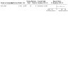
Forest plot of comparison: 1 Protons versus photons, outcome: 1.1 Local control
Overall survival (Mortality)
Four observational studies with 65 participants compared proton with photon therapy and measured mortality rather than overall survival (Jagersberg 2017; Park 2006; Sheybani 2014; Yasuda 2012). The meta‐analysis for mortality included 39 participants across two non‐randomised studies (Park 2006; Jagersberg 2017). Median follow‐up time ranged between 45.5 months and 66 months. Sheybani 2014 was not included in the meta‐analysis as analysis could not be performed on only one participant who was treated with photons with an unknown outcome. We did not include Yasuda 2012 in the meta‐analysis as this paper contained insufficient data to complete data extraction and data clarification was required, we contacted the study corresponding author by email but numeric data couldn't be extracted per each group. We re‐analysed individual participant data (IPD) for Park 2006 and Jagersberg 2017, using the Excel spreadsheet by Tierney 2007. The log HR (‐1.2) and standard error of log HR (1.34) were estimated by re‐analysis of IPD (Tierney 2007) and estimated HR was 0.03, 95% CI 0.02 to 4.19). Similarly, estimated log HR and SE(log HR) were ‐0.698 to 0.736 for Park 2006.
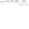
We are uncertain if proton compared to photon therapy has any effect on survival (pooled HR 0.44 to 95% CI 0.13 to 1.57; 4 observational studies, 65 participants (39 analysed); Analysis 1.2 . We assessed the certainty of evidence to be very‐low (Figure 5).
1.2. Analysis.

Comparison 1: Protons versus photons, Outcome 2: Mortality (instead of overall survival)
5.
Forest plot of comparison: 1 Protons versus photons, outcome: 1.2 Mortality (instead of overall survival)
Progression‐free survival (Recurrence)
Four observational studies including 94 participants compared proton with photon therapy and measured recurrence rather than progression‐free survival (Colli 2001; Park 2006; Takahashi 2009; Yasuda 2012). The meta‐analysis for recurrence included 81 participants across 3 non‐randomised studies (Colli 2001; Park 2006; Takahashi 2009). Median follow‐up time ranged between 3 years and 10 years. We did not include Yasuda 2012 in the meta‐analysis as we were unable to extract numeric data for each group. For Colli 2001, recurrence log HR (‐2.37) and standard error of log HR (0.75) were estimated from Kaplan Meier Curve using Excel spreadsheet by Tierney 2007, with estimated HR 0.09, 95% CI 0.02 to 0.40. Re‐analysis was also performed for Park 2006 and estimated log HR, SE(log HR) were ‐0.215 and 0.725 respectively. Similarly, for Takahashi 2009 log HR and SE(log HR) were estimated by re‐analysis of IPD ‐0.71 and 0.67.
We are uncertain if proton compared to photon therapy has any effect on recurrence (pooled HR 0.34, 95% CI 0.10 to 1.17; 4 observational studies, 94 participants (81 analysed); Analysis 1.3; ). We assessed the certainty of evidence to be very‐low (Figure 6).
1.3. Analysis.

Comparison 1: Protons versus photons, Outcome 3: Recurrence (instead of progression‐free survival)
6.

Forest plot of comparison: 1 Protons versus photons, outcome: 1.1 Recurrence (instead of progression‐free survival)
Treatment‐related toxicity
One study was included in the meta‐analysis for treatment‐related toxicity (Colli 2001). It reported that four participants treated with proton therapy developed radiation‐induced necrosis of their temporal bone, radiation‐induced damage to the brainstem, and chronic mastoiditis. One participant treated with photon therapy developed hearing loss, worsening of the seventh cranial nerve paresis, and ulcerative keratitis. The other studies did not report on this outcome.
We are uncertain of the effect of proton compared to photon therapy on treatment‐related toxicity (risk ratio (RR) 1.28, 95% CI 0.17 to 9.86; 1 observational study, 33 participants; Analysis 1.4). We assessed the certainty of evidence to be very low (Figure 7).
1.4. Analysis.

Comparison 1: Protons versus photons, Outcome 4: Treatment‐related toxicity
7.
Forest plot of comparison: 1 Protons versus photons, outcome: 1.4 Treatment‐related toxicity
Toxicity data more than 10 years old were not available, so we didn't report the incidence of chronic or late toxicities according to the RTOG/European Organisation for the Research and Treatment of Cancer Late Radiation Morbidity Scoring Schema (Cox 1995).
Stratified analysis:
We planned but were unable to conduct a stratified analysis by tumour location because all included studies involved cranial chordoma except Park 2006, which included participants with sacral chordoma.
Discussion
We conducted this systematic review to determine if there were differences in outcomes between those who received proton therapy compared to photon therapy for the treatment of chordoma. We included six observational studies in the meta‐analysis but were unable to find evidence in support of one treatment over the other.
Summary of main results
There are insufficient data to show an advantage for proton or photon therapy with respect to local control, overall survival (mortality), progression‐free survival (recurrence), and treatment‐related toxicity.
Overall completeness and applicability of evidence
Overall, we observed no difference between interventions in the outcomes measured. However, we were only able to analyse data from two studies for local control and one for treatment‐related toxicity, areas where the theoretical benefit of protons exist due to their dosimetry. In addition, overall survival is more susceptible to selection bias, as the theoretical benefits of protons could be more often reserved for people with longer life expectancy, due to the limited availability of protons worldwide.
Since the publication of the analysed studies, advancements in proton and photon therapy have continued. For example, more proton centres now implement intensity‐modulated proton therapy (IMPT), a delivery system that results in similar dosimetric advantages as intensity‐modulated radiation therapy (IMRT) over conventional radiotherapy. Advancements in imaging and planning for both proton and photon therapy have also been made in recent decades. Whether these advancements would result in a benefit of one intervention compared to the other for chordoma, warrants future study.
Quality of the evidence
Our review did not identify any randomised studies that compared proton therapy to photon therapy. All studies were retrospective or observation, therefore, did not allow baseline randomisation to either arm. Although the analysed studies were published after the year 2000, the treatment dates span decades, and therefore, some participants' work‐up and treatment would now be considered out of date, such as the lack of MRI use.
We downgraded the level of evidence to very low for all outcomes, due to high risk of bias in included studies; very serious imprecision owing to a very small sample size per study, small number of events, and very wide confidence intervals crossing the line of no effect, which failed to exclude appreciable benefit or harms; included studies for recurrence‐free survival reported variable duration of follow‐up periods. The extent of surgical resection was not consistent among studies and participants. In summary, we considered that all included studies contributed to very low‐certainty evidence, per GRADE methods.
Potential biases in the review process
The search was comprehensive and up to date, allowing us to identify all appropriate studies. We conducted duplicate, independent screening by at least two review authors, of all search results from the title and abstract phase to full‐text screening. Two review authors independently extracted data onto a redesigned data extraction form. We involved a third author to resolve any discrepancy in the extraction process. We tried to contact a study authors to retrieve more details for data extraction. We followed a strict analysis approach to extract log hazard ratio and respective variance, using different methods, and guided by Parmar 1998; Tierney 2007; and Higgins 2011. We assessed the certainty of the evidence for each outcome according to the GRADE approach, and presented a summary of findings table.
Potential limitations of the review were clinical heterogeneity, such as tumour location and completeness of resection, variable duration of follow‐up periods, as well as a limited number of included studies. Small sample sizes led to serious imprecision, hence, downgrading the certainty of the evidence. Some studied were not included in the meta‐analysis due to incomplete available data, or only one participant who was treated with photons. Also, all included studies were observational. Although we identified all relevant studies regardless of language, we did not systematically search all other language databases, therefore, we cannot rule out the possibility that some studies may have been missed.
Agreements and disagreements with other studies or reviews
This study primarily differs from other reviews, because in our efforts to reduce bias, we only considered studies that included people treated with proton or photon therapy. This is different from the Amichetti 2009 systematic review, in which they included single arm proton studies, and authors ultimately concluded that proton therapy had better results compared to conventional photon radiation. Another systematic review summarised proton outcomes without direct comparison to photon treatments (Alahmari 2019). Because these studies showed better outcomes with proton therapy, the Chordoma Global Consensus Group prefers the use of proton therapy, and considers conformal photon irradiation a viable alternative only when similar radiation doses to targets and sparing of normal tissue can be achieved (Stacchiotti 2015). The recommendation is appropriately scored as level of evidence V (studies without control group, case reports, and experts' opinions), consistent with our literature search.
Authors' conclusions
Implications for practice.
Currently, the evidence is very uncertain about the effects of proton therapy compared to photon therapy for people with chordoma, on local control, mortality, recurrence, or treatment‐related toxicity.
Implications for research.
Given the rarity of this tumour type and limited availability of proton therapy, it is unlikely a randomized trial will be performed comparing protons versus photons. With this limitation, we encourage researchers to prospectively document important standardised endpoints, such as local control, treatment‐related toxicity, and quality of life, so that as proton availability increases, informed decision‐making with future people with chordoma will be improved.
History
Protocol first published: Issue 12, 2018
Acknowledgements
We thank Robin Grant for clinical and editorial advice, Jo Platt for designing the search strategy, and Gail Quinn, Clare Jess, and Tracey Harrison for their contribution to the editorial process. We thank Dante Amelio and Maurizio Amichetti who conceived the original protocol and undertook preliminary steps, and Robin Jha and Luluel Khan for assistance in the protocol phase.
This project was supported by the National Institute for Health Research (NIHR), via Cochrane infrastructure funding to the Cochrane Gynaecological, Neuro‐oncology and Orphan Cancers Group. The views and opinions expressed therein are those of the review authors and do not necessarily reflect those of the Systematic Reviews Programme, the NIHR, the NHS, or the Department of Health.
We thank the referees for many helpful suggestions and comments, including Helen Bulbeck, Michael Brada, Andrew Bryant, and Normand Laperriere.
Appendices
Appendix 1. CENTRAL search strategy
#1 MeSH descriptor: [Chordoma] this term only #2 chordoma* or chordocarcinoma* or chordoepithelioma* or notochordoma* #3 #1 or #2 #4 MeSH descriptor: [Radiotherapy] explode all trees #5 Any MeSH descriptor in all MeSH products and with qualifier(s): [radiotherapy ‐ RT] #6 radiotherap* or irradiation* or radiation* or radiosurgery#7 MeSH descriptor: [Photons] this term only #8 MeSH descriptor: [Ions] explode all trees #9 MeSH descriptor: [Protons] this term only #10 photon* or ion* or proton* or particle* or hadron* #11 #4 or #5 or #6 or #7 or #8 or #9 or #10 #12 #3 and #11
Appendix 2. MEDLINE Ovid search strategy
1 Chordoma/ 2 (chordoma* or chordocarcinoma* or chordoepithelioma* or notochordoma*).mp. 3 1 or 2 4 exp Radiotherapy/ 5 radiotherapy.fs. 6 (radiotherap* or irradiation* or radiation* or radiosurgery).mp. 7 Photons/ 8 exp Ions/ 9 Protons/ 10 (photon* or ion* or proton* or particle* or hadron*).mp. 11 4 or 5 or 6 or 7 or 8 or 9 or 10 12 3 and 11
key: mp=title, abstract, original title, name of substance word, subject heading word, keyword heading word, protocol supplementary concept word, rare disease supplementary concept word, unique identifier fs=floating subheading
Appendix 3. Embase Ovid search strategy
1 chordoma/ 2 (chordoma* or chordocarcinoma* or chordoepithelioma* or notochordoma*).mp. 3 1 or 2 4 exp radiotherapy/ 5 rt.fs. 6 (radiotherap* or irradiation* or radiation* or radiosurgery).mp. 7 photon/ 8 exp Ion/ 9 proton/ 10 (photon* or ion* or proton* or particle* or hadron*).mp. 11 4 or 5 or 6 or 7 or 8 or 9 or 10 12 3 and 11
key:
mp=title, abstract, original title, name of substance word, subject heading word, keyword heading word, protocol supplementary concept word, rare disease supplementary concept word, unique identifier
fs=floating subheading
Data and analyses
Comparison 1. Protons versus photons.
| Outcome or subgroup title | No. of studies | No. of participants | Statistical method | Effect size |
|---|---|---|---|---|
| 1.1 Local control | 1 | Hazard Ratio (IV, Random, 95% CI) | Totals not selected | |
| 1.2 Mortality (instead of overall survival) | 2 | 39 | Hazard Ratio (IV, Random, 95% CI) | 0.44 [0.13, 1.57] |
| 1.3 Recurrence (instead of progression‐free survival) | 3 | 81 | Hazard Ratio (IV, Random, 95% CI) | 0.34 [0.10, 1.17] |
| 1.4 Treatment‐related toxicity | 1 | Risk Ratio (M‐H, Fixed, 95% CI) | Totals not selected |
Characteristics of studies
Characteristics of included studies [ordered by study ID]
Colli 2001.
| Study characteristics | ||
| Methods |
|
|
| Participants |
|
|
| Interventions |
|
|
| Outcomes |
|
|
| Notes | Study author email: bocolli@fmrp.usp.br | |
| Risk of bias | ||
| Bias | Authors' judgement | Support for judgement |
| Bias due to confounding | High risk | Study authors did not use an appropriate analysis method that adjusted for all important confounding domains |
| Bias in selection of participants into the study | High risk | Participant groups determined by enrolment time period (older: photons; more recent: protons) |
| Bias in classification of interventions | High risk | The intervention groups were not clearly defined; no information used to define intervention groups that was recorded at the start of the intervention. |
| Bias due to deviations from intended interventions | Unclear risk | Not specified |
| Bias due to missing data | High risk | Not specified, follow‐up data from outside institutions |
| Bias in measurement of outcomes | Low risk | Most outcome measures were objective; were not influenced by knowledge of the intervention received |
| Bias in selection of the reported result | Unclear risk | Progression‐free survival reported for both arms |
Jagersberg 2017.
| Study characteristics | ||
| Methods |
|
|
| Participants |
|
|
| Interventions |
|
|
| Outcomes |
|
|
| Notes | Study author email: max.jaegersberg@hcuge.ch | |
| Risk of bias | ||
| Bias | Authors' judgement | Support for judgement |
| Bias due to confounding | High risk | Study authors did not use an appropriate analysis method that adjusted for all important confounding domains. |
| Bias in selection of participants into the study | Unclear risk | Reported data for entire cohort, not by treatment group |
| Bias in classification of interventions | Unclear risk | Reported data for entire cohort, not by treatment group |
| Bias due to deviations from intended interventions | Unclear risk | Not specified |
| Bias due to missing data | High risk | Only one participant was lost follow‐up, but there was no evidence that results were robust in the presence of missing data, in light of very small study sample. |
| Bias in measurement of outcomes | Unclear risk | Reported data for entire cohort, not by treatment group |
| Bias in selection of the reported result | Unclear risk | Reported data for entire cohort, not by treatment group |
Park 2006.
| Study characteristics | ||
| Methods |
|
|
| Participants |
|
|
| Interventions |
|
|
| Outcomes |
|
|
| Notes | Study author e‐mail: lilypark@hotmail.com | |
| Risk of bias | ||
| Bias | Authors' judgement | Support for judgement |
| Bias due to confounding | High risk | Study authors mentioned potential confounders (location, dose and extent of resection) but did not use appropriate analysis method that adjusted for them in context of association between type of intervention and outcomes. |
| Bias in selection of participants into the study | High risk | Selection of participants for both arms based on participant characteristics observed after surgery. |
| Bias in classification of interventions | High risk | No information used to define intervention groups was recorded at the start of the intervention. |
| Bias due to deviations from intended interventions | Unclear risk | Not specified |
| Bias due to missing data | Low risk | Complete outcomes reported for both arms. |
| Bias in measurement of outcomes | Low risk | Most outcome measures were objective; would not be influenced by knowledge of the intervention received. |
| Bias in selection of the reported result | Low risk | Complete outcomes reported for both arms. |
Sheybani 2014.
| Study characteristics | ||
| Methods |
|
|
| Participants |
|
|
| Interventions |
|
|
| Outcomes |
|
|
| Notes | Study author e‐mail: arshin‐sheybani@uiowa.edu | |
| Risk of bias | ||
| Bias | Authors' judgement | Support for judgement |
| Bias due to confounding | High risk | Only one participant received photons, with inappropriate control of confounders in analysis. |
| Bias in selection of participants into the study | High risk | Only one participant received photons. |
| Bias in classification of interventions | High risk | Only one participant received photons. The information used to define intervention groups was not recorded at the start of the intervention |
| Bias due to deviations from intended interventions | Unclear risk | Not specified |
| Bias due to missing data | Low risk | Reported data for entire cohort, not by treatment group |
| Bias in measurement of outcomes | Unclear risk | Most outcome measures were objective; would not be influenced by knowledge of the intervention received |
| Bias in selection of the reported result | Low risk | Reported data for entire cohort, not by treatment group |
Takahashi 2009.
| Study characteristics | ||
| Methods |
|
|
| Participants |
|
|
| Interventions |
|
|
| Outcomes |
|
|
| Notes | Study author e‐mail: kawase@sc.itc.keio.ac.jp | |
| Risk of bias | ||
| Bias | Authors' judgement | Support for judgement |
| Bias due to confounding | High risk | Study authors mentioned potential confounders (location), but did not use an appropriate analysis method that adjusted for them in context of association between type of intervention and outcomes. |
| Bias in selection of participants into the study | High risk | Selection of participants for both arms based on participant characteristics observed after surgery. |
| Bias in classification of interventions | High risk | The information used to define intervention groups was not recorded at the start of the intervention. |
| Bias due to deviations from intended interventions | Unclear risk | Not specified |
| Bias due to missing data | Low risk | Outcomes reported for both arms. |
| Bias in measurement of outcomes | Low risk | Most outcome measures were objective; would not have been influenced by knowledge of the intervention received. |
| Bias in selection of the reported result | Low risk | Outcomes reported for both arms. |
Yasuda 2012.
| Study characteristics | ||
| Methods |
|
|
| Participants |
|
|
| Interventions |
|
|
| Outcomes |
|
|
| Notes | Study author email: myasuda@aichi‐med‐u.ac.jp | |
| Risk of bias | ||
| Bias | Authors' judgement | Support for judgement |
| Bias due to confounding | High risk | Study authors mentioned potential confounders (location), but did not use an appropriate analysis method that adjusted for them in context of association between type of intervention and outcomes. |
| Bias in selection of participants into the study | High risk | Selection of participants for both arms based on participant characteristics observed after surgery. |
| Bias in classification of interventions | High risk | The information used to define intervention groups was not recorded at the start of the intervention. |
| Bias due to deviations from intended interventions | Unclear risk | Not specified |
| Bias due to missing data | Low risk | No missing data among participants who received radiotherapy |
| Bias in measurement of outcomes | High risk | Most outcome measures were objective; would not be influenced by knowledge of the intervention received. However, we could not extract outcomes for each intervention. |
| Bias in selection of the reported result | High risk | Outcomes could not be extracted for each intervention. |
SD: standard deviation; HR: hazard ratio; MRI: magnetic resonance imaging; IPD: individual participant data; SRS: stereotactic radiosurgery; KPS: Karnofsky performance status; CGE: Cobalt Grey equivalent
Characteristics of excluded studies [ordered by study ID]
| Study | Reason for exclusion |
|---|---|
| McDonald 2013 | We could not extract numeric data for outcomes per group. Study tables did not give any information that could be stratified per group. There were results for overall survival, overall local control, and toxicities, without subgroup analyses by protons and photons. |
| Nowakowski 1992 | Out of 52 participants with tumours, 24 participants were treated for paraspinal chordoma or chondrosarcoma. Data for treatment with charged particles therapy and photon therapy were unavailable for participants with chordoma or chondrosarcoma, |
| Van Gompel 2015 | The study is mainly an overview of presenting characteristics, imaging, pathology, and overview of different treatment modalities for chordoma, rather than comparing efficacy between protons and photons. |
| Weber 2005 | The study mainly assesses the local control and progression‐free survival of proton therapy. Only two cases of photon therapy and no available data for those two cases. |
Differences between protocol and review
We stated in the protocol that eligible studies could include early versus delayed treatment with either particles or photons. However, for the aim of this review, and to accurately assess the effects and toxicity of particle versus photons, we restricted selection to only studies that compared particles versus photons. Furthermore, the screening process revealed several studies with people treated with one modality, but not both. These were restricted in an attempt to reduce bias. All included studies used protons, therefore we changed the name to reflect this; the comparison was protons versus photons.
Contributions of authors
I El Sayed, D Trifiletti, and S Dutta completed the screening of abstracts and full texts. I El Sayed and S Dutta conducted methodological assessment using the ROBIN‐I tool. I El Sayed conducted data synthesis, analysis and the GRADE assessment to create a summary of findings table, using GRADEpro GDT software. I El Sayed, D Trifiletti, E Lehrer, TN Showalter, and S Dutta completed the final version of the review.
Sources of support
Internal sources
None, Other
External sources
None, Other
Declarations of interest
I El Sayed: nothing to declare D Trifiletti: funding from Novocure for unrelated clinical trial research E Lehrer: nothing to declare T Showalter: nothing to declare S Dutta: nothing to declare
New
References
References to studies included in this review
Colli 2001 {published data only}
- Colli BO, Al-Mefty O. Chordomas of the skull base: follow-up review and prognostic factors. Neurosurgical Focus 2001;10:E1. [DOI] [PubMed] [Google Scholar]
Jagersberg 2017 {published data only}
- Jagersberg M, El Rahal A, Dammann P, Merkler D, Weber DC, Schaller K. Clival chordoma: a single-centre outcome analysis. Acta Neurochirurgica 2017;159:1815-23. [DOI] [PubMed] [Google Scholar]
Park 2006 {published data only}
- Park L, Delaney TF, Liebsch NJ, Hornicek F, Goldberg S, Mankin H, et al. Sacral chordomas: impact of high-dose proton/photon-beam radiation therapy combined with or without surgery for primary versus recurrent tumor. International Journal of Radiation Oncology, Biology, Physics 2006;65:1514-21. [DOI] [PubMed] [Google Scholar]
Sheybani 2014 {published data only}
- Sheybani A, Ahmed R, TenNapel M, Pennington EC, Hyer DE, Anderson KM, et al. Preliminary experience in treating skull base chordomas with high-dose hyperfractionated stereotactic photon radiation therapy. Journal of Radiation Oncology 2014;3:57-64. [Google Scholar]
Takahashi 2009 {published data only}
- Takahashi S, Kawase T, Yoshida K, Hasegawa A, Mizoe JE. Skull base chordomas: efficacy of surgery followed by carbon ion radiotherapy. Acta Neurochirurgica 2009;151:759-69. [DOI] [PubMed] [Google Scholar]
Yasuda 2012 {published data only}
- Yasuda M, Bresson D, Chibbaro S, Cornelius JF, Polivka M, Feuvret L, et al. Chordomas of the skull base and cervical spine: clinical outcomes associated with a multimodal surgical resection combined with proton-beam radiation in 40 patients. Neurosurgical Review 2012;35:171-82; discussion 182. [DOI] [PubMed] [Google Scholar]
References to studies excluded from this review
McDonald 2013 {published data only}
- McDonald MW, Linton OR, Shah MV. Proton therapy for reirradiation of progressive or recurrent chordoma. International Journal of Radiation Oncology, Biology, Physics 2013;87:1107-14. [DOI] [PubMed] [Google Scholar]
Nowakowski 1992 {published data only}
- Nowakowski VA, Castro JR, Petti PL, Collier JM, Daftari I, Ahn D, et al. Charged particle radiotherapy of paraspinal tumors. International Journal of Radiation Oncology, Biology, Physics 1992;22:295-303. [DOI] [PubMed] [Google Scholar]
Van Gompel 2015 {published data only}
- Van Gompel JJ, Janus JR. Chordoma and chondrosarcoma. Otolaryngologic Clinics of North America 2015;48:501-14. [DOI] [PubMed] [Google Scholar]
Weber 2005 {published data only}
- Weber DC, Rutz HP, Pedroni ES, Bolsi A, Timmermann B, Verwey J, et al. Results of spot-scanning proton radiation therapy for chordoma and chondrosarcoma of the skull base: the Paul Scherrer Institut experience. International Journal of Radiation Oncology, Biology, Physics 2005;63:401-9. [DOI] [PubMed] [Google Scholar]
Additional references
Alahmari 2019
- Alahmari M, Temel Y. Skull base chordoma treated with proton therapy: a systematic review. Surgical Neurology International 2019;10(96):213. [DOI] [PMC free article] [PubMed] [Google Scholar]
Alcorn 2014
- Alcorn SR, Chen MJ, Claude L, Dieckmann K, Ermoian RP, Ford EC, et al. Practice patterns of photon and proton pediatric image guided radiation treatment: results from an International Pediatric Research Consortium. Practical Radiation Oncology 2014;4(5):336-41. [DOI] [PubMed] [Google Scholar]
Amichetti 2009
- Amichetti M, Cianchetti M, Amelio D, Enrici RM, Minniti G. Proton therapy in chordoma of the base of the skull: a systematic review. Neurosurgical Review 2009;32(4):403-16. [DOI] [PubMed] [Google Scholar]
Amichetti 2012
- Amichetti M, Amelio D, Minniti G. Radiosurgery with photons or protons for benign and malignant tumours of the skull base: a review. Radiation Oncology 2012;7:210. [DOI] [PMC free article] [PubMed] [Google Scholar]
Atkins 2004
- Atkins D, Best D, Briss PA, Eccles M, Falck-Ytter Y, Flottorp S, et al, GRADE Working Group. Grading quality of evidence and strength of recommendations. BMJ 2004;328(7454):1490. [DOI] [PMC free article] [PubMed] [Google Scholar]
Baskar 2012
- Baskar R, Lee KA, Yeo R, Yeoh KW. Cancer and radiation therapy: current advances and future directions. International Journal of Medical Sciences 2012;9(3):193-9. [PMID: ] [DOI] [PMC free article] [PubMed] [Google Scholar]
Borba 1996
- Borba LA, Al-Mefty O, Mrak RE, Suen J. Cranial chordomas in children and adolescents. Journal of Neurosurgery 1996;84(4):584-91. [DOI] [PubMed] [Google Scholar]
Casali 2007
- Casali PG, Stacchiotti S, Sangalli C, Olmi P, Gronchi A. Chordoma. Current Opinion in Oncology 2007;19(4):367-70. [DOI] [PubMed] [Google Scholar]
Catton 1996
- Catton C, O'Sullivan B, Bell R, Laperriere N, Cummings B, Fornasier V, et al. Chordoma: long-term follow-up after radical photon irradiation. Radiotherapy and Oncology 1996;41(1):67-72. [DOI] [PubMed] [Google Scholar]
Cox 1995
- Cox JD, Stetz J, Pajak TF. Toxicity criteria of the Radiation Therapy Oncology Group (RTOG) and the European Organization for Research and Treatment of Cancer (EORTC). International Journal of Radiation Oncology, Biology, Physics 1995;31(5):1341-6. [DOI] [PubMed] [Google Scholar]
CTCAE 2017
- US Department of Health and Human Services, National Institutes of Health, National Cancer Institute. Common Terminology Criteria for Adverse Events (CTCAE) Version 5.0. Published November 27, 2017. ctep.cancer.gov/protocolDevelopment/electronic_applications/docs/CTCAE_v5_Quick_Reference_8.5x11.pdf accessed prior to 5 December 2018.
Cummings 1983
- Cummings BJ, Hodson DI, Bush RS. Chordoma: the results of mega voltage radiation therapy. International Journal of Radiation Oncology, Biology, Physics 1983;9(5):633-42. [DOI] [PubMed] [Google Scholar]
Deeks 2001
- Deeks JJ, Altman DG, Bradburn MJ. Chapter 15: Statistical methods for examining heterogeneity and combining results from several studies in meta-analysis. In: Egger M, Davey Smith G, Altman DG, editors(s). Systematic Reviews in Health Care: Meta-analysis in Context. 2nd edition. London (UK): BMJ Publication Group, 2001. [Google Scholar]
DerSimonian 1986
- DerSimonian R, Laird N. Meta-analysis in clinical trials. Controlled Clinical Trials 1986;7(3):177-88. [DOI] [PubMed] [Google Scholar]
Gatfield 2018
- Gatfield ER, Noble DJ, Barnett GC, Early NY, Hoole ACF, Kirkby NF, et al. Tumour volume and dose influence outcome after surgery and high-dose photon radiotherapy for chordoma and chondrosarcoma of the skull base and spine. Clinical Oncology 2018;30(4):243-53. [DOI] [PubMed] [Google Scholar]
GRADEpro GDT [Computer program]
- McMaster University (developed by Evidence Prime) GRADEpro GDT. Hamilton (ON): McMaster University (developed by Evidence Prime), accessed prior to 5 December 2018. Available at gradepro.org.
Hasselblatt 2016
- Hasselblatt M, Thomas C, Hovestadt V, Schrimpf D, Johann P, Bens S, et al. Poorly differentiated chordoma with SMARCB1/INI1 loss: a distinct molecular entity with dismal prognosis. Acta Neuropathologica 2016;132(1):149–51. [DOI] [PubMed] [Google Scholar]
Heffelfinger 1973
- Heffelfinger MJ, Dahlin DC, MacCarty CS, Beabout JW. Chordomas and cartilaginous tumors at the skull base. Cancer 1973;32(2):410-20. [PMID: ] [DOI] [PubMed] [Google Scholar]
Higgins 2011
- Higgins JP, Green S, editor(s). Cochrane Handbook for Systematic Reviews of Interventions Version 5.1.0 (updated March 2011). The Cochrane Collaboration, 2011. Available from handbook.cochrane.org.
Igaki 2004
- Igaki H, Tokuuye K, Okumura T, Sugahara S, Kagei K, Hata M, et al. Clinical results of proton beam therapy for skull base chordoma. International Journal of Radiation Oncology, Biology, Physics 2004;60(4):1120-6. [DOI] [PubMed] [Google Scholar]
Jian 2010
- Jian BJ, Bloch OG, Yang I, Han SJ, Aranda D, Tihan T, et al. Adjuvant radiation therapy and chondroid chordoma subtype are associated with a lower tumor recurrence rate of cranial chordoma. Journal of Neuro-oncology 2010;98(1):101-8. [PMID: ] [DOI] [PubMed] [Google Scholar]
Langendam 2013
- Langendam MW, Akl EA, Dahm P, Glasziou P, Guyatt G, Schünemann HJ. Assessing and presenting summaries of evidence in Cochrane Reviews. Systematic Reviews 2013;2:81. [DOI] [PMC free article] [PubMed] [Google Scholar]
McMaster 2001
- McMaster ML, Goldstein AM, Bromley CM, Ishibe N, Parry DM. Chordoma: incidence and survival patterns in the United States, 1973-1995. Cancer Causes and Control 2001;12(1):1–11. [DOI] [PubMed] [Google Scholar]
Meader 2014
- Meader N, King K, Llewellyn A, Norman G, Brown J, Rodgers M, et al. A checklist designed to aid consistency and reproducibility of GRADE assessments: development and pilot validation. Systematic Reviews 2014;3:82. [DOI] [PMC free article] [PubMed] [Google Scholar]
NCCN Guidelines 2018
- National Comprehensive Cancer Network (NCCN). NCCN clinical practice guidelines in oncology. Bone cancer. Version 2. www.nccn.org/professionals/physician_gls/f_guidelines.asp accessed prior to 5 December 2018.
Noel 2005
- Noel G, Feuvret L, Calugaru V, Dhermain F, Mannar H, Haie-Méder C, et al. Chordomas of the base of the skull and upper cervical spine. One hundred patients irradiated by a 3D conformal technique combining photon and proton beams. Acta Oncologica 2005;44(7):700-8. [DOI] [PubMed] [Google Scholar]
Parmar 1998
- Parmar MK, Torri V, Stewart L. Extracting summary statistics to perform meta-analyses of the published literature for survival endpoints. Statistics in Medicine 1998;17(24):2815-34. [DOI] [PubMed] [Google Scholar]
Review Manager 2014 [Computer program]
- Nordic Cochrane Centre, The Cochrane Collaboration Review Manager 5 (RevMan 5). Version 5.3. Copenhagen: Nordic Cochrane Centre, The Cochrane Collaboration, 2014.
Rotondo 2015
- Rotondo RL, Folkert W, Liebsch NJ, Chen YL, Pedlow FX, Schwab JH, et al. High-dose proton-based radiation therapy in the management of spine chordomas: outcomes and clinicopathological prognostic factors. Journal of Neurosurgery: Spine 2015;23(6):788-97. [DOI] [PubMed] [Google Scholar]
Sahgal 2014
- Sahgal A, Chan MW, Atenafu EG, Masson-Cote L, Bahl G, Yu E, et al. Image-guided, intensity-modulated radiation therapy (IG-IMRT) for skull base chordoma and chondrosarcoma: preliminary outcomes.. Neuro-oncology Jun 2015;17(6):889-94. [DOI] [PMC free article] [PubMed] [Google Scholar]
Shadish 2002
- Shadish WR, Cook TD, Campbell DT. Experimental and Quasi-Experimental Designs for Generalized Causal Inference. Boston: Houghton Mifflin Company, 2002. [Google Scholar]
Stacchiotti 2015
- Stacchiotti S, Sommer J. Building a global consensus approach to chordoma: a position paper from the medical and patient community. The Lancet Oncology February 2015;16(2):e71-e83. [DOI] [PubMed] [Google Scholar]
Sterne 2016
- Sterne JAC, Hernán MA, Reeves BC, Savović J, Berkman ND, Viswanathan M, et al. ROBINS-I: a tool for assessing risk of bias in non-randomized studies of interventions. BMJ 2016;355:i4919. [DOI: 10.1136/bmj.i4919] [DOI] [PMC free article] [PubMed] [Google Scholar]
Terahara 1999
- Terahara A, Niemierko A, Goitein M, Finkelstein D, Hug E, Liebsch N, et al. Analysis of the relationship between tumor dose inhomogeneity and local control in patients with skull base chordoma. International Journal of Radiation Oncology, Biology, Physics 1999;45(2):351-8. [DOI] [PubMed] [Google Scholar]
Tierney 2007
- Tierney JF, Stewart LA, Ghersi D, Burdett S, Sydes MR. Practical methods for incorporating summary time-to-event data into meta-analysis. Trials 2007;8(1):1-6. [DOI] [PMC free article] [PubMed] [Google Scholar]
Walcott 2012
- Walcott BP, Nahed BV, Mohyeldin A, Coumans JV, Kahle KT, Ferreira MJ. Chordoma: current concepts, management, and future directions. Lancet Oncology 2012;13(2):e69-76. [DOI] [PubMed] [Google Scholar]
Weber 2016
- Weber DC, Malyapa R, Albertini F, Bolsi A, Kliebsch U, Walser M, et al. Long term outcomes of patients with skull-base low-grade chondrosarcoma and chordoma patients treated with pencil beam scanning proton therapy. Radiotherapy and Oncology 2016;120(1):169-74. [PMID: ] [DOI] [PubMed] [Google Scholar]
References to other published versions of this review
Dutta 2018
- Dutta SW, El Sayed I, Lehrer EJ, Showalter TN, Jha RK, Khan L, et al. Particles versus photons for the treatment of chordoma.. Cochrane Database of Systematic Reviews 2018, Issue 12. Art. No: CD013224. [DOI: 10.1002/14651858.CD013224] [DOI] [PMC free article] [PubMed] [Google Scholar]