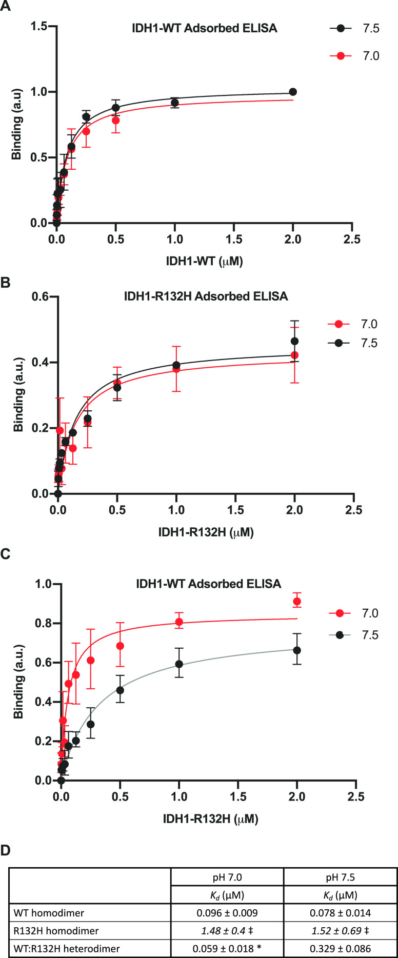
Figure 6.
IDH1 heterodimer formation is pH sensitive in an ELISA. (A–C) ELISAs for in vitro binding of IDH1 proteins (see Methods for details). (A) WT homodimer assay. IDH1-WT binding to adsorbed IDH1-WT at pH 7.0 and 7.5. (B) R132H homodimer assay. IDH1-R132H binding to adsorbed IDH1-R132H at pH 7.0 and 7.5. (C) Heterodimer assay. IDH1-R132H binding to adsorbed IDH1-WT at pH 7.0 and 7.5. For panels A–C, means ± standard deviation with binding curve fits shown. Data obtained from three replicate assays (each result is the average of two technical replicates) across two protein preparations. (D) Table of binding constants (Kd) calculated from binding curves in panels A–C. Significance was determined using Graph Pad Prism one-site specific nonlinear fitting (see Methods for details) (*p < 0.05, compared to pH 7.5). ⧧ Indicates Kd fits were made, but confidence intervals did not close.

