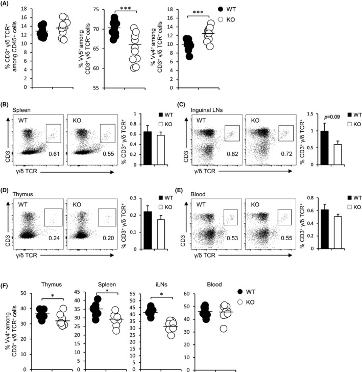
FIGURE 3.

FABP3‐deficient adult mice displayed a high percentage of Vγ4 + γ/δ T cells in the skin, but not thymus, spleen, inguinal lymph nodes, and peripheral blood. (A) Percentages of CD3+ γ/δ TCR+ cells, Vγ5+, and Vγ4+ γ/δ T cells in the ear skin of adult WT and KO mice. (B‐E) Total CD3+ γ/δ T cell profiles in the spleen (B), iLNs (C), thymus (D), and peripheral blood (E) from WT and KO mice. Numbers adjacent to outlined areas (left) indicate percentages of CD3+ and γ/δ TCR+ T cells. (F) Percentages of Vγ4+ γ/δ T cells in various tissues of WT and KO mice. *P < .05, ***P < .001 (Student's t test). Data are one experiment representative of at least three independent experiments with similar results (mean ± SEM of three replicates (A‐F))
