Abstract
Objectives/Hypothesis
To determine whether the presence of detectable upper respiratory infections (URIs) at the time of adenoidectomy/adenotonsillectomy is associated with increased morbidity, complications, and unexpected admissions.
Study Design
Prospective double‐blinded cohort.
Methods
In this prospective cohort study, nasopharyngeal swabs were obtained intraoperatively from 164 pediatric patients undergoing outpatient adenoidectomy/tonsillectomy with or without pressure equalization tubes (PETs) and were analyzed with PCR for the presence of 22 known URIs, including SARS‐CoV‐2. Surgeons and families were blinded to the results. At the conclusion of the study, rates of detectable infection were determined and intraoperative and postoperative events (unexpected admissions, length of PACU stay, rates of laryngospasm/bronchospasm, oxygen desaturation, bradycardia, and postoperative presentation to an emergency department) were compared between infected and uninfected patients.
Results
Of the 164 patients (50% male, 50% female, ages 8 mo‐18 y), 136 patients (82.9%) tested positive for one or more URI at the time of surgery. Forty one patients (25.0%) tested positive for three or more URIs concurrently, and 11 (6.7%) tested positive for five or more URIs concurrently. There were no significant differences in admission rates, length of PACU stay, rates of laryngospasm/bronchospasm, oxygen desaturation, bradycardia, or postoperative presentation to an emergency department between positive and negative patients. No patients tested positive for SARS‐CoV‐2.
Conclusions
A recent positive URI test does not confer any additional intraoperative or postoperative risk in the setting of outpatient adenoidectomy/tonsillectomy in healthy patients. There is no utility in preoperative URI testing, and delaying surgery due to a recent positive URI test is not warranted in this population.
Level of Evidence
3 Laryngoscope, 131:E2074–E2079, 2021
Keywords: Upper respiratory pathogen testing, outpatient surgery, adenoidectomy, tonsillectomy
INTRODUCTION
The preoperative evaluation for outpatient elective surgery considers the patient's overall health, including the potential of underlying acute respiratory illnesses. In the case of an overtly symptomatic child presenting with fever or a productive cough the decision to delay elective surgery is relatively straightforward. The most common outpatient elective surgery requiring endotracheal intubation at our institution is adenotonsillectomy, with or without pressure equalization tube placement, for children with recurrent otitis media, sleep apnea, and/or recurrent tonsillitis. This population can present challenges in the preoperative setting, because these children often present with chronic rhinorrhea, mild cough, and other symptoms which mimic a low‐grade viral illness. In our experience, patients scheduled for minor outpatient procedures requiring endotracheal intubation have occasionally been admitted postoperatively due to airway complications such as oxygen desaturation, need for bronchodilators, requirement of reintubation, etc., and were subsequently confirmed to be “viral positive” by nasopharyngeal swab/PCR. The goal of this study was to determine if a positive PCR test for URIs at the time of surgery could predict adverse outcomes, and if so, determine if preoperative testing might reduce complications.
At our institution, the policy is to delay elective surgery for 6 to 8 weeks after a positive URI test is obtained, which often occurs when the child presents to their pediatrician or to the emergency department with symptoms prior to surgery. The decision to delay surgery is based on several studies and the experience of our anesthesiologists, however, no blinded prospective studies in this patient population exist to support this decision. One of the earliest studies associating URIs with poor outcomes was performed in 1991 1 ; this retrospective chart review found that having a “respiratory infection” increased the risk of postoperative complications by 11‐fold, which was similar to a study by Tait et al. 2 More recent reports have cast doubt on the clinical utility of URI PCR testing (reviewed by Gill et al. 3 ), and several have retrospectively evaluated the impact of URI positivity on postoperative outcomes, citing increased length of stay (LOS) and intensive care unit LOS. 4 , 5 A recent retrospective study examining the effect of URI positivity in inpatient pediatric patients undergoing airway evaluation found no difference in postoperative complications between URI‐negative and URI‐positive patients. 6 No prospective studies to our knowledge have evaluated the prevalence of respiratory viral or bacterial infection in the routine pediatric outpatient surgery population, or in the otolaryngologic patient population where surgical indications (stertor, chronic rhinorrhea, chronic otitis, etc.) could also be considered symptoms of an active URI in an otherwise healthy cohort.
The majority of studies on outcomes of URI positive testing have by necessity focused on symptomatic patients or patients admitted to the hospital. Advani et al. 7 observed that 83% of symptomatic and 43% of asymptomatic patients admitted to an inpatient pediatric ward tested positive for one or more viral infections by RT‐PCR of nasopharyngeal aspirates. Another study compared hospitalized children to nonhospitalized, symptomatic outpatient children and found that 90% of hospitalized patients tested positive for one or more viruses compared to 52% of nonhospitalized controls. 8 One retrospective study of over 1000 general surgery patients demonstrated an overall prevalence of URI positivity of 10.5% and suggested that infection with parainfluenza virus type 3 (PIV3) may increase the likelihood of postoperative pneumonia, however, this study did not classify the patients regarding whether they underwent outpatient procedures or the presence of any underlying comorbidities.
Outpatient adenoidectomy and tonsillectomy are two of the most common procedures performed at our institution, with over 2000 procedures performed per year. Complications that can result from the procedure include difficulties with extubation, odynophagia with dehydration, bleeding, pulmonary edema, and atlantoaxial subluxation. 9 Fortunately, most of these complications are rare in occurrence but the resulting unplanned admissions can have a substantial economic impact on hospitals and patients, as well as increase their risk for nosocomial complications. It is impossible to precisely calculate this cost; an overnight stay at our institution on an inpatient floor is approximately $4500 whereas one night in the ICU can cost upwards of $10,000 depending on services provided.
In an attempt to provide evidence based recommendations in regard to perioperative management of children with detectable URIs, this study aims to demonstrate the prevalence of URI in the asymptomatic or mild symptomatic pediatric otolaryngologic population, determine the impact of a positive URI PCR test on postoperative outcomes, and evaluate the potential utility of preoperative testing as a means of reducing intraoperative and postoperative complications and unexpected admissions.
MATERIALS AND METHODS
Patient Selection
One hundred and sixty‐four pediatric patients undergoing outpatient otolaryngologic surgery requiring orotracheal intubation (adenoidectomy, tonsillectomy, or adenotonsillectomy with or without pressure equalization tube placement) were enrolled in the study which was approved by our Institutional Review Board (ACRI IRB #249953). The study took place between October 2019 and March 2020. Surgeries were performed using similar techniques by seven surgeons. Patients with underlying medical comorbidities including asthma, cardiac disease, severe OSA, cystic fibrosis, syndromic patients, or any patients planned for overnight observation were excluded. Patient histories of recent URIs, current symptoms of URIs, and preoperative vital signs were recorded by our anesthesiologists.
Testing Methods
After induction of anesthesia and orotracheal intubation, a nasopharyngeal swab was collected and placed into 3 mL of M4‐RT viral transport media (Remel, US). The FDA‐approved BioFire® FilmArray® Respiratory 2 (RP2) Panel was performed according to the manufacturer's instructions. 10 In brief, this closed test system performs sample preparation, reverse transcription, polymerase chain reaction, and detection of 21 respiratory pathogens using 300 μL of inoculated M4‐RT viral transport media. SARS‐CoV‐2 analysis was performed on the GeneXpert System using the emergency use authorization (EUA)‐approved Xpert Xpress SARS‐CoV‐2 test (Cepheid, Sunnyvale, CA) according to the manufacturer's instructions. 11 In brief, a defined volume of sample (nasopharyngeal swab specimen in 3‐mL M4‐RT) is transferred to the testing cartridge and placed on the GeneXpert System, which integrates sample preparation, nucleic acid extraction, and real‐time PCR‐based amplification and detection of target sequences (N2 and E genes). The limit of detection of this test is 250 copies/mL. SARS‐CoV‐19 analysis was only conducted on patient samples from January 2020 to March 2020 (n = 75) due to availability of previously stored samples.
Data Analysis
Patients, families, and surgeons were blinded to the results of the panel. Following the completion of the study, the charts of all patients were analyzed for postoperative complications including unexpected admission, laryngospasm, bronchospasm, oxygen desaturation below 95%, requirement of bronchodilators, bradycardia, and readmission or presentation to our emergency department for respiratory complications. The total time spent in the recovery area following the procedure was also recorded. The presence of detectable URIs and number of URIs detected were analyzed to detect differences in outcome rates using either the Fisher's exact test, t‐test, or Kruskal‐Wallis test. Statistical analyses were performed using SAS 9.4 (SAS Institute Inc., Cary, NC, USA).
RESULTS
A total of 164 patients were enrolled in this prospective, double‐blinded cohort study. The mean age of the population was 4.0 years. Thirty four (20.7%) patients reported acute rhinorrhea or nasal congestion on the day of surgery and 20 patients (12.2%) reported having a URI within 4 weeks of surgery. All patients were afebrile (temperature <99.0°F measured with tympanic thermometer). Sixteen (9.7%) underwent adenoidectomy only, 89 (54.3%) underwent adenotonsillectomy only, 49 (30%) underwent adenoidectomy/PETs, and 10 (6.1%) underwent adenotonsillectomy/PETs. The details of the cohort are illustrated in Table I.
TABLE I.
Description of Enrolled cohort of Patients, Including Male to Female Ratio and Age Information for Entire Population and by Procedure Performed.
| All Patients | n = 164 (100% of Total) |
|---|---|
| Male | n = 82 (50%) |
| Female | n = 82 (50%) |
| Average age | 4 years |
| Median age | 4 years |
| Age range | 8 months–18 years |
| Procedure: adenoidectomy | n = 16 (9.7% of total) |
| Male | n = 8 (50%) |
| Female | n = 8 (50%) |
| Average age | 2.8 years |
| Median age | 2 years |
| Age range | 15 months–4 years |
| Procedure: adenoidectomy/PETs | n = 49 (30% of total) |
| Male | n = 27 (54%) |
| Female | n = 22 (46%) |
| Average age | 2.5 years |
| Median age | 1.9 years |
| Age range | 8 months–12 years |
| Procedure: adenotonsillectomy | n = 89 (54.3% of total) |
| Male | n = 44 (49%) |
| Female | n = 45 (51%) |
| Average age | 6.7 years |
| Median age | 6 years |
| Age range | 3–9 years |
| Procedure: adenotonsillectomy + PETs | n = 10 (6.1% of total) |
| Male | n = 3 (30% |
| Female | n = 7 (70%) |
| Average age | 4 years |
| Median age | 3 years |
| Age range | 3–9 years |
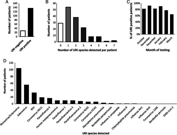
There were 28 patients (17.1%) who tested negative for all respiratory pathogens whereas 136 patients (82.9%) tested positive for one or more URI (Fig. 1A). Many patients tested positive for multiple species; 41 patients (25.0%) tested positive for 3 or more URIs, and 11 (6.7%) tested positive for five or more concurrent URIs (Fig. 1B). There was a range of infection rates over the course of several months, with the highest infection rate occurring during the month of November (n = 24/26 positive patients, 92%) and the lowest rate occurring in March (n = 9/14 positive patients, 64%) (Fig. 1C). The most commonly detected URI was rhinovirus/enterovirus (n = 104, 63%) followed by adenovirus (n = 56, 33.9%), RSV (n = 33, 20%), coronavirus HKU1 (n = 19, 11.5%), parainfluenza virus 4 (n = 17, 10.3%), human metapneumovirus (n = 10, 6.1%), parainfluenza virus 1 (n = 10, 6.1%), parainfluenza virus 3 (n = 10, 6.1%), parainfluenza virus 2 (n = 8, 4.8%), coronavirus NL63 (n = 6, 3.6%), coronavirus OC43 (n = 6, 3.6%), Mycoplasma pneumoniae (n = 3, 1.8%), Bordetella parapertussis (n = 2, 1.2%), influenza A untypeable (n = 1, 0.6%), influenza B (n = 1, 0.6%), and Chlamydophila pneumoniae (n = 1, 0.6%) (Fig. 1D). No patients tested positive for Bordetella pertussis, influenza AH1, influenza AH3, coronavirus 229E, or SARS‐CoV‐19.
Fig. 1.

Incidence of upper respiratory infections (URIs) in the outpatient otolaryngology population. (A) A total of 164 patients underwent intra‐operative URI testing by nasopharyngeal PCR. Twenty eight patients had no detectable respiratory pathogens, whereas 136 patients tested positive for one or more URIs. (B) Distribution of number of URI species detected within the cohort. (C) Infection rate per month of analysis. (D) Number of patients testing positive for each URI present on the panel or for SARS‐CoV‐2.
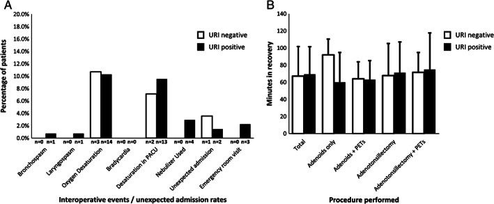
We next sought to determine whether patients with a positive URI test (“URI positive” population) experienced increased intraoperative or immediate postoperative complications, need for bronchodilators, frequency of unexpected admissions, and frequency of a return to our emergency department within 1 week of surgery (Fig. 2A) when compared to the patients with no detectable URI (“URI negative” population). In the URI positive group, one patient (0.7%) experienced intraoperative bronchospasm/laryngospasm and required bronchodilators. Three URI negative (10.7%) and 14 URI positive (10.3%) patients experienced brief oxygen desaturations to below 95% in the operating room. In the recovery area, two URI negative (7.1%) and 13 URI positive patients (9.6%) experienced brief oxygen desaturations below 95%. Two patients in the URI positive group (2.2%) and 1 patient in the URI negative group (3.6%) were admitted unexpectedly due to the need for supplementary oxygen in recovery. Three patients in the URI positive group (2.2%) were seen within 1 week of surgery in our emergency department for postoperative fever but were discharged from the ED. No patients experienced significant bradycardia.
Fig. 2.

(A) Comparison of intraoperative and post‐operative events between URI negative and URI positive patients. (B) Comparison of time spent in recovery between URI negative and URI positive patients as an entire cohort or by procedure performed.
Time spent in postoperative recovery has a direct impact on operating room efficiency, and therefore we sought to determine if URI positivity had an effect on the duration of time spent in the recovery room prior to discharge (Fig. 2B). The average time spent in recovery for our entire cohort was 68.3 minutes. We analyzed recovery time for URI positive and URI negative patients as an entire cohort as well as per procedure performed.
We next performed statistical analysis to determine if URI positivity, number of URIs detected, or procedure performed was correlated to any intraoperative or postoperative outcomes (Table II). There was no statistically significant difference in rate of unexpected admissions, bronchospasm, laryngospasm, oxygen desaturation, need for a nebulizer, or length of time in recovery between URI positive or URI negative patients either in total or within procedural groups. We also analyzed whether patient age and number of URIs detected was associated with an increased amount of time spent in recovery, and again found no significant difference between URI positive and URI negative patients (Figure S1).
TABLE II.
Statistical Analysis of Intraoperative and Post‐Operative Outcomes.
| URI Detected | Total N (%) | Admitted N (%) | Bronchospasm N (%) | Laryngospasm N (%) |
|---|---|---|---|---|
| Yes | 136 (82.9%) | 2 (1.47%) | 1 (0.74%) | 1 (0.74%) |
| No | 28 (17.1%) | 1 (3.57%) | 0 (0.00%) | 0 (0.00%) |
| P value | 0.313 | 0.782 | 0.782 | |
| Odds ratio (95% CI) | 2.032 (0.042–2.759) | 0.631 (0.024–16.635) | 0.631 (0.024–16.635) | |
| Test used | Wald Chi‐sq Test | Wald Chi‐sq Test | Wald Chi‐sq Test | |
| # species detected | ||||
| 0 | 28 (17.1%) | 1 (3.57%) | 0 (0.00%) | 0 (0.00%) |
| 1 | 57 (34.7%) | 0 (0.00%) | 1 (1.75%) | 1 (1.75%) |
| 2 | 38 (23.2%) | 1 (2.63%) | 0 (0.00%) | 0 (0.00%) |
| 3 | 22 (13.4%) | 0 (0.00%) | 0 (0.00%) | 0 (0.00%) |
| 4 | 8 (4.87%) | 1 (12.50%) | 0 (0.00%) | 0 (0.00%) |
| 5 | 8 (4.87%) | 0 (0.00%) | 0 (0.00%) | 0 (0.00%) |
| 6 | 1 (0.06%) | 0 (0.00%) | 0 (0.00%) | 0 (0.00%) |
| 7 | 2 (0.12%) | 0 (0.00%) | 0 (0.00%) | 0 (0.00%) |
| P value | 0.551 | 0.985 | 0.985 | |
| Odds Ratio (95% CI) | 1.199 (0.537–2.107) | 1.01 (0.104–2.492) | 1.01 (0.104–2.492) | |
| Test used | Wald Chi‐sq Test | Wald Chi‐sq Test | Wald Chi‐sq Test | |
| Procedure | ||||
| Adenoidectomy | 16 | 1 (6.25%) | 0 (0.00%) | 0 (0.00%) |
| Adenoidectomy/PETs | 49 | 0 (0.00%) | 1 (2.04%) | 0 (0.00%) |
| Adenotoinsillectomy/PETs | 10 | 0 (0.00%) | 0 (0.00%) | 0 (0.00%) |
| Adenotonsillectomy | 89 | 2 (2.25%) | 0 (0.00%) | 1 (1.12%) |
| P value | 0.3351 | 0.4573 | 1.00 | |
| Test used | Fisher's Exact Test | Fisher's Exact Test | Fisher's Exact Test | |
| URI Detected | Desaturation in OR N (%) | Desaturation in PACU N (%) | Nebulizer Used N (%) | Duration in Recovery Mean (SD or Q1,Q3) |
|---|---|---|---|---|
| Yes | 14 (10.29%) | 13 (9.56%) | 4 (2.94%) | 67.8382 (32.7218) |
| No | 3 (10.71%) | 2 (7.14%) | 0 (0.00%) | 69.4643 (34.4851) |
| P value | 0.977 | 0.687 | 0.666 | 0.7267 |
| Odds Ratio (95% CI) | 0.956 (0.256–3.577) | 1.374 (0.292–6.455) | 1.936 (0.097–38.80) | n/a |
| Test used | Wald Chi‐sq Test | Wald Chi‐sq Test | Wald Chi‐sq Test | T Test |
| # species detected | ||||
| 0 | 3 (10.71%) | 2 (7.14%) | 0 (0.00%) | 60.50 (49.50, 78.00) |
| 1 | 7 (12.28%) | 7 (12.28%) | 3 (5.26%) | 62.00 (48.00, 80.00) |
| 2 | 3 (7.89%) | 3 (7.89%) | 0 (0.00%) | 59.00 (48.00, 76.00) |
| 3 | 2 (9.09%) | 1 (4.55%) | 1 (4.55%) | 57.50 (46.00, 82.00) |
| 4 | 1 (12.50%) | 1 (12.50%) | 0 (0.00%) | 54.00 (41.50, 85.50) |
| 5 | 0 (0.00%) | 0 (0.00%) | 0 (0.00%) | 56.50 (51.50, 70.00) |
| 6 | 0 (0.00%) | 0 (0.00%) | 0 (0.00%) | 50.00 (50.00, 50.00) |
| 7 | 1 (50.00%) | 1 (50.00%) | 0 (0.00%) | 54.50 (39.00, 70.00) |
| P value | 0.977 | 0.943 | 0.9024 | 0.9681 |
| Odds Ratio (95% CI) | 0.995 (0.706–1.403) | 1.013 (0.708–1.451) | 0.961 (0.404–1.708) | n/a |
| Test used | Wald Chi‐sq Test | Wald Chi‐sq Test | Wald Chi‐sq Test | Kruskal‐Wallis Test |
| Procedure | ||||
| Adenoidectomy | 1 (6.25%) | 3 (18.75%) | 0 (0.00%) | 64.00 (34.80) |
| Adenoidectomy/PETs | 10 (20.41%) | 5 (10.20%) | 2 (4.08%) | 63.08 (22.51) |
| Adenotoinsillectomy/PETs | 0 (0.00%) | 1 (10.00%) | 0 (0.00%) | 72.70 (36.03) |
| Adenotonsillectomy | 6 (6.74%) | 6 (6.74%) | 2 (2.25%) | 67.36 (27.30) |
| P value | 0.0686 | 0.3515 | 0.8085 | 0.7056 |
| Test used | Fisher's Exact test | Fisher's Exact test | Fisher's Exact test | F test |
DISCUSSION
The presence of respiratory pathogens has long been anecdotally believed to be a poor prognostic indicator for outpatient surgeries, leading in theory to higher chance of admission, failure to extubate, and respiratory complications. This has led to a recent positive test for a URI as being a relative contraindication to elective surgery at many institutions which delays patient care. Our current study demonstrates that despite testing positive for a multitude of URIs on the day of surgery, relatively healthy outpatients can undergo elective surgery requiring intubation without significantly higher complication rates.
A total of 136 out of 164 (82.9%) patients undergoing routine outpatient procedures tested positive for one or more URIs on the commonly used RRP2 PCR panel. This is significantly higher than the previous studies of surgical patients 12 but is comparable to rates of symptomatic patients admitted to the pediatric wards. 7 The disparity in our results from previously published infection rates could possibly be due to the underlying pathology of our patient population; it may be that children with enlarged adenoids, large tonsils, and/or recurrent ear infections may be harboring chronic respiratory pathogens in the nasopharynx. A follow up prospective blinded study evaluating infection rates in the nonotolaryngologic population would address that question and contribute to our understanding of the etiology of adenotonsillar hypertrophy.
The double‐blinded, prospective nature of this study provides several advantages over previous work. First, by blinding the health care providers, we were unbiased in making clinical decisions during the perioperative period and during follow up care. Second, by blinding families to the results, there was less chance of patients reporting to the emergency room following surgery due to concerns over respiratory infections. We also employed a unique method to control for underlying conditions that might otherwise have confounded our data. Only patients who were cleared to enter our “Surgery Efficiency Model” (SEM) OR were enrolled. The SEM program exists to screen patients for underlying conditions that would exclude them from a typical outpatient surgery center. This determination is made by a team of physicians and nurses weeks prior to surgery and is based on the patient's history, physical exam, and surgical indications. The SEM room operates much like an outpatient surgery center with shorter turnover times, one team of providers for the entire day, and no residents or medical students participating. By only enrolling patients cleared for our SEM OR, we reduced variability between patients to the greatest extent possible.
This prospective study did have a few limitations: 1) The patient population was only otolaryngologic procedures and may not be generalizable to the surgical population as a whole as described above; 2) The study was stopped short of the anticipated 1 year of data collection due to the COVID‐19 pandemic. This led to a lower sample size and the inability to fully assess for seasonal variance.
CONCLUSION
Preoperative URI PCR panel testing is not indicated in the setting of outpatient adenoidectomy/tonsillectomy in healthy patients. Delaying elective procedures due to a recent URI or positive URI PCR panel in this population is not indicated and may delay surgical care unnecessarily.
Supporting information
Figure S1. Correlation analysis between age, length of stay, and number of viruses detected.
Editor's Note: This Manuscript was accepted for publication on October 22, 2020.
All authors confirm that they have no financial disclosures. This study was funded by NIH grant UG1OD024945 (National Institute of Child Health and Human Development, PI: Strub, G).
The authors have no other funding, financial relationships, or conflicts of interest to disclose.
BIBLIOGRAPHY
- 1. Cohen MM, Cameron CB. Should you cancel the operation when a child has an upper respiratory tract infection? Anesth Analg 1991;72:282–288. [DOI] [PubMed] [Google Scholar]
- 2. Tait AR, Knight PR. Intraoperative respiratory complications in patients with upper respiratory tract infections. Can J Anaesth 1987;34:300–303. [DOI] [PubMed] [Google Scholar]
- 3. Gill PJ, Richardson SE, Ostrow O, Friedman JN. Testing for respiratory viruses in children: to swab or not to swab. JAMA Pediatr 2017;171:798–804. [DOI] [PubMed] [Google Scholar]
- 4. Spaeder MC, Carson KA, Vricella LA, Alejo DE, Holmes KW. Impact of the viral respiratory season on postoperative outcomes in children undergoing cardiac surgery. Pediatr Cardiol 2011;32:801–806. [DOI] [PubMed] [Google Scholar]
- 5. Moynihan K, Barlow A, Alphonso N, et al. Impact of viral respiratory pathogens on outcomes after pediatric cardiac surgery. Pediatr Crit Care Med 2017;18:219–227. [DOI] [PubMed] [Google Scholar]
- 6. Niermeyer WL, Ball J, Worobetz N, et al. Respiratory viral panels and pediatric airway evaluation: the role of testing for upper respiratory infections to stratify perioperative risk. Int J Pediatr Otorhinolaryngol 2020;134:110057. [DOI] [PMC free article] [PubMed] [Google Scholar]
- 7. Advani S, Sengupta A, Forman M, Valsamakis A, Milstone AM. Detecting respiratory viruses in asymptomatic children. Pediatr Infect Dis J 2012;31:1221–1226. [DOI] [PMC free article] [PubMed] [Google Scholar]
- 8. Singleton RJ, Bulkow LR, Miernyk K, et al. Viral respiratory infections in hospitalized and community control children in Alaska. J Med Virol 2010;82:1282–1290. [DOI] [PMC free article] [PubMed] [Google Scholar]
- 9. Randall DA, Hoffer ME. Complications of tonsillectomy and adenoidectomy. Otolaryngol Head Neck Surg 1998;118:61–68. [DOI] [PubMed] [Google Scholar]
- 10. Leber AL, Everhart K, Daly JA, et al. Multicenter evaluation of BioFire FilmArray respiratory panel 2 for detection of viruses and bacteria in nasopharyngeal swab samples. J Clin Microbiol 2020;23:e00155–20. [DOI] [PMC free article] [PubMed] [Google Scholar]
- 11. Wolters F, van de Bovenkamp J, van den Bosch B, et al. Multi‐center evaluation of cepheid xpert(R) xpress SARS‐CoV‐2 point‐of‐care test during the SARS‐CoV‐2 pandemic. J Clin Virol 2020;128:104426. [DOI] [PMC free article] [PubMed] [Google Scholar]
- 12. Zhang D, Lou X, Yan H, et al. Respiratory virus associated with surgery in children patients. Respir Res 2019;20:126. [DOI] [PMC free article] [PubMed] [Google Scholar]
Associated Data
This section collects any data citations, data availability statements, or supplementary materials included in this article.
Supplementary Materials
Figure S1. Correlation analysis between age, length of stay, and number of viruses detected.


