

Highlights
-
•
Red face was seen in three patients with COVID-19.
-
•
Red face may reflect a cytokine storm.
-
•
Red face may be predictive of a sudden deterioration.
Keywords: COVID-19, SARS-CoV-2 alpha variant, Red face, Pneumonia, Cytokine storm
Abstract
Japan is currently suffering the fourth wave of the COVID-19 pandemic, with the dominant type being SARS-CoV-2 alpha variant. Patients with COVID-19 variant types show more aggressive symptoms. In the present study, three patients developed a red face during treatment. Two of them suddenly worsened shortly after. We assumed that the red face reflected a cytokine storm and conjectured that it may be a specific sign of variant type COVID-19, because we have never seen it in patients with non-variant type. Moreover, we believe that red face may be predictive of a sudden deterioration.
Introduction
We are suffering the fourth wave of the pandemic, with the dominant type being SARS-CoV-2 alpha variant [1].
Frontline workers who are treating patients with COVID-19 have noticed different symptoms in patients with variant types more than in patients with non-variant. We have experienced more treatment failure in patients with variant types, and we feel the need to adjust treatment as soon as possible when we notice changes in these patients. Therefore, it is important to identify signs of aggravation, especially in patients with variant-type COVID-19. However, the specific features of the variant types are not even known.
In the present study, three patients with variant COVID-19 showed red face. Two of them experienced sudden deterioration after this symptom appeared, while one showed no other symptoms. We believe that red face may be a specific symptom of variant type, because we have never seen it before (Table 1).
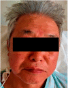
Table 1.
Patients’ characteristics.
| Case 1 | Case 2 | Case 3 | ||
|---|---|---|---|---|
| Age | 71 | 67 | 78 | |
| Sex | F | F | M | |
| Vital on admission | BP(mmHg) | 159/103 | 115/84 | 148/81 |
| HR(bpm) | 90 | 90 | 83 | |
| BT(°C) | 36.3 | 36.4 | 36.7 | |
| SpO2(%) | 97 | 97 | 97 | |
| Laboratory data | D-dimer(μg/mL) | 0.5 | 0.7 | 0.9 |
| ferritin(ng/mL) | 166 | 79 | 315 | |
| CRP(mg/L) | 0.73 | 0.29 | 3.88 | |
| KL-6(U/mL) | 136 | 337 | 556 | |
| CT on admission | 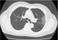 |
 |
 |
|
| Treatment | favipiravir deximesathone | favipiravir deximesathone | remdesivir methylprednisolone | |
| Vital on sudden change | BP(mmHg) | 132/83 | 144/87 | – |
| HR(bpm) | 84 | 73 | – | |
| BT(°C) | 37.9 | 37.1 | – | |
| SpO2(%) | 93 | 90 | – | |
| Red Face |  |
 |
 |
|
| CT on sudden change |  |
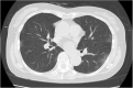 |
No sudden change | |
Case 1
A 71-year-old Japanese woman was admitted to our hospital because her husband had COVID-19 and had been hospitalized at our institution 3 days prior. She had no symptoms and had gone to the swimming pool the day before hospitalization. Although she had no symptoms, computerized tomography (CT) showed mild pneumonia. She was diagnosed with an intermediate disease with pneumonia, according to Japanese guidelines [2]. She had a history of dyslipidemia and had one aggravation risk factor: age > 65-year-old. Her body temperature was 37.2 °C and her saturation of percutaneous oxygen (SpO2) was 98 % upon arrival. She received information from the public health center that her specimen had revealed the COVID-19 variant type.
She was started on favipiravir (3600 mg on day 1, 1800 mg from day 2 onwards) and dexamethasone (6 mg from day 1 to day 10). She developed steroid-induced hyperglycemia, which was controlled using insulin. After hospitalization, she showed intermittent fever on day 4 (37.6 °C), day 7 (37.3 °C), and day 8 (37.9 °C), which was immediately resolved using antipyretics. On the morning of day 7, she noticed that her face had turned red. She felt heat on both cheeks, without sweat. There was no rash on the trunk. During the night of day 7, her SpO2 gradually decreased from 97 % to 91 %, and CT showed worsening of the pneumonia. However, she had no upper respiratory symptoms. She was started on oxygen during the night. On the morning of day 8, her SpO2 had not improved (92 % with 2 L of O2). She was transferred to an advanced medical center. Her red face continued until the transfer.
Case 2
A 68-year-old Japanese woman who developed fatigue and fever (maximum: 38.8 °C) was diagnosed with COVID-19. There had been an outbreak at her swimming club, which was the same club to which the patient in Case 1 belonged. She was diagnosed with mild disease without pneumonia upon arrival. She had two aggravation risk factors: age > 65 years and hypertension.
Her temperature was 37.1 °C and SpO2 97 % upon admission. She was started on favipiravir (3600 mg on day 1, 1800 mg from day 2 onwards) and dexamethasone (6 mg on days 1–10). Her temperature was normal on the next day, and her fatigue gradually improved. On the morning of day 5, the nurse pointed out a change in her face color. There was no rash on her trunk, and a red color was localized in her face. She did not feel heat on her cheeks, had no other symptoms, and her SpO2 level was 99 %. Her vital signs were stable, and she did not complain of any other symptoms, so we simply observed her. A few hours later, her SpO2 suddenly dropped from 98 % to 94 %. Although she did not complain of any new symptoms, we performed CT. CT revealed a new lesion of pneumonia. She was switched from favipiravir to remdesivir (200 mg/day on day 1, 100 mg from day 2 onwards) and from dexamethasone to methylprednisolone (1000 mg/day on days 1–3, 125 mg/day on days 4–5). Her SpO2 dropped to 90 % at the lowest during the night, and she was started on oxygen. The next morning, she had recovered from the hypoxia (SpO2: 98 %), and oxygen was stopped. Her red face disappeared at that time. Her pneumonia had resolved and she was discharged 12 days later.
Case 3
A 78-year-old Japanese man who developed fatigue, cough, and dysgeusia without fever was diagnosed with COVID-19. His body temperature was 36.7 °C and his SpO2 97 % upon arrival. His CT showed pneumonia, and he was diagnosed with intermediate disease with pneumonia. His medical history included hypertension, dyslipidemia, hyperuricemia, and cerebral infarction. He had two aggravation risk factors: age > 65 years and hypertension. He was started on remdesivir (200 mg/day on day 1, 100 mg from day 2 onwards) and methylprednisolone (1000 mg/day on days 1–3, 125 mg/day on days 4–5). The patient’s subjective symptoms improved. On day 5, he complained that his face was red. He felt heat on both cheeks, without sweat. His trunk did not change, he was afebrile, and his SpO2 was 97 %. The red face continued for 2 days. He showed no aggravation of the disease, and he was discharged 14 days after hospitalization.
Discussion
Several clinical manifestations have been reported since the COVID-19 pandemic emerged. However, red face has not yet been reported.
In Cases 1 and 2, a red face was observed just before the sudden deterioration. In Case 1, the patient developed intermittent fever, but the red face was not associated with any fever; instead, it was seen continuously, even when the fever was down. In Case 2, the patient had no fever or heat on the cheeks. In all cases, the red face was not restricted to the cheeks; it was on the whole face, was not necessarily accompanied by heat on the cheeks, and occurred without sweat or fever. In Cases 1 and 2, the patients developed worsening pneumonia after they developed red face, so we reasoned that the red face may have reflected cytokine release. Both showed a red face in the morning and developed pneumonia in the afternoon, so there was a time lag.
Our patients had already started steroid therapy, so we cannot exclude the influence of steroids. Flush has been reported as an adverse effect of steroids. The timing of steroid-related flush is unclear, but most adverse effects of steroids occur soon after the drug start. Our patients developed red face 5–7 days after steroid administration. In Case 3, methylprednisolone was initiated, but the patient showed red face on day 7, when the methylprednisolone had already been stopped. Therefore, the timing of red face and steroid use did not match. We believe that the red face was not related to the steroids, but rather that the steroids were masking the symptoms of a cytokine storm, which is why the patient in Case 3 did not develop severe pneumonia.
We believe that this symptom is a specific sign of the COVID-19 caused by SARS-CoV-2 alpha variant and that it may be predictive of a sudden deterioration. We hope this report will help medical workers struggling with COVID-19 on the frontline.
Declaration of Competing Interest
The authors report no declarations of interest.
Sources of funding
This study has no funding for research.
Consent
This study was approved by the institutional review board of Biwako Ohashi Hospital. Written informed consent was obtained from the patient for publication of this case report and accompanying images. A copy of the written consent is available for review by the Editor-in- Chief of this journal on request.
Authors contribution
AN: study design, data collections, data analysis, writing, EO: data analysis, YK, MY, EY, KH, ST, YT, YM, MH, MY, YK, YA, KY, SS, KO, KI: data collections.
Author statement
Aya Nakaya: Conceptualization, Writing-Original Draft, Eiji Ogura: Supervision, Yuki Katayama: Data Curation, Masami Yoshii :Data Curation, Eiko Yoshino :Data Curation, Kazuya Hozumi :Data Curation, Saori Tago: Data Curation, Yuko Teranishi: Data Curation, Yuki Minamibashi: Data Curation, Makiko Harada: Data Curation, Mami Yoshioka: Data Curation, Yuri Kawano: Data Curation, Yuka Arai: Data Curation, Keno Yoshida: Data Curation, Shozo Shimizu: Data Curation, Kazuma Ogura: Data Curation, Katsuaki Iwashita: Data Curation.
Acknowledgments
We thank Yuu Tochitani, Rika Umeda, Shizuka Morita, Yohei Takano, Masanori Fujie, Tsutomu Ito, Yuko Hirai, Ayako Igosaki, and Satomi Ozawa for their assistance.
References
- 1.Ministry of Health, Labor and Welfare . 2021. Information on Covid-19.https://www.mhlw.go.jp (Accessed 24 May 2021) [Google Scholar]
- 2.Ministry of Health, Labor and Welfare and the National Institute of Infectious Diseases . 2021. Clinical management of patients with COVID-19 (version 4.2) [Google Scholar]