Abstract
During spermatogenesis, the process in which sperm for fertilization are produced from germline cells, gene expression is spatiotemporally highly regulated. In Drosophila, successful expression of extremely large male fertility factor genes on Y-chromosome spanning some megabases due to their gigantic intron sizes is crucial for spermatogenesis. Expression of such extremely large genes must be challenging, but the molecular mechanism that allows it remains unknown. Here we report that a novel RNA-binding protein Maca, which contains two RNA-recognition motifs, is crucial for this process. maca null mutant male flies exhibited a failure in the spermatid individualization process during spermatogenesis, lacked mature sperm, and were completely sterile, while maca mutant female flies were fully fertile. Proteomics and transcriptome analyses revealed that both protein and mRNA abundance of the gigantic male fertility factor genes kl-2, kl-3, and kl-5 (kl genes) are significantly decreased, where the decreases of kl-2 are particularly dramatic, in maca mutant testes. Splicing of the kl-3 transcripts was also dysregulated in maca mutant testes. All these physiological and molecular phenotypes were rescued by a maca transgene in the maca mutant background. Furthermore, we found that in the control genetic background, Maca is exclusively expressed in spermatocytes in testes and enriched at Y-loop A/C in the nucleus, where the kl-5 primary transcripts are localized. Our data suggest that Maca increases transcription processivity, promotes successful splicing of gigantic introns, and/or protects transcripts from premature degradation, of the kl genes. Our study identified a novel RNA-binding protein Maca that is crucial for successful expression of the gigantic male fertility factor genes, spermatogenesis, and male fertility.
Author summary
Sperm is produced from germline cells via the process called spermatogenesis, during which a special gene expression program dedicated to this process operates. In fruit fly Drosophila, extremely large male fertility factor genes on Y-chromosome containing mega-base-sized introns are expressed only during spermatogenesis, and their successful expression is crucial for spermatogenesis. However, expressing such large genes must be very challenging for cells and the molecular mechanisms that ensure their successful expression remain unknown. In this study, we identified a novel RNA-binding protein encoded in the gene CG5213 that is required for this process. We created mutant flies lacking this protein using a genome-editing technique, and investigated them using genetic, microscopic, molecular, transcriptomic, and proteomic approaches. We found that this RNA-binding protein is crucial for successful expression of the extremely large male fertility factor genes, spermatogenesis, and male fertility. We named the CG5213 gene maca, after the plant maca grown in the high Andes mountains whose root has been traditionally believed and used to increase sperm quality and promote male fertility. By identifying the novel RNA-binding protein Maca, our studies give significant insight into how the special gene expression program operates during spermatogenesis.
Introduction
Spermatogenesis is the highly conserved and tightly regulated process where diploid germline stem cells develop into mature, haploid sperm capable of fertilizing oocytes. Spatiotemporally highly regulated gene expression is crucial during spermatogenesis, and transcriptional and post-transcriptional regulation by RNA-binding proteins play key roles. Human male sterility results from abnormal spermatogenesis and is mostly due to chromosomal alterations, Y chromosome microdeletions, and related gene mutations. Mutations in the germline-specific DAZ (Deleted in AZoospermia) family of RNA-binding proteins, which contain a highly conserved RNA recognition motif (RRM) and are crucial regulators of gene expression during spermatogenesis, impair spermatogenesis and cause human male sterility [1–12].
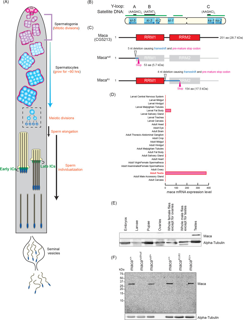
Genetically well-tractable Drosophila has been an excellent model system for spermatogenesis studies [13–16]. The Drosophila testis, where spermatogenesis occurs, is a coiled tube with a closed apical end and a basal end that connects to the seminal vesicle. The germline stem cells (GSCs) reside at the apical tip attached to the hub cells and differentiating germ cells are gradually displaced distally (Fig 1A). GSCs divide asymmetrically to produce a self-renewed GSC and a daughter cell that undergoes differentiation and becomes 16 spermatogonia by four rounds of synchronous mitotic divisions. The 16-cell spermatogonia then enter the meiotic S phase and become spermatocytes. Spermatocytes grow in size during their G2 growth phase, which spans as long as ~90 hours. During the spermatocyte growth, the homologous chromosomes pair and partition into individual chromosome territories, and almost all genes encoding proteins for later meiotic and post-meiotic processes (spermiogenesis) are transcribed. These transcripts are stored without being efficiently translated until their protein activities are required later for meiosis and spermiogenesis. Thus, transcriptional and post-transcriptional events during the spermatocyte growth phase are crucial and must be spatiotemporally highly regulated.
Fig 1. Maca domain structure, mutant alleles, and expression pattern.
(A) Diagram of spermatogenesis in Drosophila testis. At the apical tip of testis, germline stem cells (GSCs) are attached to the hub cells (Hub). GSCs produce daughter cell gonialblasts, which become mitotically-amplifying spermatogonia that then become spermatocytes. Spermatocytes grow in size for ~90 hours and then undergo the meiotic divisions to become 64 interconnected spermatids, which are then separated into individual spermatids by individualization complexes (ICs). Individualized mature motile sperm are stored in seminal vesicles. (B) Diagram of the Drosophila Y chromosome. Locations of the 6 male fertility factor genes (cyan. The transcription directions of the encoded genes are indicated by blue arrows), regions enriched for satellite DNA (green bars), and the Y-loop forming regions (black bars) with associated satellite DNA sequences are shown. (C) Domain structures of Drosophila Maca (Maca/CG5213), and its mutants, Macanull and MacaR1. (D) maca mRNA expression pattern. Data are obtained from http://flybase.org/reports/FBgn0038345. (E) Western blots of dissected fly tissues. (F) Western blots of testis lysates.
After their G2 growth phase, spermatocytes divide twice by meiosis and then differentiate into 64 interconnected haploid spermatids. After 64 interconnected spermatids elongate, they are separated into individual spermatids by individualization complexes (ICs) in the sperm individualization process (Fig 1A). Individualization complexes form around the sperm nuclei and move processively in a highly coordinated manner from the heads to the tips of the sperm tails along the spermatid bundle, removing excess cytoplasm and unneeded organelles and encasing each sperm cell in its own plasma membrane. The individualized, mature motile sperm are stored in the seminal vesicles for fertilization.
The ~40 Mb Y chromosome of Drosophila is required only for male fertility, but not fly viability, mostly (~80%) comprised of repetitive sequences (primarily short tandem repeat satellite DNAs), and entirely heterochromatic [17–19]. Because of the extremely repetitive nature, the genome DNA sequence of most of the Y chromosome is not fully determined yet and available genome sequences of the Y chromosome contain many gaps. Despite the considerable size of the Y chromosome, it has only 16 known protein-coding genes. The Y chromosome contains six loci called male fertility factors that are essential for spermatogenesis and male fertility (Fig 1B) [18,20–22]. Four (kl-5, kl-3, kl-2, and kl-1) are on the long arm of the Y and two (ks-1 and ks-2) are on the short arm. kl-2, kl-3, and kl-5 encode dynein proteins that are crucial components of the axoneme, the microtubule-based cytoskeletal structure that forms the core of a sperm flagellum [23–28]. The kl-2, kl-3, and kl-5 genes are transcribed during the spermatocyte growth and their mRNAs form cytoplasmic ribonucleoprotein (RNP) granules called kl-granules in late-stage spermatocytes [29]. kl-2, kl-3, and kl-5 mRNAs are suggested to start being efficiently translated only when axoneme elongates in the sperm elongation process in spermiogenesis. The male fertility factor genes including kl-2, kl-3, and kl-5 contain gigantic, megabase-sized introns rich in repetitive, satellite DNA. For example, kl-3 gene spans at least 4.3 Mb while its coding sequence is only ~14 kb [18,30,31]. Their gene sizes and structures are reminiscent of those of human Dystrophin, a causative gene for Duchenne Muscular Dystrophy, which spans ~2.2 Mb with gigantic introns rich in repetitive DNA and only ~11 kb coding sequence [32]. Transcription of these extremely large genes and processing their transcripts including splicing must be challenging for cells. There must be unknown special molecular mechanisms and factors that enable their efficient, precise, and regulated transcription and processing.
In spermatocytes, kl-3, kl-5, and ks-1 form lampbrush-like nucleoplasmic structures named Y-loops [denoted as loops A (kl-5), B (kl-3), and C (ks-1) (Fig 1B), analogous to the lampbrush loops of amphibian oocytes [33]. Y-loop structures reflect the robust transcription of underlying genes in spermatocytes. Each loop consists of a DNA axis associated with huge repetitive RNA transcripts, which are in turn associated with large amounts of proteins [33–35].
In this study, we hypothesized that there may be a previously uncharacterized RNA-binding protein that is important for the testis-specific gene expression program. We predicted that such an RNA-binding protein, if any, is expressed exclusively in testes. We found that the gene CG5213, located on the right of the third chromosome, meets these criteria: it encodes a novel RNA-binding protein with two RNA recognition motifs (RRMs) (Fig 1C) and its mRNA is expressed almost exclusively in adult testes (Fig 1D). We genetically investigated the functions of CG5213 and revealed that it is required for spermatogenesis and male fertility in Drosophila. We discovered that the CG5213 protein is expressed in spermatocytes, resides in the nucleus enriched at Y-loop A/C, and is required for successful expression and splicing of the kl-2, kl-3, and kl-5 transcripts. We named the CG5213 gene maca, after the plant maca grown in the high Andes mountains whose root has been traditionally believed and used to increase sperm quality and promote male fertility.
Results
Maca is exclusively expressed in testes
We generated a polyclonal anti-Maca antibody against a recombinant full-length Maca protein and examined Maca protein expression in Drosophila tissues using hand-dissected control (w1118) fly samples. We found that Maca protein is expressed exclusively in adult testes (Fig 1E).
maca mutant flies
To study biological and molecular functions of Maca in vivo, we created two maca mutant alleles, macanull and macaR1 by introducing deletions within the Maca coding region using a CRISPR/Cas9 genome editing system (Fig 1C) [36]. The macanull allele has a 5-nt long deletion before the first RRM (deletion of 135–139 nucleotide residues in the Maca coding sequence (CDS)), which caused a translation frameshift and produced a premature stop codon resulting in encoding an N-terminal 45 amino acid (aa) fragment of Maca followed by additional 8 aa (total 53 aa, 5.7 kDa. Fig 1C). This short fragment is unlikely to have any functions and we consider this allele as null. The macaR1 allele has a 4-nt long deletion after the first RRM (deletion of 353–356 nucleotide residues in the Maca CDS), causing a translation frameshift and producing a premature stop codon. The macaR1 allele encodes an N-terminal 117 aa fragment of Maca followed by additional 37 aa (total 154 aa, 17.5 kDa. Fig 1C). Both macanull and macaR1 homozygous mutant flies were viable, showing that Maca is dispensable for fly viability.
To validate the maca mutant fly strains and the Maca antibody that we created, we performed Western blots of testis lysates from the maca mutant flies using the anti-Maca antibody. In testis lysates of wild-type (maca+/+) and heterozygous controls of macanull and macaR1 (macanull/+ and macaR1/+), the full-length Maca protein was detected as expected (Fig 1F). The full-length Maca protein was not detected in the testis lysates from the homozygous mutants of macanull and macaR1 (macanull/null and macaR1/R1). These results validated our maca mutant fly strains and anti-Maca antibody. No smaller proteins corresponding to the Maca fragments were detected in the macanull and macaR1 strains (macanull/+, macanull/null, macaR1/+, and macaR1/R1). Therefore, the Maca fragments encoded in these two mutant alleles are likely unstable or not expressed.
maca is essential for male fertility
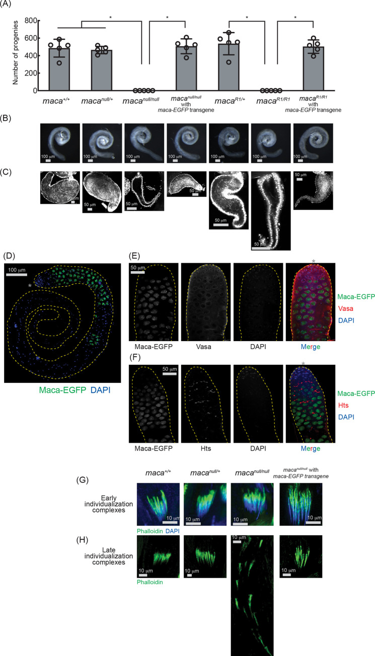
Maca’s testis-specific expression (Fig 1D and 1E) suggested that Maca plays an important role in testes. To test this, we performed fertility assays to determine whether maca is required for male fertility. Hundreds of progenies were obtained when the control flies (maca+l+, macanull/+, and macaR1/+) were crossed with wild-type virgin females (Fig 2A). In contrast, no progenies were obtained at all when macanull/null or macaR1/R1 male flies were crossed with wild-type virgin females. Trans-heterozygous mutant flies macanull/Df and macaR1/Df, which have the macanull or macaR1 allele and the Df(3R)Exel6174 chromosomal deficiency allele uncovering the maca gene, also showed complete male sterility (S1A Fig). These results indicated that maca is strictly required for male fertility. To confirm that male sterility is due to loss of maca, not due to any unintended secondary mutations, we tested if a maca transgene can rescue male fertility in the macanull/null and macaR1/R1 backgrounds. We created maca transgenic rescue fly strains using a maca transgene that expresses C-terminally EGFP-fused Maca protein under control of a maca promoter. This maca-EGFP transgenic allele fully rescued male fertility in the macanull/null and macaR1/R1 backgrounds (Fig 2A). macanull/null, macanull/Df, macaR1/R1, and macaR1/Df did not exhibit any significant changes in female fertility compared with the controls (S1B and S1C Fig). We concluded that maca is strictly required for male fertility, but not for female fertility. This is consistent with the observation that Maca is expressed almost exclusively in testes but not in female flies (Fig 1D and 1E).
Fig 2. maca mutant flies are male-sterile, lack mature motile sperm, and have disorganized individualization complexes.
(A) Male fertility assay. The numbers of the progeny flies obtained from the crosses between test males and OregonR wild-type virgin females are shown. Mean +/- SD (n = 5 biological replicates). P-value <0.05 (Student’s t-test, unpaired, two-tailed) are indicated by *. (B) Stereomicroscope images of dissected whole testes. Scale bars are 100 μm. (C) Confocal images of dissected seminal vesicles stained with DAPI. Mature motile sperm is absent in macanull/null and macaR1/R1. Scale bars are 50 μm. (D) Confocal images of dissected whole testis from the maca-EGFP transgenic fly. Maca-EGFP (green) and DAPI (blue). The apical tip of the testis, where the hub cells reside, is marked with an asterisk (*). Scale bar is 100 μm. (E) Confocal images of the apical region of the testis from the maca-EGFP transgenic fly. Maca-EGFP (green), Vasa (red), and DAPI (blue). The apical tip of the testis, where the hub cells reside, is marked with an asterisk (*) in the merged panel. Scale bar is 100 μm. (F) Confocal images of the apical region of the testis from the maca-EGFP transgenic fly. Maca-EGFP (green), Hts (red), and DAPI (blue). The apical tip of the testis, where the hub cells reside, is marked with an asterisk (*) in the merged panel. Scale bar is 100 μm. (G) Confocal images of the early-stage individualization complexes. Phalloidin (Actin, green), DAPI (blue). Scale bars are 10 μm. (H) Confocal images of the late-stage individualization complexes. Phalloidin (Actin, green). Scale bars are 10 μm.
maca is essential for spermatogenesis
To explore the mechanism underlying the complete male sterility in the maca mutant flies (Figs 2A and S1), we first examined whole testis morphology. We did not observe any major defects in whole testis morphology in the maca mutant flies compared with the controls (Fig 2B). Next, using DAPI staining of needle-shaped sperm nucleus and confocal imaging, we examined mature motile sperm, which are produced in testes and stored in seminal vesicles. As expected, we observed plenty of mature motile sperm in the seminal vesicles of the control flies (maca+l+, macanull/+, and macaR1/+) (Fig 2C). In contrast, the seminal vesicles of macanull/null and macaR1/R1 flies were empty lacking mature motile sperm. Plenty of mature motile sperm were observed in the seminal vesicles of macanull/null and macaR1/R1 flies with the maca-EGFP rescue transgene. Using the DJ-GFP allele, which labels mature motile sperm with bright GFP signal in the control seminal vesicles, we further confirmed the loss of mature motile sperm in the seminal vesicles of macanull/null and macaR1/R1 (S2 Fig). We concluded that maca is essential for spermatogenesis.
Maca is expressed in spermatocytes and resides in the nucleus
We investigated an expression pattern of Maca protein in testes using the maca-EGFP transgenic flies. We found that Maca-EGFP is expressed in spermatocytes residing in the nucleus and becomes undetectable after spermatocytes complete their growth (Fig 2D). Next, we performed immunostaining of maca-EGFP transgenic fly testes with Vasa and Hu-li tai shao (Hts) antibodies. Maca-EGFP was expressed in spermatocytes and was localized in the nucleus while Vasa was expressed in germline stem cells and developing germline cells including spermatogonia and spermatocytes and resided in the cytoplasm and at the nuclear periphery (Fig 2E). Hts labels filamentous fusome structures connecting the dividing spermatogonia and spermatocytes (Fig 2F). Maca-EGFP started to be expressed soon after the Hts filamentous structures were formed. These results together showed that Maca-EGFP is expressed in spermatocytes and resides in the nucleus. We also transiently expressed EGFP-Maca in cultured S2 cells and found that EGFP-Maca protein is localized in the S2 cell nucleus (S3 Fig), confirming that Maca is a nuclear protein.
maca is essential for sperm individualization
To examine which step in spermatogenesis is impaired in macanull/null testes, we examined the sperm individualization process [37]. Individualization complexes were properly organized in the early stage of the sperm individualization process in macanull/null testes as in the control testes (Fig 2G). However, unlike in the control testes, individualization complexes were disorganized and scattered in the late stage of the individualization process in macanull/null testes, showing axoneme formation defects (Fig 2H). Proper individualization complex organization was rescued by the transgenic maca-EGFP in the macanull/null background. We concluded that maca is required for proper individualization complex organization in the late stage of the sperm individualization process. Loss of proper sperm individualization explains the loss of mature sperm and the complete male sterility in macanull/null.
Kl-2, Kl-3, and Kl-5 protein levels are reduced in macanull/null testes
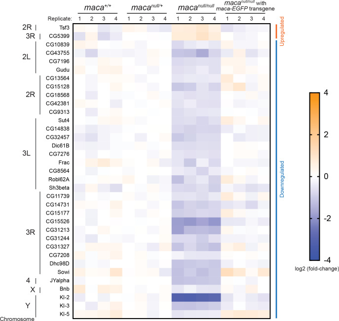
To start to understand the molecular mechanism by which the sperm individualization complexes are impaired in macanull/null testes, we examined protein levels in a proteome-wide manner in four biological replicates each of the testes of maca+/+, macanull/+, macanull/null, and macanull/null with maca-EGFP rescue transgene, using mass spec with TMT-labeling (see Materials and Methods). Among the 6,559 proteins we detected, two proteins were significantly upregulated (relative abundance >1.3, adjusted P-value <0.05) and 33 proteins were significantly downregulated (relative abundance <0.77, adjusted P-value <0.05) in macanull/null compared with all the other three genotypes consistently (Figs 3 and S4A). Among these consistently dysregulated proteins, male fertility factor Kl-2 showed the strongest and most significant downregulation in macanull/null (relative abundance 0.12 and adjusted P-value 1.7 x 10⁻⁵ in macanull/null compared with macanull/+) (Figs 3 and S4A). Interestingly, two other male fertility factors Kl-3 and Kl-5 were also among the 33 consistently downregulated proteins. Kl-2 is an inner dynein arm (IDA) dynein heavy chain protein and Kl-3 and Kl-5 are outer dynein arm (ODA) dynein heavy chain proteins [23–28]. The male fertility factor genes kl-2, kl-3, and kl-5 encoding these proteins are all located in the Y chromosome long arm and span several megabases containing gigantic introns rich in repetitive sequences (Fig 1B).
Fig 3. Proteins dysregulated in macanull/null testes including downregulated Kl-2, Kl-3, and Kl-5 revealed by mass spec.
Heatmap of protein levels that were significantly dysregulated in the macanull/null testes compared with the testes from maca+/+, macanull/+, and macanull/null with maca-EGFP rescue transgene determined by mass spec with TMT labeling. Log2(fold-change compared with the means of macanull/+) are shown. Four biological replicates for each genotype. Two proteins (at the top) were significantly upregulated (relative abundance >1.3, adjusted P-value <0.05) and 33 proteins were significantly downregulated (relative abundance <0.77, adjusted P-value <0.05) in macanull/null compared with all the other three genotypes. Male fertility factor Kl-2 was most strongly and significantly downregulated (relative abundance 0.12 and adjusted P-value 1.7 x 10⁻⁵ in macanull/null compared with macanull/+). Proteins are sorted based on the chromosomal positions of the corresponding genes.
No significant change in small RNA levels in macanull/null testes
Next, we examined testis small RNAs (miRNAs, siRNA, and piRNAs) from three biological replicates each of maca+/+, macanull/+, macanull/null, macanull/+ with maca-EGFP rescue transgene, and macanull/null with maca-EGFP rescue transgene in a transcriptome-wide manner using high throughput sequencing. No miRNAs, siRNA, or piRNAs (including Y-linked Suppressor of Stellate (Su(Ste) piRNAs, which are crucial for male fertility[38]) showed significantly changed levels in macanull/null testes compared with the other four genotypes (S5 Fig).
kl-2, kl-3, and kl-5 mRNA levels are reduced in macanull/null testes
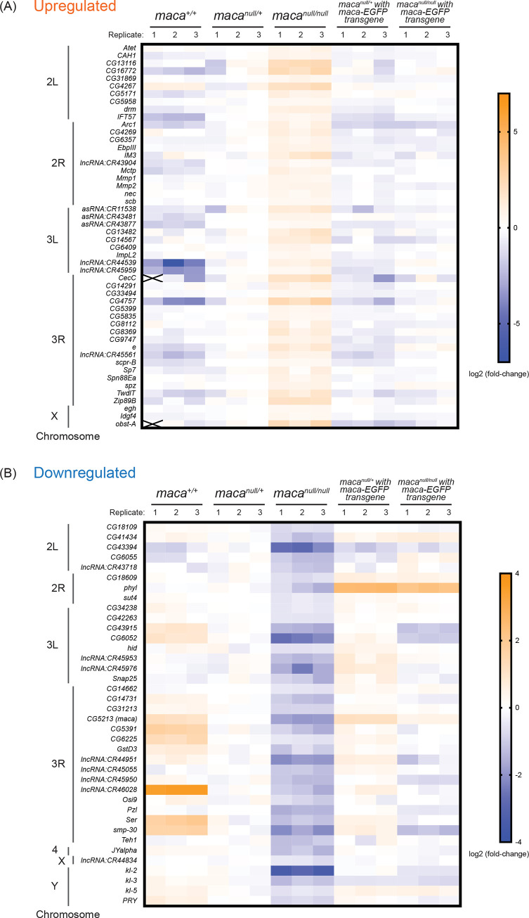
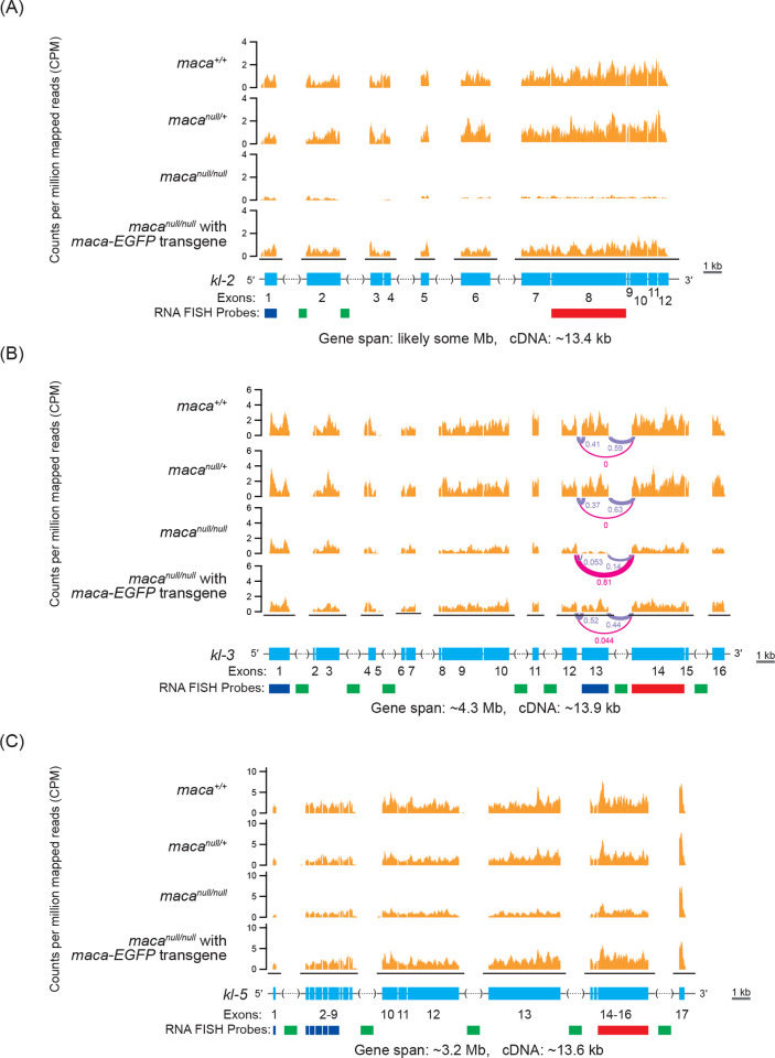
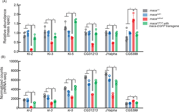
Considering that Maca resides in the nucleus (Figs 2D, 2E and S3) and has RNA-recognition motifs, if any observed protein level dysregulation in macanull/null testes is directly caused by maca loss of function, then it is likely through dysregulation of their corresponding transcripts rather than translational or post-translational dysregulation. Therefore, we examined testis poly-A+ RNAs in three biological replicates each of maca+/+, macanull/+, macanull/null, macanull/+ with maca-EGFP transgene, and macanull/null with maca-EGFP transgene in a transcriptome-wide manner using high throughput sequencing (poly-A+ RNA-seq). Among the 17,137 genes we examined, we detected reads for 15,937 genes in at least one sample. Among them, 50 genes showed significant upregulation (relative abundance >1.32, adjusted P-value <0.01) and 38 genes showed significant downregulation (relative abundance <0.76, adjusted P-value <0.01) in mRNA levels in macanull/null testes compared with all the other four genotypes consistently (Fig 4A and 4B). Interestingly, male fertility factor kl-2 mRNA showed the most significant downregulation in macanull/null testes among all the detected mRNAs (relative abundance 0.10 and adjusted P-value 1.65 x 10−196 in macanull/null compared with macanull/+) (Figs 4B, 6A, S4B and S5). Moreover, kl-3 and kl-5 mRNAs were also among the 38 consistently downregulated (Figs 4B, 6B, 6C, S4B, S7 and S8). Thus, kl-2, kl-3, and kl-5 exhibited significant downregulation in both protein and mRNA levels in macanull/null testes compared with all the other genotypes (Fig 5). CG31213 and JYalpha also showed both protein and mRNA level downregulation and CG5399 showed upregulation in macanull/null testes compared with the all other genotypes (Figs 5 and S4). kl-2, kl-3, and kl-5 are known to be essential for male fertility [27,29,39] and thus their downregulation in macanull/null testes can nicely explain the male sterility in macanull/null. Therefore, we decided to focus on kl-2, kl-3, and kl-5.
Fig 4. Poly-A+ RNA-seq analysis revealed RNAs dysregulated in macanull/null testes including downregulated kl-2, kl-3, and kl-5 mRNAs.
(A, B) Heatmaps of RNA levels that are significantly upregulated or downregulated in the macanull/null testes compared with the testes from maca+/+, macanull/+, macanull/+ with maca-EGFP rescue transgene, and macanull/null with maca-EGFP rescue transgene, determined by poly-A+ RNA-seq. Log2(fold-change compared with the means of macanull/+) are shown. Three biological replicates for each genotype. (A) Fifty genes were significantly upregulated (relative abundance >1.32, adjusted P-value <0.01) and (B) 38 genes were significantly downregulated (relative abundance <0.76, adjusted P-value <0.01) in macanull/null compared with all the other four genotypes. Male fertility factor kl-2 mRNA was most strongly and significantly downregulated (relative abundance 0.10 and adjusted P-value 1.65 x 10−196 in macanull/null compared with macanull/+). Genes are sorted based on their chromosomal positions.
Fig 6. Poly-A+ RNA-seq read mapping counts for kl-2, kl-3, and kl-5 mRNAs show their reduction in macanull/null testes.
Poly-A+ RNA-seq normalized read counts (counts per million mapped reads, CPM) in the (A) kl-2, (B) kl-3, and (C) kl-5 gene regions. For kl-3, splicing ratios among (1) exon 12—exon 13, (2) exon 13—exon 14, and (3) exon 12—exon 14 (= exon 13-skipped) determined by LeafCutter [39] are shown. Only one replicate out of 3 is shown for each genotype. All replicates are shown in S5–S7 Figs. Gene structures are shown with exons (cyan), introns (black line), intronic satellite DNA repeats (dashed line in parentheses. Contain gaps in the publically available Drosophila genome sequence). Regions targeted by RNA FISH probes used in Figs 8, 10, S10, S13, S14, S15 and S16 (blue, green, and red).
Fig 5. Abundance of both protein and mRNA of kl-2, kl-3, and kl-5 are decreased in macanull/null testes.
(A) Relative abundance of dysregulated proteins determined by mass spec whose mRNAs were also dysregulated in macanull/null compared with all the other tested genotypes. Mean +/- SD (n = 4 biological replicates). Adjusted P-values <0.05 are indicated by *. (B) Normalized counts (reflecting relative abundance) of dysregulated mRNAs determined by poly-A+ RNA-seq whose protein products were also dysregulated in macanull/null compared with all the other tested genotypes. Mean +/- SD (n = 3 biological replicates). Adjusted P-values <0.01 are indicated by *.
kl-3 exon 13 skipping occurs in macanull/null testes
We wondered if splicing dysregulation occurs in any transcripts in macanull/null testes. To test this, we analyzed our poly-A+ RNA-seq data using LeafCutter [39]. Curiously, we found that kl-3 mRNA exon 13 skipping occurs in macanull/null testes, but not in the other four genotypes (Figs 6B and S7). No other mRNA showed consistent splicing dysregulation in macanull/null compared with the other four genotypes. We also found that read mapping count was reduced rather evenly along the kl-2 and kl-5 gene regions in macanull/null compared with the other genotypes (Figs 6A, 6C, S6 and S8). In contrast, read mapping was reduced more severely in the kl-3 exon 13 region than in the other kl-3 coding regions in macanull/null, which is consistent with the exon 13 skipping (Figs 6B and S7). Thus, our poly-A+ RNA-seq data revealed kl-3 exon 13 skipping in macanull/null testes. No major mRNA level reduction, exon skipping, or protein level reduction for ks-1 (ory) was observed in macanull/null compared with the controls (S9 Fig).
maca mutation phenocopies kl-2, kl-3, and kl-5 RNAi knockdown
Gene mutation or RNAi-mediated knockdown of kl-2, kl-3, or kl-5 in testis germline cells causes spermatogenesis defects including a scattering of the individualization complexes and male sterility [27,29,39]. We first confirmed that kl-2, kl-3, or kl-5 RNAi knockdown depleted corresponding cytoplasmic mature mRNAs, but not the nuclear precursor mRNA (pre-mRNA) transcripts in spermatocytes (S10 Fig). kl-2, kl-3, or kl-5 RNAi knockdown caused loss of mature motile sperm in seminal vesicles (S11A Fig) and scattering of the individualization complexes in the late stage of the sperm individualization process (S11B and S11C Fig), similar to maca mutation (Fig 2C, 2G and 2H). Thus, maca mutation and kl-2, kl-3, or kl-5 RNAi knockdown depleting their cytoplasmic mRNAs phenocopy each other.
kl-2 transcript level reduction in macanull/null testes detected with RT-qPCR
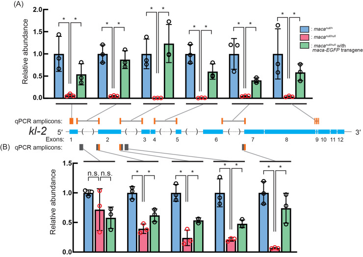
To validate the strong kl-2 mRNA level reduction observed in poly-A+ RNA-seq (Figs 4B, 5B, S4B and S6), we quantitated kl-2 transcripts in testis using RT-qPCR. We performed the reverse transcription (RT) step using random primers so that precursor transcripts including transcription intermediates before poly-A addition as well as mature poly-A+ mRNAs can be detected in the RT-qPCR assays. When qPCR primer set that amplifies a region in kl-2 exon 1 was used, macanull/null showed a dramatic reduction compared with the control macanull/+ (became 0.07 fold) (Fig 7A). Similarly, when qPCR primer sets spanning large kl-2 introns and that spanning normal size kl-2 introns were used, macanull/null showed a severe reduction (became 0.01–0.05 fold). Mature kl-2 mRNAs can be detected by all of these primer sets.
Fig 7. Quantification of kl-2 transcripts by RT-qPCR show their reduction in macanull/null testes.
Quantification of kl-2 transcripts in testis RNA by RT-qPCR. The RT step was performed with random hexamer primers. (A) qPCR amplicons targeted by designed primer sets are indicated by orange bars. The primers span exon-exon junctions to amplify only spliced transcripts, except for the first amplicon, which targets exon 1. Black lines indicate intron regions not included in the amplicons. (B) qPCR amplicons targeted by designed primer sets are indicated by gray and orange bars, which indicate intron and exons regions, respectively. At least one primer in each primer set targets an intron and therefore the primer sets target kl-2 transcripts before splicing (pre-mRNAs). Data were normalized to actin5C mRNA and the means of macanull/+. Mean +/- SD (n = 3 biological replicates). P-values <0.05 (Student’s t-test, unpaired, two-tailed) are indicated by *.
Next, we used qPCR primer sets to amplify regions in kl-2 introns and regions located at kl-2 exon-intron or intron-exon boundaries. kl-2 pre-mRNA transcripts before splicing, but not mature kl-2 mRNAs after splicing, can be detected by these qPCR primer sets. With these primer sets, the reduction degree in macanull/null compared with the control became gradually stronger as a primer set target location is shifted from the 5′ end to the 3′ end of the kl-2 gene (Fig 7B). Yet, the reduction degrees in macanull/null detected using these primer sets targeting kl-2 pre-mRNAs (Fig 7B) were not as strong as those obtained using the primer sets targeting the kl-2 exon regions (Fig 7A). The kl-2 transcript level was rescued in macanull/null with maca-EGFP rescue transgene in most cases (Fig 7A and 7B). Thus, in macanull/null testes, the kl-2 pre-mRNA transcript levels are more strongly reduced toward the 3′ end and the mature kl-2 mRNA levels are dramatically reduced, suggesting that later steps in the kl-2 mRNA transcription/processing/stability is more strongly affected than early steps.
Severe reduction of cytoplasmic mature kl-2 mRNA in macanull/null spermatocytes revealed by RNA FISH
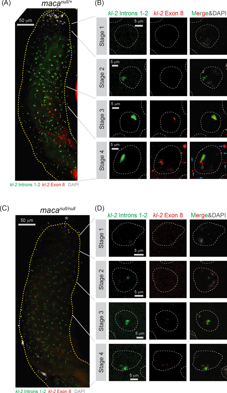
Next, we performed RNA in-situ hybridization (FISH) to visualize a spatiotemporal expression pattern of kl-2 transcripts in testes. In the control macanull/+ testes, we found that transcription of the kl-2 gene progresses in a highly spatiotemporal manner as previously shown for the kl-3 and kl-5 genes [40]. kl-3 and kl-5 expression during spermatocyte growth was subdivided into four stages based on their transcription progress from the 5′ end to the 3′ end of the genes [40]. Here we subdivided kl-2 expression in four stages as well. The transcription initiation occurs in early spermatocytes where neither kl-2 introns 1–2 nor exon 8 was detectable yet in the nucleus (stage 1) (Fig 8A and 8B). In the next stage, kl-2 introns 1–2 were expressed (stage 2). Then, kl-2 exon 8 was additionally expressed (stage 3). Finally, while the intron and exon signals were continued to be detected in the nucleus, mature kl-2 mRNAs that contain only exons, but not introns, were exported to the cytoplasm and reside as cytoplasmic kl-granules in late spermatocytes (stage 4). Thus, the gigantic kl-2 gene is transcribed gradually during the ~90-hour spermatocyte growth, and only in the late stage, kl-2 gene transcription is completed and spliced mature kl-2 mRNAs are exported to the cytoplasm.
Fig 8. RNA FISH for kl-2 transcripts shows their reduction in macanull/null testes.
RNA FISH to visualize kl-2 transcripts expression in (A, B) macanull/+ and (C, D) macanull/null testes. Introns 1–2 (green), exon 8 (red), and DAPI (white). (A, C) Apical regions of testes including the spermatocyte growth region are shown. The apical tip of testis is marked by *. Scale bars are 50 μm. (B, D) Single spermatocyte nuclei (white dashed line) at each stage of kl-2. Cytoplasmic mRNA granules are indicated by cyan arrows. Scale bars are 5 μm.
In macanull/null testes, FISH signal for kl-2 pre-mRNA transcripts in the spermatocyte nucleus was weaker than in macanull/+ testes (Fig 8C and 8D). FISH signal for kl-2 exon 8 seemed more severely reduced than those for kl-2 introns 1–2. Furthermore, cytoplasmic mature kl-2 mRNA granule in stage 4 spermatocyte was almost completely absent in macanull/null testes. We concluded that kl-2 pre-mRNA transcript levels are detectably and cytoplasmic kl-2 mature mRNA levels are severely reduced in macanull/null testes, consistent with our RT-qPCR results (Fig 7).
kl-3 mRNA reduction and kl-3 exon 13 skipping in macanull/null testes confirmed by RT-PCR
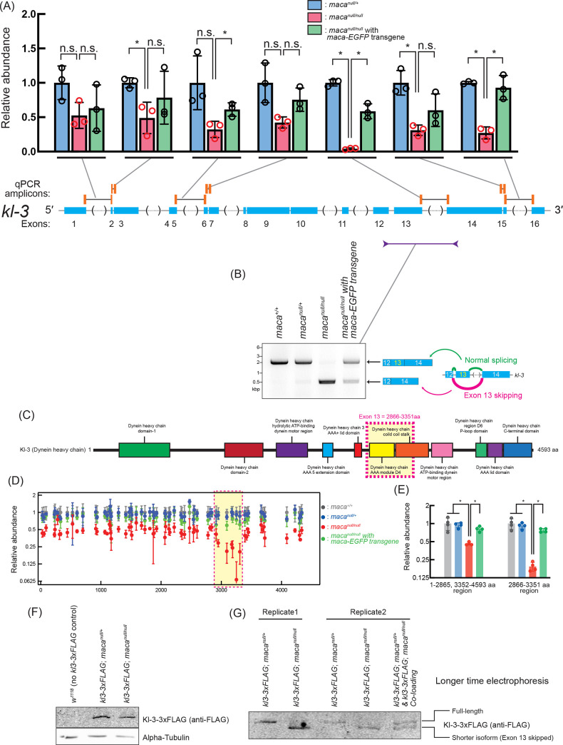
To validate the kl-3 mRNA level reduction and the kl-3 exon 13 skipping observed in the poly-A+ RNA-seq analysis (Figs 4B, 5B, 6B, S4B and S7), we performed RT-qPCR to measure kl-3 mRNA levels in testes. Primer sets spanning kl-3 introns to amplify mature kl-3 mRNAs revealed a reduction to 0.27–0.53 fold in macanull/null testes compared with macanull/+, except for one primer set (Fig 9A). The exception was the qPCR primer set with a forward primer targeting a region in exon 13 and a reverse primer targeting the junction between exon 13 and exon 14 (= spanning intron 13); it showed a much stronger reduction in macanull/null testes compared with macanull/+ (became 0.04 fold). The kl-3 mRNA levels were rescued in macanull/null with maca-EGFP rescue transgene. These results confirm the kl-3 mRNA level reduction and the kl-3 exon 13 skipping, consistent with the poly-A+ RNA-seq results.
Fig 9. kl-3 mRNA reduction and exon 13 skipping in macanull/null testes revealed by RT-(q)PCR.
(A) Quantification of kl-3 transcripts in testis RNA by RT-qPCR. The RT step was performed with random hexamer primers. qPCR amplicons targeted by designed primer sets are indicated by orange bars. The primers span exon-exon junctions to amplify only spliced transcripts. Black lines indicate intron regions not included in the amplicons. Data were normalized to actin5C mRNA and the mean of macanull/+. Mean +/- SD (n = 3 biological replicates). P-values <0.05 (Student’s t-test, unpaired, two-tailed) are indicated by *. (B) Agarose gel electrophoresis of RT-PCR products using the indicated primers to examine kl-3 exon 13 skipping in testis RNA. (C) Diagram of Kl-3 protein domains. Amino acid residues 2866–3351 corresponding to the exon 13 is indicated with a magenta dash-lined yellow box. (D) Abundance of peptide fragments derived from Kl-3 protein determined by mass spec with TMT labeling plotted along the Kl-3 amino acid residue numbers. Data were normalized to the means of maca+/+. Mean +/- SD (n = 4 biological replicates). The amino acid residues 2866–3351 corresponding to the kl-3 mRNA exon 13 is indicated with a magenta dash-lined yellow box. (E) Abundance of Kl-3 protein determined using peptide fragments data (left) from the 1–2865 or 3352–4593 aa region or (right) from the 2866–3351 aa region, of Kl-3 in mass spec with TMT labeling. Data were normalized to the means of maca+/+. Mean +/- SD (n = 4 biological replicates). P-values <0.05 (Student’s t-test, unpaired, two-tailed) are indicated by *. (F, G) Western blots of testis lysates for Kl-3-3xFLAG using (F) a standard protocol and (G) an extended SDS-PAGE gel electrophoresis time to achieve better separation. The FLAG tag was inserted at the C-terminal end of Kl-3 at the endogenous kl-3 locus on the Y-chromosome [40].
To further confirm the kl-3 exon 13 skipping, we performed RT-PCR using testis RNA with a forward PCR primer targeting a region in exon 12 and a reverse PCR primer targeting a region in exon 14 and ran the PCR products on an agarose gel. A ~2.1 kb band showing exon 13 retention was predominantly obtained from the testis RNA of the controls (maca+/+ and macanull/+) and macanull/null with maca-EGFP rescue transgene, while a ~0.6 kb band showing exon 13 skipping was predominantly obtained from the testis RNA of macanull/null (Fig 9B). These results confirmed the kl-3 exon 13 skipping in macanull/null testes.
Similarly, we detected the kl-3 exon 13 skipping when whole adult male RNA was used for RT-PCR analysis (S12A Fig). The ~2.1 kb PCR product showing kl-3 exon 13 retention was obtained from maca+/+ and macanull/+ males while the ~0.6 kb PCR product showing exon 13 skipping was obtained from macanull/null males. Consistent with the exclusive expression of maca in testes (Fig 1D and 1E), no PCR bands were obtained when whole adult female RNA was used (S12A Fig). In contrast, sex-specific alternative splicing of sxl, tra, and msl2, was not dysregulated in macanull/null males and females compared with maca+/+ and macanull/+ (S12B–S12D Fig). These results demonstrated that splicing dysregulation in macanull/null is specific to kl-3 exon 13 skipping, consistent with the poly-A+ RNA-seq analysis.
kl-3 exon 13 skipping causes an internal deletion of Kl-3 protein in macanull/null testes
The kl-3 mRNA exon 13 skipping would result in an internal deletion of amino acid residues 2,866–3,351 of Kl-3 protein without causing a translation frameshift (Fig 9C). We wondered if the internally deleted Kl-3 protein is expressed in macanull/null testes. To test this, we first searched our TMT-labeled testis-proteome mass spec data for peptide fragments containing the 2,865–2,866 residues or the 3,351–3,352 residues of the full-length Kl-3 protein, which would derive from the full-length Kl-3 protein produced from kl-3 mRNA with the exon 13-retained, or for peptide fragments containing the residue 2,865 followed by the residue 3,352, which would derive from the internally deleted Kl-3 protein variant produced from the exon 13-skipped mRNA. However, none of these fragments was detected in our data, which has ~20% coverage for Kl-3 protein.
Next, we took an alternative approach. The aa residues 1–2,865 and 3,352–4,593 are common between the full-length Kl-3 and the internally-deleted Kl-3 proteins while the aa residues 2,866–3,351 are specific to the full-length Kl-3. We analyzed the relative abundance of detected Kl-3 peptide fragments along the full-length Kl-3 amino acid sequence. Relative abundances of peptide fragment levels derived from the 2,866–3,351 amino acid residues were more strongly reduced in macanull/null compared with the peptide fragment levels derived from the other Kl-3 region (Fig 9D). The Kl-3 protein level was calculated to be 0.46 fold in macanull/null testes compared in maca+/+ when the peptide fragment data derived from the aa residues 1–2,865 and 3,352–4,593 were analyzed, while it was 0.19 fold when the peptide fragment data derived from the aa residues 2,866–3,351 were analyzed (Fig 9E). These mass spec data supported that kl-3 exon 13 skipping causes an expression of the internally deleted Kl-3 protein variant in macanull/null testes.
Furthermore, we attempted to detect the internally deleted Kl-3 protein variant using Western blot. We utilized a C-terminally 3xFLAG tagged kl-3 gene at its endogenous locus in the Y-chromosome [40]. While the size difference of the C-terminally 3xFLAG tagged Kl-3 protein between in macanull/+ and in macanull/null was not apparent in a standard protocol Western blot (Fig 9F), the size difference became apparent when gel electrophoresis time was much extended to achieve better separation (Fig 9G). The detected Kl-3 protein size was smaller in macanull/null than in macanull/+, supporting the idea that the full-length protein is predominantly expressed in macanull/+ testes while the internally deleted Kl-3 protein is predominant in macanull/null testes. When testis protein lysate from macanull/+ and that from macanull/null were mixed and loaded together on a Western blot gel, it produced double bands, confirming the existence of two different size Kl-3 proteins (Fig 9G). Taken together, we concluded that the kl-3 mRNA exon 13 skipping in macanull/null testes causes expression of the Kl-3 protein variant with an internal deletion of the corresponding amino acid residues. The deleted region contains the entire Dynein heavy chain AAA module D4 domain and the N-terminal half of the Dynein heavy chain coiled coil stalk domain of the full-length Kl-3 (Fig 9C). The Kl-3 protein activity is likely to be significantly impaired by this deletion.
kl-3 exon 13 skipping in macanull/null spermatocytes revealed by RNA FISH
We performed RNA FISH for kl-3. First, as shown previously [40], we observed gradual transcription of kl-3 pre-mRNA transcripts from the 5′ end to the 3′ end during the spermatocyte growth using FISH probes against exon 1, introns, and exon 14, in the control macanull/+ testis (S13A and S13B Fig). As previously performed [40], to visualize kl-3 introns, we probed the (AAUAU)n repeat sequences, which are produced from the (AATAT)n satellite DNA found in multiple kl-3 introns including intron 1 (the intron between exon 1 and exon 2) (Fig 1B). (AATAT)n is the only satellite DNA found in Y-loop B [41,42]. Only in late-stage spermatocytes, transcription of the entire kl-3 gene is completed and cytoplasmic mature kl-3 mRNA containing signal from both exon 1 and exon 14, but lacking signal from introns, were observed in kl-granules (S13B Fig, stage 4). Similarly, in macanull/null also, we observed the gradual expression of the kl-3 transcripts during stages 1–3 and cytoplasmic mature kl-3 mRNA granules with overlapping signals from exon 1 and exon 14 lacking introns in stage 4 (S13C and S13D Fig).
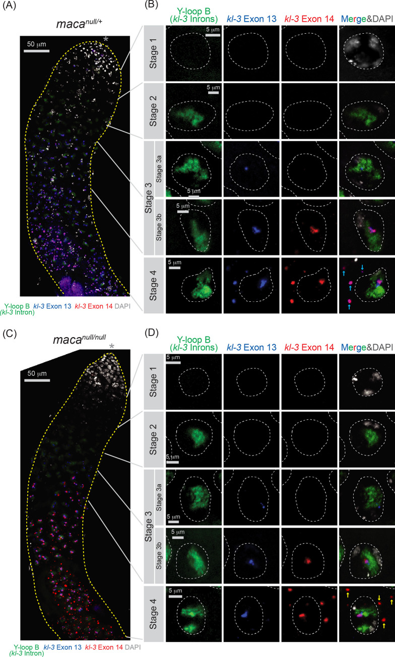
Next, to examine the exon 13 skipping, we used FISH probes for Y-loop B (kl-3 introns), exon 13, and exon 14. Again, we observed gradual expression of the kl-3 pre-mRNA transcripts. In stage 1 spermatocytes in the control macanull/+ testes, we did not observe any probe signal (Fig 10A and 10B). In stage 2, we observed only Y-loop B (kl-3 introns) signal. Signal from Y-loop B (kl-3 introns) and that from exon 13 were detected in the nucleus in stage 3a, while exon 14 signal was additionally observed in stage 3b. Finally, in stage 4, in addition to all these signals that were still observed in the nucleus, mature kl-3 mRNA signal containing both exons 13 and 14, but lacking the Y-loop B (kl-3 introns) signal, was observed in cytoplasm as kl-granules. In macanull/null, signal in stages 1–3 spermatocytes was similar to what was observed in macanull/+ (Fig 10C and 10D). In contrast, in the macanull/null stage 4 spermatocytes, while both exon 13 and exon 14 signals were observed in the nucleus, cytoplasmic mRNA granules contained only exon 14 signal but not exon 13 signal (Fig 10C and 10D). These results together showed that cytoplasmic mature kl-3 mRNAs in macanull/null spermatocytes contain exons 1 and 14 but not exon 13, consistent with the exon 13 skipping. We note that because FISH is not a very quantitative assay, we did not reliably detect kl-3 mRNA level reduction other than exon 13 region in macanull/null spermatocytes, while our poly-A+ RNA-seq and qRT-PCR showed significant kl-3 mRNA level reduction in macanull/null testes.
Fig 10. RNA FISH for kl-3 transcripts shows exon 13 skipping in macanull/null testes.
RNA FISH to visualize kl-3 transcripts in (A, B) macanull/+ and (C, D) macanull/null testes. Introns (Y-loop B, Alexa488-(AATAT)6, green), exon 13 (blue), exon 14 (red), and DAPI (white). (A, C) Apical regions of testes including the spermatocyte growth region are shown. The apical tip of testis is marked by *. Scale bars are 50 μm. (B, D) Single spermatocyte nuclei (white dashed line) at each stage of kl-3 expression. Cytoplasmic mRNA granules are indicated by cyan arrows for macanull/+, which contains signal from both exon 13 and exon 14, and by yellow arrows for macanull/null, which contains signal from exon 14 but not exon 13. Scale bars are 5 μm.
kl-5 RT-qPCR and RNA FISH
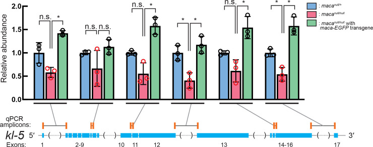
We also performed RT-qPCR to measure kl-5 mRNA levels and detected a moderate reduction in macanull/null testes compared with the control testes using some of the primer sets tested (Fig 11). This is consistent with the statistically significant and consistent but rather mild reduction of kl-5 mRNA level in macanull/null testes revealed by poly-A+ RNA-seq (Figs 4B, 5B, 6C and S8). We also performed RNA FISH for kl-5 with probes against exons 1–6, introns, and exons 16–17. As previously performed [40], for kl-5 introns, we probed the (GUCUU)n repeat sequences, which are produced from the (AAGAC)n satellite DNA and found in Y-loops A and C (Fig 1B) [34]. Gradual transcription of kl-5 gene during the spermatocytes growth and cytoplasmic mature kl-5 mRNA with signal from both exons 1–6 and exons 16–17 as kl-granules were observed in both macanull/+ and macanull/null (S14 Fig). Again, because of the limited quantitativeness of the FISH assay, we did not reliably detect kl-5 transcript level reduction in macanull/null spermatocytes.
Fig 11. RT-qPCR of kl-5 transcripts shows their reduction in macanull/null testes.
Quantification of kl-5 transcripts in testis RNA by RT-qPCR. The RT step was performed with random hexamer primers. qPCR amplicons targeted by designed primer sets are indicated by orange bars. The primers span exon-exon junctions to amplify only spliced transcripts. Black lines indicate intron regions not included in the amplicons. Data were normalized to actin5C mRNA and the mean of macanull/+. Mean +/- SD (n = 3 biological replicates). P-values <0.05 (Student’s t-test, unpaired, two-tailed) are indicated by *.
Maca is enriched at Y-loop A/C in spermatocyte nucleus
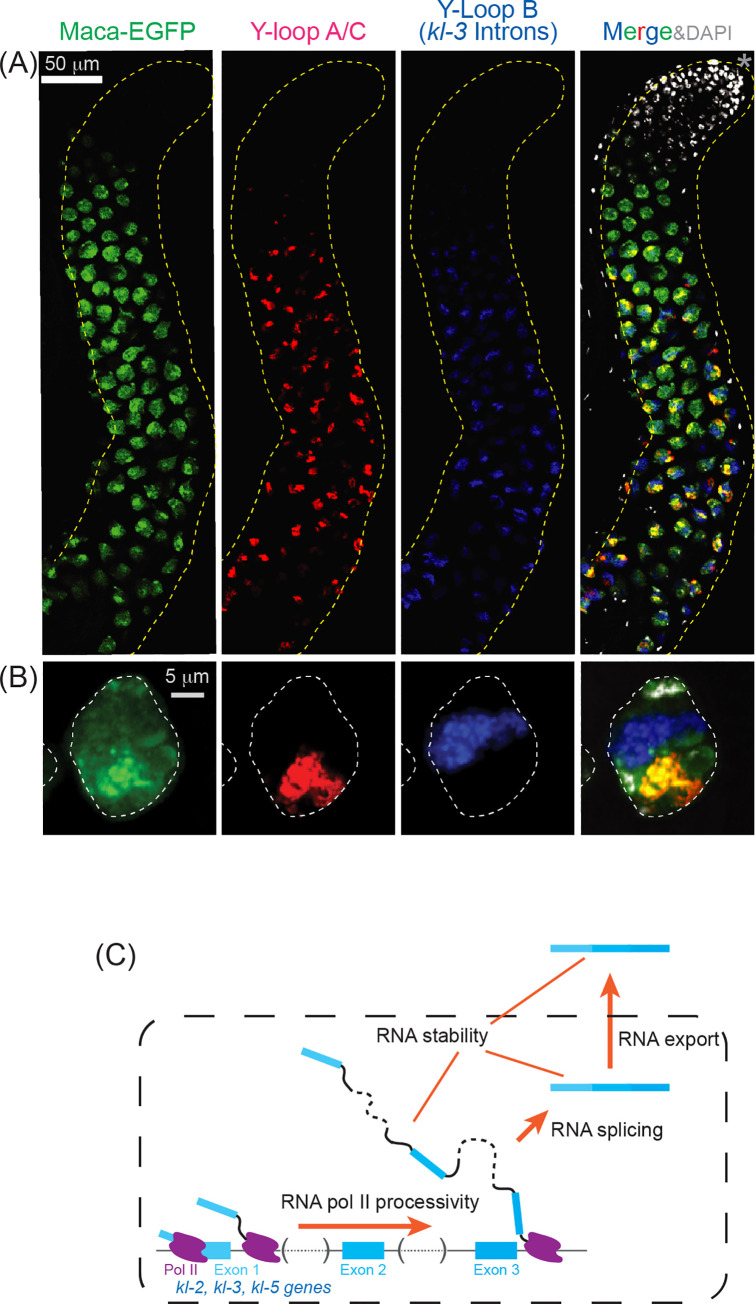
Considering the effects of maca mutation on the kl-2, kl-3, and kl-5 transcript levels and splicing, we wondered if Maca colocalizes with kl-2, kl-3, and/or kl-5 transcripts in the spermatocyte nucleus. To test this, we performed RNA FISH using the maca-EGFP transgenic fly testes in the otherwise wild-type background probing different regions within the kl-2, kl-3, and kl-5 transcripts including the Y-loops (Figs 12A, 12B, S15A, S15B and S15E–S15L). Interestingly, while Maca-EGFP resides in the entire region within the spermatocyte nucleus, it was enriched at Y-loop A/C (Fig 12A and 12B). When the dyes on the probes for Y-loop A/C and Y-loop B (kl-3 introns) were swapped, Maca-EGFP still showed an enriched localization at Y-loop A/C (S15A and S15B Fig). Maca-EGFP and Y-loop A/C exhibited similar spatiotemporal expression patterns during spermatocyte growth; they started to be expressed in early spermatocytes, exhibited colocalization during the spermatocyte growth phase, and became undetectable prior to the first meiotic metaphase (Fig 12A and 12B). The localization of Y-loop A/C in the spermatocyte nucleus was distinct from those of Y-loop B and the kl-2 and kl-3 transcripts in the control testes (Figs 12A, 12B and S15A–S15D). The distinct localizations of Y-loop A/C and Y-loop B were observed in macanull/null as well (S16A–S16D Fig), revealing that maca is not essential for their distinct localizations. ks-1 mRNA resides at the cytoplasmic granule that is different from the kl-granules, where kl-5 mRNA resides, in the late-stage spermatocytes in the control testes (S16E Fig). The distinct ks-1 mRNA cytoplasmic granule was observed in macanull/null as well (S16F Fig), revealing that maca is not essential for the distinct cytoplasmic localization of ks-1 mRNA.
Fig 12. Maca is enriched at Y-loop A/C in the spermatocyte nucleus.
(A, B) RNA FISH to visualize Y-loops in maca-EGFP testis. Maca-EGFP (green), Y-loop A/C (Cy3-(AAGAC)6, red), Y-loop B (kl-3 introns, Cy5-(AATAT)6, blue), and DAPI (white). (A) Apical regions of testes including spermatocyte growth region are shown. The apical tip of testis is marked by *. Scale bar is 50 μm. (B) Single spermatocyte nuclei (white dashed line) at late stage. Scale bar is 5 μm. (C) Model for Maca functions on kl-2, kl-3, and kl-5 transcripts.
Discussion
In this study, we identified a novel RNA-binding protein Maca in Drosophila. We showed that Maca is specifically expressed in spermatocytes in testes, resides in the nucleus enriched at Y-loop A/C, and is required for a properly orchestrated organization of late-stage individualization complexes, mature motile sperm, and male fertility. The maca mutant phenotypes were reminiscent of those of male fertility factor genes kl-2, kl-3, and kl-5 (kl genes) which have gigantic introns. Unbiased genome-wide proteome and transcriptome analyses revealed that maca is required for proper expression of the mRNAs and proteins of kl genes. kl-2 was particularly strongly downregulated in maca mutant testes at both mRNA and protein levels. RT-qPCR confirmed the severe kl-2 mRNA level reduction and also showed a reduction of kl-2 pre-mRNA. maca was also required for proper splicing of the kl-3 transcript.
The kl genes span megabases with gigantic introns containing satellite DNAs. Their transcription gradually progresses in spermatocytes as the cells grow for ~90 hours. The extended time period required for the complete transcription of the kl-3 and kl-5 genes due to the presence of the gigantic introns was proposed to function as a ‘developmental timer’ for the spermatocyte growth [40]. In this study, we also showed that the complete transcription of the kl-2 gene also takes an extended time during the spermatocyte growth. Thus, the kl-2 transcription may function as a developmental timer as well where the presence of the gigantic introns is important.
Considering that Maca is a nuclear RNA-binding protein, it likely regulates nuclear RNA processes rather than cytoplasmic ones such as translation. Poly-A+ RNA-seq, RT-qPCR, and RNA FISH together showed that in macanull/null testes, (1) spliced kl-2 transcript level was severely reduced, more severely than unspliced kl-2 transcripts, (2) the degree of reduction of unspliced kl-2 transcripts gradually increased from the 5′ end region to the 3′ end region of the kl-2 pre-mRNA, (3) spliced kl-3 and kl-5 transcript levels were reduced, (4) kl-3 splicing was dysregulated with exon 13 skipped, and (5) cytoplasmic mRNA granules of kl-2, kl-3, and kl-5 were absent in late-stage spermatocytes. Based on these results, we propose models for Maca functions below (Fig 12C).
Complete transcription of extremely long genes like the kl genes requires a high processivity of RNA polymerase II (pol II). High processivity of pol II is more important for the kl genes since they contain highly repetitive sequences such as satellite DNAs. Repetitive sequences can form high-order structures in DNA, RNA, and DNA-RNA hybrids, which may inhibit transcription elongation of pol II. In fact, repeat expansion (as in trinucleotide expansion diseases) and tandem arrays slow an elongating pol II and cause premature dissociation [43–46]. We propose that Maca helps high processive transcription of the kl genes, allowing pol II to transcribe completely through their extremely long, repetitive DNA sequences (Fig 12C).
It must be also challenging to properly splice extremely long transcripts like the kl transcripts, where exons are separated by megabase-sized introns and a 5′ splice site and a 3′ splice site that are extremely distant in a primary sequence need to be brought together. Furthermore, high-order structures in RNA caused by repetitive sequences are thought to pose a big challenge in splicing. Thus, we also propose that Maca plays important roles in the splicing of the kl pre-mRNAs (Fig 12C). The observed dysregulation of kl-3 splicing (exon 13 skipping), in maca mutant spermatocytes supports this model. Kl-3 gene has the gigantic intron 13 (between exon 13 and exon 14) while its intron 12 is only 274 nt long. Properly splicing out the gigantic intron 13 and joining the exons 13 and 14 requires Maca functions.
The extremely long kl transcripts are thought to be more easily degraded since they have more access points for degradation enzymes/chemicals than normal length transcripts. We propose that Maca may protect the kl transcripts from degradation (Fig 12C). Finally, Maca might be required for the export of kl mRNAs from the nucleus to the cytoplasm, which can also explain the observed reduction of kl mRNAs (in the cytoplasm). These proposed molecular functions of Maca are not mutually exclusive. Maca likely plays roles in multiple of these functions.
We found that Maca is enriched at Y-loop A/C in the spermatocyte nucleus. Considering that kl-5 mRNA levels were reduced in the maca mutant testes, this colocalization may suggest that Maca directly binds the kl-5 transcripts and thereby promote their transcription and/or processing or prevent their premature degradation. We note that while Maca-EGFP signal is stronger at Y-loop A/C, it was detectable in the entire nucleus (Figs 12A, 12B and S15). Thus, Maca may directly bind the kl-2 and kl-3 transcripts and promote their transcription and/or processing or prevent their premature degradation as well, which is supported by our results that the kl-2 and kl-3 mRNA levels were reduced and the kl-3 splicing was dysregulated in the maca mutant testes. It will be important to determine direct RNA-binding targets of Maca in future studies.
Some other RNA-binding proteins are enriched at the Y-loops in the spermatocyte nucleus and are required for male fertility. Blanks [40], Hephaestus (Heph)[40], Boule [47], Hrb98DE [48], Pasilla [48,49], and Rb97D [50] are among them. Only two of them, Blanks and Heph, seem to play roles in Y-loop gene expression in spermatocytes [40]. Blanks colocalizes with Y-loop B (kl-3 introns) and is required for kl-3 mRNA expression, proper individualization complex organization in late-stage spermatocytes, and mature motile sperm formation [40,51]. In our previous study, we performed poly-A+ RNA-seq of blanks mutant testes, which revealed downregulation of kl-3 mRNA [51]. In our blanks mutant poly-A+ RNA-seq results, we did not observe splicing dysregulation of kl-3 or any other fertility factor gene transcripts, unlike in maca mutant. Since both kl-3 nuclear transcript levels and cytoplasmic mature kl-3 mRNA levels in spermatocytes were reduced in blanks mutant spermatocytes judged by RNA FISH, it was proposed that Blanks may increase the pol II processivity by binding to kl-3 nascent transcripts in the nucleus [40]. Heph colocalizes with Y-loop A/C and is required for kl-3, kl-5, and ks-1 (ory) mRNA expression, proper individualization complex organization in late-stage spermatocytes, and mature motile sperm formation. Since cytoplasmic mature kl-5 mRNA granules were diminished without a large decrease in the nuclear kl-5 transcript levels in heph mutant spermatocytes, it was proposed that Heph may play crucial roles in kl-5 transcript processing such as splicing and/or preventing premature degradation rather than in the transcription step. The protein and mRNA abundance of Blanks and Heph were not significantly changed in the macanull/null testes compared with the controls in our mass spec and poly-A+ RNA-seq data, excluding the possibility that the macanull/null phenotypes are mediated through changes in Blanks and Heph. Thus, multiple RNA-binding proteins seem to regulate and be required for the successful expression of the gigantic male fertility factor genes. As far as we know, Maca is the first protein that is demonstrated to be required for proper splicing of a male fertility factor gene and for the kl-2 transcript abundance.
Human Dystrophin gene is a causative gene for Duchenne Muscular Dystrophy and spans megabases with gigantic introns rich in repetitive DNA as in the Drosophila kl genes. Exon-skipping induced by antisense oligonucleotides to skip the exons containing premature stop codon mutations or translation reading-frame shift mutations is used as a therapeutic approach for Duchenne Muscular Dystrophy[52]. Our work showing that Maca is crucial for proper splicing of the kl gene (without Maca exon-skipping occurs) gives us useful insight into the splicing regulation of genes containing gigantic introns.
BLAST Homology search against Drosophila proteins using the Maca amino acid sequence identified Sex lethal (Sxl) as the top hit. Like Maca, Sxl has two RRMs, and Maca and Sxl share 41% (71/175) identify and 61% (106/175) similarity in their amino acid sequences of the regions containing the two RRMs. Sxl regulates alternative splicing of specific mRNAs including sxl itself, tra, and msl-2, and is a crucial regulator for sex determination [53]. We showed that Maca is dispensable for proper alternative splicing of these three mRNAs (S12 Fig). Sxl and Maca might have been produced by duplication of an ancestral gene and have evolved to have their own specific functions in mRNA regulation. BLAST Homology search against human proteins using Maca identified ELAV-like protein 4 (ELAV4, Hu antigen D) as the top hit. ELAV4 has three RRMs, and Maca and ELAV4 share 37% (79/211) identity and 55% (118/211) similarity in the regions containing the first two RRMs of ELAV4. ELAV4 is neuron-specific and ELAV4-binding stabilizes bound mRNAs [54]. Evolutional and functional links between these two proteins are unknown.
In summary, by identifying and characterizing the novel RNA-binding protein Maca, which colocalizes with the Y-loop A/C and is required for successful expression of Y-chromosome male fertility factor genes and spermatogenesis, our studies provide insights into the molecular mechanism in the spatiotemporally highly regulated gene expression program during spermatogenesis.
Materials and methods
Fly strains
We generated the macanull and macaR1 strains by introducing deletions within the maca coding region using genome editing with a CRISPR/Cas9 system as we previously reported [36,55–57]. The transgenic maca-EGFP fly strain was produced as we previously reported [58]. Genomic fragment containing the maca cDNA flanked by ~2.5 kbp upstream and ~0.9 kbp downstream genomic sequences was cloned. EGFP gene was fused to the C-terminus of the maca coding sequence in-flame within the genomic fragment. This fragment was then inserted in a pattB plasmid vector [59,60]. The resulting P(change before submission)maca-EGFP) was integrated site-specifically into the 51C1 landing site within fly genome using the BDSC:24482 strain and the PhiC31 system [59,60]. The mini-white gene (w+mC) was removed by using the Cre-Lox system.
kl-3-3xFLAG fly strain [40] was a kind gift from Dr. Yukiko Yamashita. Df(3R)Exel6174 (BDSC: 7653), dj-GFP (BDSC: 5417), bam-Gal4-VP16 (BDSC: 80579), UAS-kl-3TRiP.HMC03546 (BDSC: 53317), UAS-kl-5TRiP.HMC03747 (BDSC: 55609), and UAS-whiteTRiP.HMS00004 (BDSC: 33613) were obtained from the Bloomington Stock Center (BDSC). UAS-kl-2GD8807 (VDRC: v19181) and UAS-whiteGD14981 (VDRC: v30033) were obtained from the Vienna Drosophila Resource Center.
Fertility assay
For the male fertility assay, one test male was mated with five virgin wild-type (OregonR) females in a vial [56,61]. After 3 days, the five OregonR females were transferred to a new vial (vial 1). Every two days, the five OregonR females were transferred to a new vial until a total of 4 vials were obtained. The five OregonR females were removed from the last vial (vial 4) after two days. The total number of progenies in these four vials was counted. At least five individual males were tested per genotype. For the female fertility assay, five test virgin females were mated with three wild-type (OregonR) males in a cage with a 6-cm grape juice agar plate with wet yeast paste (Genesee) [56,61,62]. The grape juice agar plate was exchanged with a fresh one every day. The number of eggs laid on the third grape juice agar plate (set up on Day 3 and recovered on Day 4) was counted. Then, this grape juice agar plate was kept for one more day at 25C and the number of hatched eggs was counted. At least three individual cages were tested per genotype.
Maca antibodies
We expressed a recombinant full-length Maca protein as an N-terminally 6xHis-MBP-fused form using a modified pET vector in E. coli [63] and 6xHis-MBP-HRV3Csite-Maca was purified using Ni-sepharose (GE Healthcare) as we previously performed for other proteins [56,64]. Then the 6xHis-MBP tag was cleaved off with HRV3C protease and Maca protein was further purified using HiTrap SP (GE Healthcare). Using the purified Maca protein as an antigen, we produced poly-clonal anti-Maca sera in rabbits (Pocono Rabbit Farm & Laboratory, Inc.). The rabbit poly-clonal anti-Maca antibodies in the sera were affinity purified using the purified His-MBP-HRV3Csite-Maca protein described above and Affigel-10 (Bio-rad) as instructed in the product manual.
Mass-spec
Testes from 2–3 day old flies were hand-dissected in 1X PBS, flash-frozen in liquid nitrogen, and stored in -80°C until use. The frozen testes were placed in RIPA buffer (50 mM Tris-HCl [pH 7.4], 150 mM NaCl, 1% [v/v] IGEPAL CA-630, 0.1% [w/v] sodium dodecyl sulfate (SDS), 0.5% [w/v] sodium deoxycholate, 1 mM ethylenediaminetetraacetic acid (EDTA), and 0.5 mM phenylmethylsulfonyl fluoride (PMSF)) on ice, homogenized using a pestle, and kept on ice for 30 min. The homogenates were clarified by centrifugation at 21000g at 4°C for 10 min. The protein concentration was determined using the BCA protein assay kit (Pierce) and the homogenates were diluted with RIPA buffer to final 0.5 mg/mL protein concentration. The protein homogenate quality was examined using SDS-PAGE followed by Coomassie staining. The homogenates were aliquoted into 100 uL per tube, flash-frozen in liquid nitrogen, and stored in -80°C until use.
Proteins in the homogenates were reduced with 2.5 μL of 15 mg/mL Dithiothreitol (DTT) in 100 mM triethyl ammonium bicarbonate (TEAB) for 1 hr at 57°C with mixing. After the pH was adjusted to 8.0 with 500 mM TEAB, proteins were alkylated using 3 μL of 36 mg/mL Iodoacetomide for 10–15 min at room temperature in the dark. Proteins were precipitated by adding 8 x volume of TCA/acetone on ice and incubating at -20°C overnight. Protein pellets were washed once with iced acetone, dried, re-constituted in 95 μL 100 mM TEAB in 5% acetonitrile, sonicated for 20 min, and proteolyzed with 3 μg Trypsin (Pierce) at 37°C overnight. Peptides in each sample (50 μg) were labeled with Isobaric mass tags TMT16pro reagents (Thermo Fisher-Pierce, LOT # UH290430) by adding a unique TMT 16 plex pro reagent in 20 μL acetonitrile to each sample. The TMT labeled peptide from all samples were combined into total 800 μg combined TMT-labeled peptides, aliquoted into 3 aliquots (266 μg each), and dried.
Fractionation by basic reverse phase (bRP) chromatography: 1 aliquot 266 μg of combined TMT labeled dry peptides was re-constituted in 50 μl 50 mM TEAB buffer, cleaned on 500 μL Pierce Detergent Removal Spin Columns (Pierce) and eluted in 50 μL TEAB Buffer. 1950 μL 10 mM TEAB in water were added to the eluted peptides. Peptides were fractionated by basic reverse phase (bRP) chromatography using 85 min gradient from 100% solvent A (10mM TEAB in water) to 100% solvent B (10 mM TEAB in 90% acetonitrile) at a 250 μl/min flow rate on a XBridge C18 Column, 5 μm, 2.1 x 100 mm column (Waters) with a XBridge C18 Guard Column, 5 μm, 2.1 x 10 mm (Waters), using an Agilent HPLC system 1200 series binary capillary pumps. Collected 84 fractions were re-combined into 24 fractions for LC-MS/MS analysis.
LC-MS/MS analysis: Peptides in 24 fractions with calculated average amount per fraction 11 μg were re-constituted in 125 μL 2% acetonitrile/0.1% formic acid and 6 μL of calculated average amount / fraction 533 ng was analyzed by nano-LC-MS/MS. Peptides were analyzed on a nano-LC-Orbitrap-Lumos-Fusion-ETD (Thermo Fisher Scientific) in FTFT, interfaced with an EasyLC1200 series using reverse-phase chromatography (2%–90% acetonitrile/0.1% formic acid gradient over 100 min at 300 nl/min) on a 75 μm x 150 mm ProntoSIL-120-5-C18 H column 3 μm, 120 Å (BISCHOFF). Eluted peptiMSs were sprayed into an Orbitrap-Lumos-Fusion mass spectrometer through a 1 μm emitter tip (New Objective) at 2.4 kV. Survey scans (Full ms) were acquired on an Orbi-trap within 375–1600 Da m/z using a Data dependent Top 15 method with dynamic exclusion of 15 s. Precursor ions were individually isolated with 0.7 Da, fragmented (MS/MS) using an HCD activation collision energy 38. Precursor and fragment ions were analyzed at a resolution of 120,000 and 60,000, respectively, and the client setting were used for the other parameters in TMT experiment
MSMS data analysis: Tandem MS2 spectra (signal/noise >2) were processed by Proteome Discoverer (v2.4 ThermoFisher Scientific) using Files RC option (recalibration with appropriate database). MS/MS spectra were searched with Mascot v.2.6.2 (Matrix Science, London, UK) against RefSeq2017_83_Drosophila Melanogaster database, a small database containing enzymes, BSA. And a database containing kl3 proteins with deletions. Trypsin as an enzyme, missed cleavage 2, precursor mass tolerance 5ppm, fragment mass tolerance 0.01Da; Carbamidomethyl on C, TMT 16pro on N-terminus and on K as fixed, Oxidation on M, Deamidation on NQ as variable modifications. Peptide identifications from the Mascot searches were processed within the Proteome Discoverer2.4 and Percolator to identify peptides with a confidence threshold of 1% False Discovery Rate, based on an auto-concatenated decoy database search, and calculate the protein and peptide ratios. Only Peptide Rank 1 were considered. Peptides used for quantifications: unique; Only non-modified peptides were used for normalization. All peptides used for protein roll-up. Precursor sn>1.5, Reporter SN >3, Isolation Interference <30.
Database search against RefSeq2017_83_Drosophila Melanogaster identified 6559 proteins at high, medium, and low confidence with at least 1 peptide identified at 1% FDR, Rank1 (Percolator FDR).
Western blot
Lysates of hand-dissected testes and tissues were prepared by homogenizing in RIPA buffer (50 mM Tris-HCl [pH 7.4], 150 mM NaCl, 1% [v/v] IGEPAL CA-630, 0.1% [w/v] sodium dodecyl sulfate (SDS), 0.5% [w/v] sodium deoxycholate, 1 mM ethylenediaminetetraacetic acid (EDTA), 5 mM dithiothreitol, and 0.5 mM phenylmethylsulfonyl fluoride (PMSF)) [56,57,65,66]. The homogenates were clarified by centrifugation at 21000g at 4°C for 10 min, and the protein concentration was determined using the BCA protein assay kit (Pierce) as needed. Fifteen μg of total protein was loaded per lane for Western blot. The sources and dilutions of the primary antibodies were as below. Rabbit anti-Maca (1/10000, generated in this study), rabbit anti-Tubulin (1/1000, Sigma, T3526), mouse anti-alpha-Tubulin (1/1500, Sigma, T9026), mouse anti-FLAG (1/1000, Sigma, F1804). IRDye 800CW goat anti-mouse IgG, IRDye 800CW goat anti-rabbit IgG, IRDye 680RD goat anti-mouse, and IgG IRDye 680RD goat anti-rabbit were used as secondary antibodies. The membranes were scanned on an Odyssey imaging system (Licor).
RT-qPCR and RT-PCR
RNA from testes and whole adult flies was prepared using miRVana (Thermo Fisher Scientific) and TRIzol (Thermo Fisher Scientific), respectively. RNAs were treated with Turbo DNase (Thermo Fisher Scientific) and were reverse-transcribed into cDNA using a random hexamer primer and AMV Reverse Transcriptase (NEB). qPCR was performed using SsoAdvanced Universal SYBR Green Supermix on CFX96 (Biorad). PCR was performed using GoTaq Green Master Mix (Promega) or Phusion DNA polymerase (Thermo Fisher Scientific). The primers used are listed in S1 Table.
Immunostaining
Testes hand-dissected from 2–5 day old flies were placed in a drop of ~5 ul of PBS (137 mM NaCl, 2.7 mM KCl, 1.5 mM KH2PO4, 8.1 mM Na2HPO4, pH 7.4) on a square glass cover slip. A glass microscope slide was placed gently placed over the cover slip to squash the testes. Slides containing squashed testes were snap-frozen in liquid nitrogen and then immediately transferred to a prechilled slide rack filled with ice-cold 95% ethanol (spectrophotometric grade, methanol-free) and incubated at -20°C for 10 min. Slides were transferred to a slide rack filled with PBS containing 0.1% Triton X-100 (PBS-T) and 4% formaldehyde (Methanol-free, Pierce) and incubated at room temperature for 7 min. Slides were washed in a slide rack filled with PBS. Slides were transferred to PBS-T and incubated for 30 min at room temperature to permeabilize cell membranes. Slides were washed in PBS for 5 min at room temperature. Positions of squashed testes on slides were marked with a hydrophobic barrier pen. After 30–40 ul of primary antibody diluted in PBS-T (rat anti-Vasa (DSHB, AB_760351, 1/100; mouse anti-Hts (1B1) (DSHB, AB_528070, 1/100)) was applied to testes, the slides were incubated in a moist, dark chamber for 2 hr at room temperature. Slides were washed with PBS for 5 min at room temperature three times. After 30–40 ul of fluorophore-conjugated secondary antibody diluted in PBS (Alexa Fluor 594 Donkey anti-rat Igg (ThermoFisher, A21209), 1/100; Alexa Fluor 488 Donkey anti-mouse Igg (ThermoFisher, A21202), 1/100) was applied to testes, the slides were incubated in a moist, dark chamber for 2 hr at room temperature. After the slides were washed with PBS for 5 min at room temperature twice, 30–40 ul of VECTASHIELD with DAPI (Vector Labs) was applied to testes. Testes Images were collected with the Zeiss LSM700 confocal microscope at the Johns Hopkins University School of Medicine Microscope Facility.
RNA fluorescent in situ hybridization
Testes from 2–5 day old flies were hand-dissected in PBS and fixed in 4% formaldehyde (Methanol-free, Pierce) in PBS for 30 minutes at room temperature. Testes were washed briefly in PBS and permeabilized in 1 mL 70% ethanol overnight on a rocker in a 4°C cold room. After rinsed with 300 ul wash buffer (2X saline-sodium citrate (SSC. 300 mM NaCl, 30 mM trisodium citrate, pH 7.0), 10% formamide), testes were hybridized with 100 uL of 50 nM (for satellite repeat. Integrated DNA Technologies) or 100 nM (for all the others. Biosearch Technologies, Inc) probes in hybridization buffer (wash buffer, 10% dextran sulfate (Sigma, D8906), 1 mg/mL E. coli tRNA (Sigma, R8759), 2 mM Vanadyl Ribonucleoside complex (NEB, S1402), 0.5% BSA (Ambion, AM2618)) overnight at 37°C. After washed with 200 ul wash buffer three times for 20 minutes each at 37°C, testes were mounted in 30–40 ul of VECTASHIELD with DAPI (Vector Labs). During these procedures, sample tubes and slides containing testes with FISH probes were covered with aluminium foil to minimize photo-bleaching. Testes Images were collected with the Zeiss LSM700 confocal microscope at the Johns Hopkins University School of Medicine Microscope Facility. The FISH probes used are listed in S2 Table.
S2 cell imaging
N-terminally EGFP-fused Maca and mCherry coding sequences were inserted in pAc5.1B/V5-HisB plasmid. S2 cells were co-transfected with these two plasmids using Effectene transfection reagent kit (Qiagen, 301425). Three days after transfection, cells were transferred on a glass slide in a 6 well plate and were grown overnight. After the cell medium was removed, cells were fixed with 1 ml 4% formaldehyde in PBS at room temperature for 30 min. After washed with 1 ml PBS, cells were mounted on glass slides with 40 ul of VECTASHIELD with DAPI (Vector Labs). Images were acquired using the Zeiss LSM700 confocal microscope at the Johns Hopkins University School of Medicine Microscope Facility.
Small RNA and mRNA sequencing
Poly-A+ mRNA purification was performed as previously reported[67]. small RNA libraries and poly-A+ mRNA libraries were prepared, sequenced on Hiseq2500 (Illumina), and were analyzed, as previously reported [68,69].
Supporting information
(A) Male fertility assay. The numbers of the progeny flies obtained from the crosses between test males and OregonR wild-type virgin females are shown. Mean +/- SD (n = 5 biological replicates). P-value <0.05 (Student’s t-test) are indicated by *. Trans-heterozygous mutant flies macanull/Df and macaR1/Df have the macanull or macaR1 allele and the Df(3R)Exel6174 chromosomal deficiency allele uncovering the maca gene. (B, C) Female fertility assay. (B) Numbers of eggs laid by test virgin females crossed with OregonR wild-type males and (C) hatching rates from the eggs. Mean +/- SD (n = 3 biological replicates). P-value <0.05 (Student’s t-test) are indicated by *.
(TIF)
Confocal imaging of whole testes and seminal vesicles from indicated genotypes in the dj-GFP background. DJ-GFP (green), DAPI (blue). DJ-GFP is expressed in sperm. The apical tip of testis is marked by *. Seminal vesicles are indicated by white dashed line. Scale bars are 100 μm.
(TIF)
Confocal imaging of S2 cells transiently expressing EGFP-Maca. mCherry signal marks the cytoplasm while DAPI signal marks the nucleus. EGFP-Maca resides in the nucleus. Scale bar is 5 μm.
(TIF)
(A) Volcano plot showing log2(fold-change) and -log10(adjusted P-value) of protein abundance in macanull/null testes compared with macanull/+ testes determined by mass-spec. Light blue: Proteins with adjusted P-value (= FDR) <0.05 and fold-change < 1 in macanull/null testes compared with macanull/+. Dark blue: Proteins with adjusted P-value <0.05 and fold-change > 1 in macanull/null testes compared with maca+/+, macanull/+, and macanull/null with maca-EGFP rescue transgene, consistently. Full list shown in Fig 3. Pink: Proteins with adjusted P-value <0.05 and fold-change > 1 in macanull/null testes compared with macanull/+. Red: Proteins with adjusted P-value <0.05 and fold-change > 1 in macanull/null testes compared with maca+/+, macanull/+, and macanull/null with maca-EGFP rescue transgene, consistently. Full list shown in Fig 3. Gray: Proteins with adjusted P-value > = 0.05 and fold-change < 1 in macanull/null testes compared with macanull/+. (B) Volcano plot showing log2(fold-change) and -log10(adjusted P-value) of each mRNA in macanull/null testes compared with macanull/+ testes determined by poly-A+ RNA-seq. Light blue: Proteins with adjusted P-value (= FDR) <0.01 and fold-change < 1 in macanull/null testes compared with macanull/+. Dark blue: Proteins with adjusted P-value <0.01 and fold-change > 1 in macanull/null testes compared with maca+/+, macanull/+, macanull/+ with maca-EGFP rescue transgene, and macanull/null with maca-EGFP rescue transgene, consistently. Full list shown in Fig 4B. Pink: Proteins with adjusted P-value <0.01 and fold-change > 1 in macanull/null testes compared with macanull/+. Red: Proteins with adjusted P-value <0.01 and fold-change > 1 in macanull/null testes compared with maca+/+, macanull/+, macanull/+ with maca-EGFP rescue transgene, and macanull/null with maca-EGFP rescue transgene, consistently. Full list shown in Fig 4A. Gray: Proteins with adjusted P-value > = 0.01 and fold-change < 1 in macanull/null testes compared with macanull/+. kl-2, kl-3, kl-5, JYalpha, CG5399, and CG31213, which exhibit significant dysregulation in both protein and mRNA levels in macanull/null testes compared with all the other genotypes consistently, are labeled.
(TIF)
Scattered plots of normalized number of reads (reads per million non-rRNA-mapping genome-mapping reads) of (A) miRNAs and endo-siRNA (esi-1.1, esi-1.2, and esi-2.1) and (B) sense and antisense piRNAs in testis small RNA-seq. Each dot represents a unique miRNA, endo-siRNA, or piRNA cluster (transposon-sense or transposon-antisense mapping). Mean +/- SD (n = 3 biological replicates).
(TIF)
Poly-A+ RNA-seq normalized read counts (counts per million mapped reads, CPM) in the kl-2 gene region. Gene structures are shown with exons (cyan), introns (black line), intronic satellite DNA repeats (dashed line in parentheses). Regions targeted by RNA FISH probes used in Figs 8, S10 and S15 (green and red).
(TIF)
Poly-A+ RNA-seq normalized read counts (counts per million mapped reads, CPM) in the kl-3 gene region. Splicing ratios among (1) exon 12—exon 13, (2) exon 13—exon 14, and (3) exon 12—exon 14 (= exon 13-skipped) in determined by LeafCutter are shown. Gene structures are shown with exons (cyan), introns (black line), intronic satellite DNA repeats (dashed line in parentheses). Regions targeted by RNA FISH probes used in Figs 10, S9, S13 and S15 (blue, green, and red).
(TIF)
Poly-A+ RNA-seq normalized read counts (counts per million mapped reads, CPM) in the kl-5 gene region. Gene structures are shown with exons (cyan), introns (black line), intronic satellite DNA repeats (dashed line in parentheses). Regions targeted by RNA FISH probes used in S9, S14, S15 and S16 Figs (blue, green, and red).
(TIF)
(A) Relative abundance of Ks-1 (ORY) protein in testes determined by mass spec with TMT labeling. Mean +/- SD (n = 4 biological replicates). Adjusted P-values <0.05 are indicated by *. (B) Normalized counts (reflecting relative abundance) of ks-1 (ory) mRNA determined by poly-A+ RNA-seq. Mean +/- SD (n = 3 biological replicates). Adjusted P-values <0.01 are indicated by *. (C) poly-A+ RNA-seq normalized read counts (counts per million mapped reads, CPM) in the ks-1 (ory) gene region. Gene structures are shown with exons (cyan), introns (black line), intronic satellite DNA repeats (dashed line in parentheses).
(TIF)
RNA FISH to visualize (A) kl-2, (B) kl-3, and (C) kl-5 transcripts in late-stage spermatocytes. Single spermatocyte nuclei (white dashed line) at stage 4 of their transcript expression. Scale bars are 5 μm. Cytoplasmic mRNA granules (kl-granules) are indicated by cyan arrows. (A) kl-2 introns 1–2 (green), exon 8 (red), and DAPI (white). (B) kl-3 exon 1 (blue), introns (Y-loop B, Alexa488-(AATAT)6, green), exon 14 (red), and DAPI (white). (C) kl-5 exons 1–6 (blue), introns (Y-loop A/C, Alexa488-(AAGAC)6, green), exons 16–17 (red), and DAPI (white).
(TIF)
(A) Confocal images of dissected seminal vesicles stained with DAPI. Motile sperm is absent in RNAikl-2, RNAikl-3, and RNAikl-5. Scale bars are 50 μm. (B) Confocal images of the early-stage individualization complexes. Phalloidin (Actin, green), DAPI (blue). Scale bars are 10 μm. (C) Confocal images of the late-stage individualization complexes. Phalloidin (Actin, green). Scale bars are 10 μm.
(TIF)
Agarose gel electrophoresis of RT-PCR products to test (A) kl-3 mRNA exon 13 skipping and alternative splicing of (B) sxl, (C) tra, and (D) msl2 mRNAs. Whole fly total RNAs were used for RT-PCR.
(TIF)
RNA FISH to visualize kl-3 transcripts expression in (A, B) macanull/+ and (C, D) macanull/null testes. Exon 1 (blue), Y-loop B (kl-3 introns, Alexa488-(AATAT)6, green), exon 14 (red), and DAPI (white). (A, C) Apical regions of testes including the spermatocyte growth region are shown. The apical tip of testis is marked by *. Scale bars are 50 μm. (B, D) Single spermatocyte nuclei (white dashed line) at each stage of kl-3 expression. Cytoplasmic mRNA granules are indicated by cyan arrows. Scale bars are 5 μm.
(TIF)
RNA FISH to visualize kl-5 transcripts expression in (A, B) macanull/+ and (C, D) macanull/null testes. Exons 1–6 (blue), Y-loop A/C (kl-5 introns, Alexa488-(AAGAC)6, green), exons 16–17 (red), and DAPI (white). (A, C) Apical regions of testes including the spermatocyte growth region are shown. The apical tip of testis is marked by *. Scale bars are 50 μm. (B, D) Single spermatocyte nuclei (white dashed line) at each stage of kl-5 expression. Cytoplasmic mRNA granules are indicated by cyan arrows. Scale bars are 5 μm.
(TIF)
(A, B) RNA FISH to visualize Y-loops in maca-EGFP testis. Maca-EGFP (green), Y-loop A/C (Cy5-(AAGAC)6, blue), Y-loop B (kl-3 introns, Cy3-AATAT)6, red), and DAPI (white). (A) Apical regions of testes including spermatocyte growth region are shown. The apical tip of testis is marked by *. Scale bar is 20 μm. (B) Single spermatocyte nuclei (white dashed line) at late stage. Scale bar is 5 μm. (C, D) RNA FISH to visualize Y-loop A/C and kl-2 and kl-3 transcripts in control testis. Single spermatocyte nuclei (white dashed line) at late stage. Scale bar is 5 μm. (C) Y-loop A/C (Cy5-(AAGAC)6, green), kl-2 introns (blue), kl-2 exon 8 (red), and DAPI (white). (D) Y-loop A/C (Cy5-(AAGAC)6, green), kl-3 exon 1 (blue), kl-3 exons 16–17 (red), and DAPI (white). (E-L) RNA FISH to visualize kl-2, kl-3, kl-5, and ks-1 transcripts in maca-EGFP testis. Single spermatocyte nuclei (white dashed line) at late stage. Scale bar is 5 μm. (E) Maca-EGFP (green), kl-2 introns (blue), kl-2 exon 8 (red), and DAPI (white). (F) Maca-EGFP (green), kl-3 exon 1 (blue), kl-3 exon 14 (red), and DAPI (white). (G) Maca-EGFP (green), kl-3 exon 13 (blue), kl-3 exon 14 (red), and DAPI (white). (H) Maca-EGFP (green), kl-5 exons 1–6 (blue), kl-5 exons 16–17 (red), and DAPI (white). (I) Maca-EGFP (green), kl-5 exons 1–6 (blue), ks-1 exons all (red), and DAPI (white). (J) Maca-EGFP (green), Y-loop B (kl-3 introns, Cy5-(AATAT)6, blue), kl-3 exon 14 (red), and DAPI (white). (K) Maca-EGFP (green), kl-5 exons 1–6 (blue), Y-loop A/C (Cy3-(AAGAC)6, red), and DAPI (white). (L) Maca-EGFP (green), ks-1 exons all (blue), Y-loop A/C (Cy3-(AAGAC)6, red), and DAPI (white).
(TIF)
(A-D) RNA FISH to visualize Y-loops in (A, C) macanull/+ and (B, D) macanull/null testes. Y-loop A/C (Cy3-(AAGAC)6, red), Y-loop B (kl-3 introns, Alexa488-(AATAT)6, green), and DAPI (blue). (A, B) Apical regions of testes including spermatocyte growth region. The apical tip of testis is marked by *. Scale bar is 50 μm. (C, D) Single spermatocyte nuclei (white dashed line) at late stage. Scale bar is 5 μm. (E, F) RNA FISH to visualize Y-loop A/C and kl-5 and ks-1 transcripts in (E) macanull/+ and (F) macanull/null testes. Y-loop A/C (Alexa488-(AAGAC)6, green), kl-5 exons 1–6 (blue), ks-1 exons all (red), and DAPI (white). Single spermatocyte nuclei (white dashed line) at late stage. Cytoplasmic kl-5 and ks-1 mRNA granules are indicated by cyan and magenta arrows, respectively. Scale bar is 5 μm.
(TIF)
(DOCX)
(DOCX)
Acknowledgments
We thank Dr. Yukiko Yamashita (MIT) for her gift of fly strain. We thank Bloomington Drosophila Stock Center and Vienna Drosophila Resource Center for fly strain stocks. We thank the Johns Hopkins University School of Medicine Microscope Facility for use of the Zeiss LSM700, supported by NIH grant S10OD016374 awarded to Dr. Scot C. Kuo. We thank Dr. Tatiana Boronina and Dr. Bob Cole at the Johns Hopkins University School of Medicine Mass Spectrometry and Proteomics Core Facility for the TMT- mass-spec analysis.
Data Availability
All high throughput sequence read files are available from the SRA database (accession number PRJNA687271).
Funding Statement
This work was supported by the grants from the National Institutes of Health [R01GM116841] and Johns Hopkins University Catalyst Award to RF. The funders had no role in study design, data collection and analysis, decision to publish, or preparation of the manuscript.
References
- 1.Vangompel MJ, Xu EY. The roles of the DAZ family in spermatogenesis: More than just translation? Spermatogenesis. 2011;1(1):36–46. Epub 2011/01/01. doi: 10.4161/spmg.1.1.14659 ; PubMed Central PMCID: PMC3329454. [DOI] [PMC free article] [PubMed] [Google Scholar]
- 2.Reijo R, Lee TY, Salo P, Alagappan R, Brown LG, Rosenberg M, et al. Diverse spermatogenic defects in humans caused by Y chromosome deletions encompassing a novel RNA-binding protein gene. Nature genetics. 1995;10(4):383–93. Epub 1995/08/01. doi: 10.1038/ng0895-383 . [DOI] [PubMed] [Google Scholar]
- 3.VanGompel MJ, Xu EY. A novel requirement in mammalian spermatid differentiation for the DAZ-family protein Boule. Human molecular genetics. 2010;19(12):2360–9. Epub 2010/03/26. doi: 10.1093/hmg/ddq109 ; PubMed Central PMCID: PMC2876882. [DOI] [PMC free article] [PubMed] [Google Scholar]
- 4.Ruggiu M, Speed R, Taggart M, McKay SJ, Kilanowski F, Saunders P, et al. The mouse Dazla gene encodes a cytoplasmic protein essential for gametogenesis. Nature. 1997;389(6646):73–7. Epub 1997/09/04. doi: 10.1038/37987 . [DOI] [PubMed] [Google Scholar]
- 5.Ambulkar P, Chuadhary A, Waghmare J, Tarnekar A, Pal A. Prevalence of Y Chromosome Microdeletions in Idiopathic Azoospermia Cases in Central Indian Men. J Clin Diagn Res. 2015;9(9):GC01–4. Epub 2015/10/27. doi: 10.7860/JCDR/2015/15249.6515 ; PubMed Central PMCID: PMC4606250. [DOI] [PMC free article] [PubMed] [Google Scholar]
- 6.Han S, Cho H, Bak C, Lim H, Hwang S, Lee K. Genetic Screening for Chromosomal Abnormalities and Y Chromosome Microdeletions in 992 Korean Infertile Men. J Mol Diagn. 2017;19(2):S4–S. WOS:000568304400008. [Google Scholar]
- 7.Fu L, Xiong DK, Ding XP, Li C, Zhang LY, Ding M, et al. Genetic screening for chromosomal abnormalities and Y chromosome microdeletions in Chinese infertile men. J Assist Reprod Gen. 2012;29(6):521–7. doi: 10.1007/s10815-012-9741-y WOS:000305247700011. [DOI] [PMC free article] [PubMed] [Google Scholar]
- 8.Girardi SK, Mielnik A, Schlegel PN. Submicroscopic deletions in the Y chromosome of infertile men. Hum Reprod. 1997;12(8):1635–41. doi: 10.1093/humrep/12.8.1635 WOS:A1997XU52700010. [DOI] [PubMed] [Google Scholar]
- 9.Mascarenhas M, Thomas S, Kamath MS, Ramalingam R, Kongari AM, Yuvarani S, et al. Prevalence of chromosomal abnormalities and Y chromosome microdeletion among men with severe semen abnormalities and its correlation with successful sperm retrieval. J Hum Reprod Sci. 2016;9(3):187–93. Epub 2016/11/03. doi: 10.4103/0974-1208.192065 ; PubMed Central PMCID: PMC5070401. [DOI] [PMC free article] [PubMed] [Google Scholar]
- 10.Nakahori Y, Kuroki Y, Komaki R, Kondoh N, Namiki M, Iwamoto T, et al. The Y chromosome region essential for spermatogenesis. Horm Res. 1996;46 Suppl 1:20–3. Epub 1996/01/01. doi: 10.1159/000185175 . [DOI] [PubMed] [Google Scholar]
- 11.Simoni M, Gromoll J, Dworniczak B, Rolf C, Abshagen K, Kamischke A, et al. Screening for deletions of the Y chromosome involving the DAZ (Deleted in AZoospermia) gene in azoospermia and severe oligozoospermia. Fertil Steril. 1997;67(3):542–7. Epub 1997/03/01. doi: 10.1016/s0015-0282(97)80083-0 . [DOI] [PubMed] [Google Scholar]
- 12.Vogt PH, Edelmann A, Kirsch S, Henegariu O, Hirschmann P, Kiesewetter F, et al. Human Y chromosome azoospermia factors (AZF) mapped to different subregions in Yq11. Human molecular genetics. 1996;5(7):933–43. Epub 1996/07/01. doi: 10.1093/hmg/5.7.933 . [DOI] [PubMed] [Google Scholar]
- 13.Demarco RS, Eikenes AH, Haglund K, Jones DL. Investigating spermatogenesis in Drosophila melanogaster. Methods. 2014;68(1):218–27. Epub 2014/05/07. doi: 10.1016/j.ymeth.2014.04.020 ; PubMed Central PMCID: PMC4128239. [DOI] [PMC free article] [PubMed] [Google Scholar]
- 14.Fabian L, Brill JA. Drosophila spermiogenesis: Big things come from little packages. Spermatogenesis. 2012;2(3):197–212. Epub 2012/10/23. doi: 10.4161/spmg.21798 ; PubMed Central PMCID: PMC3469442. [DOI] [PMC free article] [PubMed] [Google Scholar]
- 15.Matunis EL, Stine RR, de Cuevas M. Recent advances in Drosophila male germline stem cell biology. Spermatogenesis. 2012;2(3):137–44. Epub 2012/10/23. doi: 10.4161/spmg.21763 ; PubMed Central PMCID: PMC3469437. [DOI] [PMC free article] [PubMed] [Google Scholar]
- 16.Lim C, Tarayrah L, Chen X. Transcriptional regulation during Drosophila spermatogenesis. Spermatogenesis. 2012;2(3):158–66. Epub 2012/10/23. doi: 10.4161/spmg.21775 ; PubMed Central PMCID: PMC3469439. [DOI] [PMC free article] [PubMed] [Google Scholar]
- 17.Peacock WJ, Lohe AR, Gerlach WL, Dunsmuir P, Dennis ES, Appels R. Fine structure and evolution of DNA in heterochromatin. Cold Spring Harbor symposia on quantitative biology. 1978;42 Pt 2:1121–35. Epub 1978/01/01. doi: 10.1101/sqb.1978.042.01.113 . [DOI] [PubMed] [Google Scholar]
- 18.Gatti M, Pimpinelli S. Cytological and Genetic Analysis of the Y Chromosome of Drosophila Melanogaster .1. Organization of the Fertility Factors. Chromosoma. 1983;88(5):349–73. doi: 10.1007/Bf00285858 WOS:A1983RV19800005. [DOI] [Google Scholar]
- 19.Voelker RA, Kojima KI. Fertility and Fitness of Xo Males in Drosophila I. Qualitative Study. Evolution. 1971;25(1):119–28. Epub 1971/03/01. doi: 10.1111/j.1558-5646.1971.tb01865.x . [DOI] [PubMed] [Google Scholar]
- 20.Brosseau GE. Genetic Analysis of the Male Fertility Factors on the Y-Chromosome of Drosophila-Melanogaster. Genetics. 1960;45(3):257–74. WOS:A1960WE11000002. [DOI] [PMC free article] [PubMed] [Google Scholar]
- 21.Hazelrigg T, Fornili P, Kaufman TC. A Cytogenetic Analysis of X-Ray-Induced Male Steriles on the Y-Chromosome of Drosophila-Melanogaster. Chromosoma. 1982;87(5):535–59. doi: 10.1007/Bf00333474 WOS:A1982QS35500009. [DOI] [Google Scholar]
- 22.Kennison JA. The Genetic and Cytological Organization of the Y-Chromosome of Drosophila-Melanogaster. Genetics. 1981;98(3):529–48. WOS:A1981MW35500004. [DOI] [PMC free article] [PubMed] [Google Scholar]
- 23.Carvalho AB, Lazzaro BP, Clark AG. Y chromosomal fertility factors kl-2 and kl-3 of Drosophila melanogaster encode dynein heavy chain polypeptides. Proceedings of the National Academy of Sciences of the United States of America. 2000;97(24):13239–44. doi: 10.1073/pnas.230438397 WOS:000165476300063. [DOI] [PMC free article] [PubMed] [Google Scholar]
- 24.Goldstein LSB, Hardy RW, Lindsley DL. Structural Genes on the Y-Chromosome of Drosophila-Melanogaster. P Natl Acad Sci-Biol. 1982;79(23):7405–9. doi: 10.1073/pnas.79.23.7405 WOS:A1982PT40700068. [DOI] [PMC free article] [PubMed] [Google Scholar]
- 25.Hardy RW, Tokuyasu KT, Lindsley DL. Analysis of Spermatogenesis in Drosophila-Melanogaster Bearing Deletions for Y-Chromosome Fertility Genes. Chromosoma. 1981;83(5):593–617. doi: 10.1007/BF00328522 WOS:A1981MD29100002. [DOI] [PubMed] [Google Scholar]
- 26.zur Lage P, Newton FG, Jarman AP. Survey of the Ciliary Motility Machinery of Drosophila Sperm and Ciliated Mechanosensory Neurons Reveals Unexpected Cell-Type Specific Variations: A Model for Motile Ciliopathies. Frontiers in genetics. 2019;10. ARTN 24 doi: 10.3389/fgene.2019.00024 WOS:000457405100001. [DOI] [PMC free article] [PubMed] [Google Scholar]
- 27.Zhang J, Luo J, Chen J, Dai J, Montell C. The Role of Y Chromosome Genes in Male Fertility in Drosophila melanogaster. Genetics. 2020;215(3):623–33. Epub 2020/05/15. doi: 10.1534/genetics.120.303324 ; PubMed Central PMCID: PMC7337068. [DOI] [PMC free article] [PubMed] [Google Scholar]
- 28.Gepner J, Hays TS. A Fertility Region on the Y-Chromosome of Drosophila-Melanogaster Encodes a Dynein Microtubule Motor. Proceedings of the National Academy of Sciences of the United States of America. 1993;90(23):11132–6. doi: 10.1073/pnas.90.23.11132 WOS:A1993MK09400052. [DOI] [PMC free article] [PubMed] [Google Scholar]
- 29.Fingerhut JM, Yamashita YM. mRNA localization mediates maturation of cytoplasmic cilia in Drosophila spermatogenesis. The Journal of cell biology. 2020;219(9). Epub 2020/07/25. doi: 10.1083/jcb.202003084 ; PubMed Central PMCID: PMC7480094. [DOI] [PMC free article] [PubMed] [Google Scholar]
- 30.Pimpinelli S, Bonaccorsi S, Gatti M, Sandler L. The Peculiar Genetic Organization of Drosophila Heterochromatin. Trends Genet. 1986;2(1):17–20. doi: 10.1016/0168-9525(86)90163-0 WOS:A1986A056400011. [DOI] [Google Scholar]
- 31.Bonaccorsi S, Pisano C, Puoti F, Gatti M. Y -Chromosome Loops in Drosophila-Melanogaster. Genetics. 1988;120(4):1015–34. WOS:A1988R286500015. [DOI] [PMC free article] [PubMed] [Google Scholar]
- 32.Pozzoli U, Sironi M, Cagliani R, Comi GP, Bardoni A, Bresolin N. Comparative analysis of the human dystrophin and utrophin gene structures. Genetics. 2002;160(2):793–8. Epub 2002/02/28. ; PubMed Central PMCID: PMC1461978. [DOI] [PMC free article] [PubMed] [Google Scholar]
- 33.Bonaccorsi S, Pisano C, Puoti F, Gatti M. Y chromosome loops in Drosophila melanogaster. Genetics. 1988;120(4):1015–34. Epub 1988/12/01. ; PubMed Central PMCID: PMC1203565. [DOI] [PMC free article] [PubMed] [Google Scholar]
- 34.Bonaccorsi S, Gatti M, Pisano C, Lohe A. Transcription of a satellite DNA on two Y chromosome loops of Drosophila melanogaster. Chromosoma. 1990;99(4):260–6. Epub 1990/08/01. doi: 10.1007/BF01731701 . [DOI] [PubMed] [Google Scholar]
- 35.Redhouse JL, Mozziconacci J, White RAH. Co-transcriptional architecture in a Y loop in Drosophila melanogaster. Chromosoma. 2011;120(4):399–407. doi: 10.1007/s00412-011-0321-1 WOS:000292973900007. [DOI] [PubMed] [Google Scholar]
- 36.Kondo S, Ueda R. Highly improved gene targeting by germline-specific Cas9 expression in Drosophila. Genetics. 2013;195(3):715–21. doi: 10.1534/genetics.113.156737 ; PubMed Central PMCID: PMC3813859. [DOI] [PMC free article] [PubMed] [Google Scholar]
- 37.Tokuyasu KT, Peacock WJ, Hardy RW. Dynamics of spermiogenesis in Drosophila melanogaster. I. Individualization process. Z Zellforsch Mikrosk Anat. 1972;124(4):479–506. Epub 1972/01/01. doi: 10.1007/BF00335253 . [DOI] [PubMed] [Google Scholar]
- 38.Adashev VE, Kotov AA, Bazylev SS, Shatskikh AS, Aravin AA, Olenina LV. Stellate Genes and the piRNA Pathway in Speciation and Reproductive Isolation of Drosophila melanogaster. Frontiers in genetics. 2020;11:610665. Epub 2021/02/16. doi: 10.3389/fgene.2020.610665 ; PubMed Central PMCID: PMC7874207. [DOI] [PMC free article] [PubMed] [Google Scholar]
- 39.Li YI, Knowles DA, Humphrey J, Barbeira AN, Dickinson SP, Im HK, et al. Annotation-free quantification of RNA splicing using LeafCutter. Nature genetics. 2018;50(1):151–8. Epub 2017/12/13. doi: 10.1038/s41588-017-0004-9 ; PubMed Central PMCID: PMC5742080. [DOI] [PMC free article] [PubMed] [Google Scholar]
- 40.Fingerhut JM, Moran JV, Yamashita YM. Satellite DNA-containing gigantic introns in a unique gene expression program during Drosophila spermatogenesis. PLoS genetics. 2019;15(5):e1008028. Epub 2019/05/10. doi: 10.1371/journal.pgen.1008028 ; PubMed Central PMCID: PMC6508621. [DOI] [PMC free article] [PubMed] [Google Scholar]
- 41.Lohe AR, Hilliker AJ, Roberts PA. Mapping Simple Repeated DNA-Sequences in Heterochromatin of Drosophila-Melanogaster. Genetics. 1993;134(4):1149–74. WOS:A1993LP87300017. [DOI] [PMC free article] [PubMed] [Google Scholar]
- 42.Piergentili R. Multiple Roles of the Y Chromosome in the Biology of Drosophila melanogaster. Thescientificworldjo. 2010;10:1749–67. doi: 10.1100/tsw.2010.168 WOS:000281544800008. [DOI] [PMC free article] [PubMed] [Google Scholar]
- 43.Li YJ, Lu Y, Polak U, Lin K, Shen JJ, Farmer J, et al. Expanded GAA repeats impede transcription elongation through the FXN gene and induce transcriptional silencing that is restricted to the FXN locus. Human molecular genetics. 2015;24(24):6932–43. doi: 10.1093/hmg/ddv397 WOS:000368372600007. [DOI] [PMC free article] [PubMed] [Google Scholar]
- 44.Kumari D, Biacsi RE, Usdin K. Repeat Expansion Affects Both Transcription Initiation and Elongation in Friedreich Ataxia Cells. Journal of Biological Chemistry. 2011;286(6):4209–15. doi: 10.1074/jbc.M110.194035 WOS:000286975700019. [DOI] [PMC free article] [PubMed] [Google Scholar]
- 45.Punga T, Buhler M. Long intronic GAA repeats causing Friedreich ataxia impede transcription elongation. EMBO molecular medicine. 2010;2(4):120–9. doi: 10.1002/emmm.201000064 WOS:000277235900004. [DOI] [PMC free article] [PubMed] [Google Scholar]
- 46.Mason PB, Struhl K. Distinction and relationship between elongation rate and processivity of RNA polymerase II in vivo. Molecular cell. 2005;17(6):831–40. doi: 10.1016/j.molcel.2005.02.017 WOS:000227912400011. [DOI] [PubMed] [Google Scholar]
- 47.Cheng MH, Maines JZ, Wasserman SA. Biphasic subcellular localization of the DAZL-related protein boule in Drosophila spermatogenesis. Developmental biology. 1998;204(2):567–76. Epub 1999/01/12. doi: 10.1006/dbio.1998.9098 . [DOI] [PubMed] [Google Scholar]
- 48.Lowe N, Rees JS, Roote J, Ryder E, Armean IM, Johnson G, et al. Analysis of the expression patterns, subcellular localisations and interaction partners of Drosophila proteins using a pigP protein trap library. Development. 2014;141(20):3994–4005. Epub 2014/10/09. doi: 10.1242/dev.111054 ; PubMed Central PMCID: PMC4197710. [DOI] [PMC free article] [PubMed] [Google Scholar]
- 49.Redhouse JL, Mozziconacci J, White RA. Co-transcriptional architecture in a Y loop in Drosophila melanogaster. Chromosoma. 2011;120(4):399–407. Epub 2011/05/11. doi: 10.1007/s00412-011-0321-1 . [DOI] [PubMed] [Google Scholar]
- 50.Heatwole VM, Haynes SR. Association of RB97D, an RRM protein required for male fertility, with a Y chromosome lampbrush loop in Drosophila spermatocytes. Chromosoma. 1996;105(5):285–92. Epub 1996/12/01. doi: 10.1007/BF02524646 . [DOI] [PubMed] [Google Scholar]
- 51.Liao SE, Ai Y, Fukunaga R. An RNA-binding protein Blanks plays important roles in defining small RNA and mRNA profiles in Drosophila testes. Heliyon. 2018;4(7):e00706. doi: 10.1016/j.heliyon.2018.e00706 ; PubMed Central PMCID: PMC6074722. [DOI] [PMC free article] [PubMed] [Google Scholar]
- 52.Verhaart IEC, Aartsma-Rus A. Therapeutic developments for Duchenne muscular dystrophy. Nat Rev Neurol. 2019;15(7):373–86. Epub 2019/05/31. doi: 10.1038/s41582-019-0203-3 . [DOI] [PubMed] [Google Scholar]
- 53.Penalva LO, Sanchez L. RNA binding protein sex-lethal (Sxl) and control of Drosophila sex determination and dosage compensation. Microbiology and molecular biology reviews: MMBR. 2003;67(3):343–59, table of contents. Epub 2003/09/11. doi: 10.1128/MMBR.67.3.343-359.2003 ; PubMed Central PMCID: PMC193869. [DOI] [PMC free article] [PubMed] [Google Scholar]
- 54.Bronicki LM, Jasmin BJ. Emerging complexity of the HuD/ELAVl4 gene; implications for neuronal development, function, and dysfunction. Rna. 2013;19(8):1019–37. Epub 2013/07/19. doi: 10.1261/rna.039164.113 ; PubMed Central PMCID: PMC3708524. [DOI] [PMC free article] [PubMed] [Google Scholar]
- 55.Zhu L, Kandasamy SK, Fukunaga R. Dicer partner protein tunes the length of miRNAs using base-mismatch in the pre-miRNA stem. Nucleic acids research. 2018. doi: 10.1093/nar/gky043 . [DOI] [PMC free article] [PubMed] [Google Scholar]
- 56.Zhu L, Kandasamy SK, Liao SE, Fukunaga R. LOTUS domain protein MARF1 binds CCR4-NOT deadenylase complex to post-transcriptionally regulate gene expression in oocytes. Nature communications. 2018;9(1):4031. doi: 10.1038/s41467-018-06404-w . [DOI] [PMC free article] [PubMed] [Google Scholar]
- 57.Zhu L, Liao SE, Fukunaga R. Drosophila Regnase-1 RNase is required for mRNA and miRNA profile remodelling during larva-to-adult metamorphosis. RNA biology. 2019;16(10):1386–400. doi: 10.1080/15476286.2019.1630799 ; PubMed Central PMCID: PMC6779395. [DOI] [PMC free article] [PubMed] [Google Scholar]
- 58.Fukunaga R, Han BW, Hung JH, Xu J, Weng Z, Zamore PD. Dicer partner proteins tune the length of mature miRNAs in flies and mammals. Cell. 2012;151(3):533–46. doi: 10.1016/j.cell.2012.09.027 ; PubMed Central PMCID: PMC3609031. [DOI] [PMC free article] [PubMed] [Google Scholar]
- 59.Bischof J, Maeda RK, Hediger M, Karch F, Basler K. An optimized transgenesis system for Drosophila using germ-line-specific phiC31 integrases. Proceedings of the National Academy of Sciences of the United States of America. 2007;104(9):3312–7. doi: 10.1073/pnas.0611511104 ; PubMed Central PMCID: PMC1805588. [DOI] [PMC free article] [PubMed] [Google Scholar]
- 60.Takeo S, Swanson SK, Nandanan K, Nakai Y, Aigaki T, Washburn MP, et al. Shaggy/glycogen synthase kinase 3beta and phosphorylation of Sarah/regulator of calcineurin are essential for completion of Drosophila female meiosis. Proceedings of the National Academy of Sciences of the United States of America. 2012;109(17):6382–9. doi: 10.1073/pnas.1120367109 ; PubMed Central PMCID: PMC3340032. [DOI] [PMC free article] [PubMed] [Google Scholar]
- 61.Liao SE, Kandasamy SK, Zhu L, Fukunaga R. DEAD-box RNA helicase Belle post-transcriptionally promotes gene expression in an ATPase activity-dependent manner. Rna. 2019. doi: 10.1261/rna.070268.118 . [DOI] [PMC free article] [PubMed] [Google Scholar]
- 62.Zhu L, Liao SE, Ai Y, Fukunaga R. RNA methyltransferase BCDIN3D is crucial for female fertility and miRNA and mRNA profiles in Drosophila ovaries. PloS one. 2019;14(5):e0217603. doi: 10.1371/journal.pone.0217603 ; PubMed Central PMCID: PMC6542536. [DOI] [PMC free article] [PubMed] [Google Scholar]
- 63.Fukunaga R, Doudna JA. dsRNA with 5’ overhangs contributes to endogenous and antiviral RNA silencing pathways in plants. The EMBO journal. 2009;28(5):545–55. doi: 10.1038/emboj.2009.2 ; PubMed Central PMCID: PMC2657584. [DOI] [PMC free article] [PubMed] [Google Scholar]
- 64.Fukunaga R. Loquacious-PD removes phosphate inhibition of Dicer-2 processing of hairpin RNAs into siRNAs. Biochemical and biophysical research communications. 2018;498(4):1022–7. doi: 10.1016/j.bbrc.2018.03.108 ; PubMed Central PMCID: PMC5881606. [DOI] [PMC free article] [PubMed] [Google Scholar]
- 65.Kandasamy SK, Fukunaga R. Phosphate-binding pocket in Dicer-2 PAZ domain for high-fidelity siRNA production. Proceedings of the National Academy of Sciences of the United States of America. 2016;113(49):14031–6. doi: 10.1073/pnas.1612393113 . [DOI] [PMC free article] [PubMed] [Google Scholar]
- 66.Kandasamy SK, Zhu L, Fukunaga R. The C-terminal dsRNA-binding domain of Drosophila Dicer-2 is crucial for efficient and high-fidelity production of siRNA and loading of siRNA to Argonaute2. Rna. 2017;23(7):1139–53. doi: 10.1261/rna.059915.116 . [DOI] [PMC free article] [PubMed] [Google Scholar]
- 67.Wang L, Si Y, Dedow LK, Shao Y, Liu P, Brutnell TP. A low-cost library construction protocol and data analysis pipeline for Illumina-based strand-specific multiplex RNA-seq. PloS one. 2011;6(10):e26426. Epub 2011/11/01. doi: 10.1371/journal.pone.0026426 ; PubMed Central PMCID: PMC3198403. [DOI] [PMC free article] [PubMed] [Google Scholar]
- 68.Han BW, Wang W, Li C, Weng Z, Zamore PD. Noncoding RNA. piRNA-guided transposon cleavage initiates Zucchini-dependent, phased piRNA production. Science. 2015;348(6236):817–21. Epub 2015/05/16. doi: 10.1126/science.aaa1264 ; PubMed Central PMCID: PMC4545291. [DOI] [PMC free article] [PubMed] [Google Scholar]
- 69.Zhang Z, Theurkauf WE, Weng Z, Zamore PD. Strand-specific libraries for high throughput RNA sequencing (RNA-Seq) prepared without poly(A) selection. Silence. 2012;3(1):9. Epub 2013/01/01. doi: 10.1186/1758-907X-3-9 ; PubMed Central PMCID: PMC3552703. [DOI] [PMC free article] [PubMed] [Google Scholar]
Associated Data
This section collects any data citations, data availability statements, or supplementary materials included in this article.
Supplementary Materials
(A) Male fertility assay. The numbers of the progeny flies obtained from the crosses between test males and OregonR wild-type virgin females are shown. Mean +/- SD (n = 5 biological replicates). P-value <0.05 (Student’s t-test) are indicated by *. Trans-heterozygous mutant flies macanull/Df and macaR1/Df have the macanull or macaR1 allele and the Df(3R)Exel6174 chromosomal deficiency allele uncovering the maca gene. (B, C) Female fertility assay. (B) Numbers of eggs laid by test virgin females crossed with OregonR wild-type males and (C) hatching rates from the eggs. Mean +/- SD (n = 3 biological replicates). P-value <0.05 (Student’s t-test) are indicated by *.
(TIF)
Confocal imaging of whole testes and seminal vesicles from indicated genotypes in the dj-GFP background. DJ-GFP (green), DAPI (blue). DJ-GFP is expressed in sperm. The apical tip of testis is marked by *. Seminal vesicles are indicated by white dashed line. Scale bars are 100 μm.
(TIF)
Confocal imaging of S2 cells transiently expressing EGFP-Maca. mCherry signal marks the cytoplasm while DAPI signal marks the nucleus. EGFP-Maca resides in the nucleus. Scale bar is 5 μm.
(TIF)
(A) Volcano plot showing log2(fold-change) and -log10(adjusted P-value) of protein abundance in macanull/null testes compared with macanull/+ testes determined by mass-spec. Light blue: Proteins with adjusted P-value (= FDR) <0.05 and fold-change < 1 in macanull/null testes compared with macanull/+. Dark blue: Proteins with adjusted P-value <0.05 and fold-change > 1 in macanull/null testes compared with maca+/+, macanull/+, and macanull/null with maca-EGFP rescue transgene, consistently. Full list shown in Fig 3. Pink: Proteins with adjusted P-value <0.05 and fold-change > 1 in macanull/null testes compared with macanull/+. Red: Proteins with adjusted P-value <0.05 and fold-change > 1 in macanull/null testes compared with maca+/+, macanull/+, and macanull/null with maca-EGFP rescue transgene, consistently. Full list shown in Fig 3. Gray: Proteins with adjusted P-value > = 0.05 and fold-change < 1 in macanull/null testes compared with macanull/+. (B) Volcano plot showing log2(fold-change) and -log10(adjusted P-value) of each mRNA in macanull/null testes compared with macanull/+ testes determined by poly-A+ RNA-seq. Light blue: Proteins with adjusted P-value (= FDR) <0.01 and fold-change < 1 in macanull/null testes compared with macanull/+. Dark blue: Proteins with adjusted P-value <0.01 and fold-change > 1 in macanull/null testes compared with maca+/+, macanull/+, macanull/+ with maca-EGFP rescue transgene, and macanull/null with maca-EGFP rescue transgene, consistently. Full list shown in Fig 4B. Pink: Proteins with adjusted P-value <0.01 and fold-change > 1 in macanull/null testes compared with macanull/+. Red: Proteins with adjusted P-value <0.01 and fold-change > 1 in macanull/null testes compared with maca+/+, macanull/+, macanull/+ with maca-EGFP rescue transgene, and macanull/null with maca-EGFP rescue transgene, consistently. Full list shown in Fig 4A. Gray: Proteins with adjusted P-value > = 0.01 and fold-change < 1 in macanull/null testes compared with macanull/+. kl-2, kl-3, kl-5, JYalpha, CG5399, and CG31213, which exhibit significant dysregulation in both protein and mRNA levels in macanull/null testes compared with all the other genotypes consistently, are labeled.
(TIF)
Scattered plots of normalized number of reads (reads per million non-rRNA-mapping genome-mapping reads) of (A) miRNAs and endo-siRNA (esi-1.1, esi-1.2, and esi-2.1) and (B) sense and antisense piRNAs in testis small RNA-seq. Each dot represents a unique miRNA, endo-siRNA, or piRNA cluster (transposon-sense or transposon-antisense mapping). Mean +/- SD (n = 3 biological replicates).
(TIF)
Poly-A+ RNA-seq normalized read counts (counts per million mapped reads, CPM) in the kl-2 gene region. Gene structures are shown with exons (cyan), introns (black line), intronic satellite DNA repeats (dashed line in parentheses). Regions targeted by RNA FISH probes used in Figs 8, S10 and S15 (green and red).
(TIF)
Poly-A+ RNA-seq normalized read counts (counts per million mapped reads, CPM) in the kl-3 gene region. Splicing ratios among (1) exon 12—exon 13, (2) exon 13—exon 14, and (3) exon 12—exon 14 (= exon 13-skipped) in determined by LeafCutter are shown. Gene structures are shown with exons (cyan), introns (black line), intronic satellite DNA repeats (dashed line in parentheses). Regions targeted by RNA FISH probes used in Figs 10, S9, S13 and S15 (blue, green, and red).
(TIF)
Poly-A+ RNA-seq normalized read counts (counts per million mapped reads, CPM) in the kl-5 gene region. Gene structures are shown with exons (cyan), introns (black line), intronic satellite DNA repeats (dashed line in parentheses). Regions targeted by RNA FISH probes used in S9, S14, S15 and S16 Figs (blue, green, and red).
(TIF)
(A) Relative abundance of Ks-1 (ORY) protein in testes determined by mass spec with TMT labeling. Mean +/- SD (n = 4 biological replicates). Adjusted P-values <0.05 are indicated by *. (B) Normalized counts (reflecting relative abundance) of ks-1 (ory) mRNA determined by poly-A+ RNA-seq. Mean +/- SD (n = 3 biological replicates). Adjusted P-values <0.01 are indicated by *. (C) poly-A+ RNA-seq normalized read counts (counts per million mapped reads, CPM) in the ks-1 (ory) gene region. Gene structures are shown with exons (cyan), introns (black line), intronic satellite DNA repeats (dashed line in parentheses).
(TIF)
RNA FISH to visualize (A) kl-2, (B) kl-3, and (C) kl-5 transcripts in late-stage spermatocytes. Single spermatocyte nuclei (white dashed line) at stage 4 of their transcript expression. Scale bars are 5 μm. Cytoplasmic mRNA granules (kl-granules) are indicated by cyan arrows. (A) kl-2 introns 1–2 (green), exon 8 (red), and DAPI (white). (B) kl-3 exon 1 (blue), introns (Y-loop B, Alexa488-(AATAT)6, green), exon 14 (red), and DAPI (white). (C) kl-5 exons 1–6 (blue), introns (Y-loop A/C, Alexa488-(AAGAC)6, green), exons 16–17 (red), and DAPI (white).
(TIF)
(A) Confocal images of dissected seminal vesicles stained with DAPI. Motile sperm is absent in RNAikl-2, RNAikl-3, and RNAikl-5. Scale bars are 50 μm. (B) Confocal images of the early-stage individualization complexes. Phalloidin (Actin, green), DAPI (blue). Scale bars are 10 μm. (C) Confocal images of the late-stage individualization complexes. Phalloidin (Actin, green). Scale bars are 10 μm.
(TIF)
Agarose gel electrophoresis of RT-PCR products to test (A) kl-3 mRNA exon 13 skipping and alternative splicing of (B) sxl, (C) tra, and (D) msl2 mRNAs. Whole fly total RNAs were used for RT-PCR.
(TIF)
RNA FISH to visualize kl-3 transcripts expression in (A, B) macanull/+ and (C, D) macanull/null testes. Exon 1 (blue), Y-loop B (kl-3 introns, Alexa488-(AATAT)6, green), exon 14 (red), and DAPI (white). (A, C) Apical regions of testes including the spermatocyte growth region are shown. The apical tip of testis is marked by *. Scale bars are 50 μm. (B, D) Single spermatocyte nuclei (white dashed line) at each stage of kl-3 expression. Cytoplasmic mRNA granules are indicated by cyan arrows. Scale bars are 5 μm.
(TIF)
RNA FISH to visualize kl-5 transcripts expression in (A, B) macanull/+ and (C, D) macanull/null testes. Exons 1–6 (blue), Y-loop A/C (kl-5 introns, Alexa488-(AAGAC)6, green), exons 16–17 (red), and DAPI (white). (A, C) Apical regions of testes including the spermatocyte growth region are shown. The apical tip of testis is marked by *. Scale bars are 50 μm. (B, D) Single spermatocyte nuclei (white dashed line) at each stage of kl-5 expression. Cytoplasmic mRNA granules are indicated by cyan arrows. Scale bars are 5 μm.
(TIF)
(A, B) RNA FISH to visualize Y-loops in maca-EGFP testis. Maca-EGFP (green), Y-loop A/C (Cy5-(AAGAC)6, blue), Y-loop B (kl-3 introns, Cy3-AATAT)6, red), and DAPI (white). (A) Apical regions of testes including spermatocyte growth region are shown. The apical tip of testis is marked by *. Scale bar is 20 μm. (B) Single spermatocyte nuclei (white dashed line) at late stage. Scale bar is 5 μm. (C, D) RNA FISH to visualize Y-loop A/C and kl-2 and kl-3 transcripts in control testis. Single spermatocyte nuclei (white dashed line) at late stage. Scale bar is 5 μm. (C) Y-loop A/C (Cy5-(AAGAC)6, green), kl-2 introns (blue), kl-2 exon 8 (red), and DAPI (white). (D) Y-loop A/C (Cy5-(AAGAC)6, green), kl-3 exon 1 (blue), kl-3 exons 16–17 (red), and DAPI (white). (E-L) RNA FISH to visualize kl-2, kl-3, kl-5, and ks-1 transcripts in maca-EGFP testis. Single spermatocyte nuclei (white dashed line) at late stage. Scale bar is 5 μm. (E) Maca-EGFP (green), kl-2 introns (blue), kl-2 exon 8 (red), and DAPI (white). (F) Maca-EGFP (green), kl-3 exon 1 (blue), kl-3 exon 14 (red), and DAPI (white). (G) Maca-EGFP (green), kl-3 exon 13 (blue), kl-3 exon 14 (red), and DAPI (white). (H) Maca-EGFP (green), kl-5 exons 1–6 (blue), kl-5 exons 16–17 (red), and DAPI (white). (I) Maca-EGFP (green), kl-5 exons 1–6 (blue), ks-1 exons all (red), and DAPI (white). (J) Maca-EGFP (green), Y-loop B (kl-3 introns, Cy5-(AATAT)6, blue), kl-3 exon 14 (red), and DAPI (white). (K) Maca-EGFP (green), kl-5 exons 1–6 (blue), Y-loop A/C (Cy3-(AAGAC)6, red), and DAPI (white). (L) Maca-EGFP (green), ks-1 exons all (blue), Y-loop A/C (Cy3-(AAGAC)6, red), and DAPI (white).
(TIF)
(A-D) RNA FISH to visualize Y-loops in (A, C) macanull/+ and (B, D) macanull/null testes. Y-loop A/C (Cy3-(AAGAC)6, red), Y-loop B (kl-3 introns, Alexa488-(AATAT)6, green), and DAPI (blue). (A, B) Apical regions of testes including spermatocyte growth region. The apical tip of testis is marked by *. Scale bar is 50 μm. (C, D) Single spermatocyte nuclei (white dashed line) at late stage. Scale bar is 5 μm. (E, F) RNA FISH to visualize Y-loop A/C and kl-5 and ks-1 transcripts in (E) macanull/+ and (F) macanull/null testes. Y-loop A/C (Alexa488-(AAGAC)6, green), kl-5 exons 1–6 (blue), ks-1 exons all (red), and DAPI (white). Single spermatocyte nuclei (white dashed line) at late stage. Cytoplasmic kl-5 and ks-1 mRNA granules are indicated by cyan and magenta arrows, respectively. Scale bar is 5 μm.
(TIF)
(DOCX)
(DOCX)
Data Availability Statement
All high throughput sequence read files are available from the SRA database (accession number PRJNA687271).


