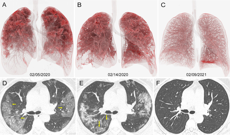
A 55-year-old woman presented on February 4, 2020 with chills, fever and weakness of the extremities, and was subsequently diagnosed with severe acute respiratory syndrome coronavirus 2 (SARS-CoV-2) using nucleic acid testing. Unenhanced chest CT showed diffuse groundglass opacities in the bilateral lungs, mainly distributed in the upper lungs (Figure, parts A, D). After antiviral (Ritonavir, AbbVie) and antibiotic (Moxifloxacin, Bayer) treatment, the CT scan at day 9 (Figure, part E) showed mild improvement. She was transferred to the local official isolation center for continued treatment for 30 days and was discharged. One year later, the patient reported no specific discomfort, and a repeated chest CT scan showed normal lung parenchyma (Figure, parts C, F).
Images in 55-year-old woman diagnosed with severe COVID-19 with oxygen saturation of 73% at presentation. A–C, Cinematic renderings with, D–F, original CT scans help show evolution of her infection. A, D, Admission CT scans show confluent ground-glass opacities in both lungs (arrows in D), primarily distributed in upper lobes. B, E, CT scans 9 days later show progressive disease in lower lobes (arrows in E). C, F, Follow-up CT scans 1 year later show resolution of all previously seen lung findings.
The long-term sequelae of severe SARS-CoV-2 pneumonia in the lung parenchyma are not known. It is possible that for some patients, irreversible lung damage occurs with scarring, as is seen after other causes of acute respiratory distress syndrome (1,2). However, as was found here, a return to normal lung density and morphologic condition may also be observed.
Footnotes
Disclosures of Conflicts of Interest: L.T. disclosed no relevant relationships. X.Z. disclosed no relevant relationships.
References
- 1. Han X, Fan Y, Alwalid O, et al. Six-month follow-up Chest CT Findings after Severe COVID-19 Pneumonia. Radiology 2021;299(1):E177–E186. [DOI] [PMC free article] [PubMed] [Google Scholar]
- 2. Wells AU, Devaraj A, Desai SR. Interstitial Lung Disease after COVID-19 Infection: A Catalog of Uncertainties. Radiology 2021;299(1):E216–E218. [DOI] [PMC free article] [PubMed] [Google Scholar]


