Abstract
Cathepsin D (cathD) is traditionally regarded as a lysosomal protease that degrades substrates in acidic compartments. Here we report cathD plays an unconventional role as a cofilin phosphatase orchestrating actin remodeling. In neutral pH environments, the cathD precursor directly dephosphorylates and activates the actin-severing protein cofilin independent of its proteolytic activity, whereas mature cathD degrades cofilin in acidic pH conditions. During development, cathD complements the canonical cofilin phosphatase slingshot and regulates the morphogenesis of actin-based structures. Moreover, suppression of cathD phosphatase activity leads to defective actin organization and cytokinesis failure. Our findings identify cathD as a dual-function molecule, whose functional switch is regulated by environmental pH and its maturation state, and reveal a novel regulatory role of cathD in actin-based cellular processes.
Subject terms: Actin, Cytokinesis
Introduction
Cathepsin D (cathD) is a ubiquitous lysosomal aspartyl protease widely expressed in eukaryotic cells. Depending on the acidic pH-sensitive protonation of two critical aspartic residues, cathD cleaves various substrates in lysosomal compartments, thereby regulating a variety of physiological and pathological processes.1 However, emerging evidence has revealed potential roles of cathD outside lysosomes2–4 in modulating cellular processes such as apoptosis, cell cycle and autophagy after translocation to the cytosol and other subcellular compartments beyond optimal pH range of its proteolytic activity.2,5–7 Moreover, proteolytically inactive cathD promotes cell proliferation, tumorigenesis and tumor invasion as potently as wild-type cathD.8–11 These findings suggest an unknown non-canonical role of cathD independent of its traditional degradation properties.
The protein level of cathD is closely associated with actin-based cellular processes, such as cell motility and proliferation. In most motile immune cells, cathD is constantly expressed at high levels.12,13 Increased cathD level enhances the proliferation and invasion of cancer cells, which is positively correlated with poor prognosis.14–16 We recently found that cathD interacted with the actin-severing factor cofilin and regulated actin-based fast cell migration after translocation to cytosol.17 Intriguingly, although cofilin is widely expressed, only two specific cofilin phosphatases, chronophin (CIN) and slingshot (ssh), have been identified so far. CIN only exists in vertebrates, while ssh is predominantly expressed in epithelial tissues and absent in several species.18 Therefore, other cofilin phosphatases likely exist to control cofilin activity.19 Based on these findings, we speculated that cathD might coordinate actin remodeling by direct regulation of cofilin activity in neutral pH environments.
In the present study, we provide evidence that cathD regulates actin-remodeling by direct dephosphorylation of cofilin independent of acidic pH or its proteolytic activity. Downregulation of cathD level or mutation in cathD disrupts the development of actin-based structures and impairs mitosis and cytokinesis. Our study identifies cathD as a dual-function molecule under different pH conditions, and provides mechanistic insights into cathD-mediated biological processes such as actin-based structure development, mitosis, and cytokinesis.
Results
CathD regulates organization of actin-based extensions in vivo
Actin cytoskeleton dynamics is tightly coordinated by various key factors during development.20 Recently, we identified the late endosome-derived cathD as a novel modulator of actin remodeling after its cytosolic translocation.17 CathD is evolutionarily conserved with a high homology from Drosophila to humans.21 To reveal whether cathD regulates actin organization in vivo, we examined actin-based sensory organs of Drosophila. Compared with wild-type controls (w1118), homozygous cathD-depleted flies (cathD1) exhibited developmental defects in actin-based structures, including extra scutellar bristles, macrochaete malformations, over-branched antenna, forked sensory hairs, and excess ocellar bristles (Fig. 1a–e). Visualized by fluorescent phalloidin staining, massive short protrusions or microvillar-like structures were observed at tips of pupal bristles in cathD1 mutant files, suggesting defects in actin patterning in the pupal stage (Fig. 1f).
Fig. 1. Loss of cathD leads to morphological defects in actin-based sensory organs.
a–e Representative scanning electron microscope (SEM) images reveal morphological defects of actin-based sensory organs (indicated by arrows) in cathD1 mutant flies, including: ectopic bristles on the scutellum (a), disrupted actin organization on the bristle surface (b), aberrant split ends of antenna (c), microchaete with split tips (d), and ectopic ocellar bristles (e). Homozygous cathD1 mutants (a–e) and hemizygous mutants carrying Df(2R)CA53 that deletes the entire cathD gene region on the second chromosome (e) were examined. f Representative images of notum bristles stained for F-actin (red) at 34 h after pupal formation (APF). cathD1 bristles exhibit disorganized microvillar-like short protrusions at the tips, whereas the wild-type bristles consist of organized parallel actin bundles. Lower panels, magnified views of dashed areas. g Scutellar bristles in adult flies were classified as follows based on severity: normal (gray, 4 bristles per scutellum), weak (orange, 5 bristles per scutellum), strong (green, 6 bristles per scutellum), and severe (dark red, > 6 bristles per scutellum). n = 120–423 flies per genotype from five independent experiments. cathD1 flies exhibit a significantly higher percentage of ectopic bristle number in both males and females, which could be restored by ubiquitous expression of wild-type human cathD (cathD1; da > cathDwt) or proteolytically inactive cathD (cathD1; da > cathDD231N). Scale bars, 50 μm for a, e, 10 μm for b, and 5 μm for c, d, f.
As dysregulated actin dynamics promote the formation of ectopic bristles,22–25 scutellar bristles of homozygous cathD1 mutants were further analyzed to evaluate the developmental defects in actin-based structures. Compared with the wild-type controls, loss of cathD increased the number of ectopic bristles by ~10 fold (Fig. 1g). Driven by the Daughterless (da) promoter, ubiquitous expression of wild-type human cathD (cathDwt) completely abrogated ectopic bristle formation in cathD-depleted flies, whereas expressing the proteolytically inactive human cathDD231N mutant also suppressed the formation of ectopic bristles (Fig. 1g), suggesting that cathD is necessary for the development of actin-based structures independent of its proteolytic activity.
CathD directly dephosphorylates cofilin
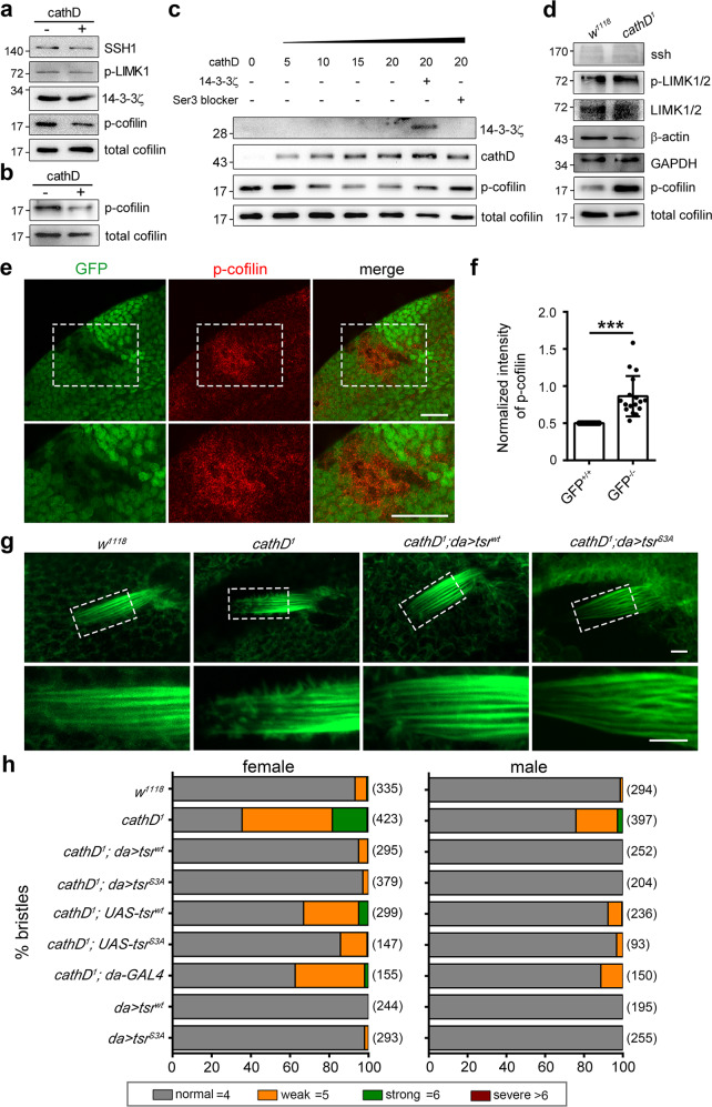
CathD shows a similar protein structure to the well-documented cofilin phosphatase CIN (Supplementary information, Fig. S1a), featuring a substrate-binding groove with paired aspartic residues responsible for nucleophilic attacks and group transfer reactions.1,18 This similarity drove us to ask whether cathD could directly activate cofilin as a CIN-like phosphatase in neutral pH environment. Therefore, we performed in vitro phosphatase assay using recombinant human cathD to interact with purified cofilin or whole-cell extracts. Our results showed that cathD directly decreased the phosphorylation level of cofilin in neutral pH buffer without affecting total cofilin or other regulatory elements (Fig. 2a, b), in a concentration-dependent manner (Fig. 2c). Furthermore, addition of recombinant scaffold protein 14-3-3ζ or exogenous Ser3-blocking peptide, which specifically binds with cofilin and protects the Ser3 residue from phosphatase attack,26 attenuated cathD-mediated decrease in phosphorylated cofilin (p-cofilin) levels (Fig. 2c). These results suggest that cathD exhibits cofilin phosphatase activities in neutral pH, under which cytosolic cathD likely executes its cofilin phosphatase function to mediate actin remodeling.
Fig. 2. CathD dephosphorylates cofilin both in vitro and in vivo.
a In vitro phosphatase assays showing that recombinant human cathD reduces phosphorylation levels of cofilin at Ser3 in whole-cell extracts from primary microglia. Levels of SSH1, 14-3-3ζ, p-LIMK, or total cofilin are comparable between groups (three independent experiments were performed). b In vitro phosphatase assays showing that recombinant human cathD exhibits phosphatase activity on purified human cofilin (1 h at 37 °C; five independent experiments were performed). c Recombinant cathD (5, 10, 15, and 20 nM) dephosphorylates purified cofilin (50 nM) in a dosage-dependent manner. Addition of recombinant 14-3-3ζ or Ser3-blocking peptide blocks cathD-mediated cofilin dephosphorylation (three independent experiments were performed). d Representative immunoblot images of total proteins extracted from wild-type (w1118) or cathD-depleted (cathD1) adult flies. p-cofilin levels are increased in cathD1 mutants. Three independent experiments were performed. e, f cathD1 mutant clones (GFP negative) were generated using FRT recombination in the 3rd instar larval wing discs. Representative images (e) and quantification (f) showing that p-cofilin levels are increased in cathD1mutant clones (GFP negative), compared with adjacent twinspot wild-type clones (GFP positive). n = 16 flies per group. Scale bar, 20 μm. g Representative images of actin-based fly bristles labeled with rhodamine-conjugated phalloidin. Dashed boxes are magnified in lower panels. Note that ubiquitous expression of either tsrwt or tsrS3A in the cathD1 mutant background (cathD1; da > tsrwt or cathD1; DA > tsrS3A) rescues actin bristle defects in cathD1 mutants at 34 h APF. Scale bar, 5 μm. h Scutellar bristles in adult flies were classified as follows based on severity: normal (gray, 4 bristles per scutellum), weak (orange, 5 bristles per scutellum), strong (green, 6 bristles per scutellum), and severe (dark red, > 6 bristles per scutellum). n = 93–423 flies per genotype from six independent experiments. Ubiquitous expression of wild-type tsrwt (cathD1; da > tsrwt) or constitutively active tsrS3A (cathD1; da > tsrS3A) restores the bristle duplication phenotype in cathD1 flies. Data are means ± SEM, unpaired student t-test. ***P < 0.001. See Supplementary information, Table S3 for detailed statistics.
To unravel the cofilin-regulating role of cathD in vivo, we next detected the phosphorylation level of the Drosophila cofilin homolog twinstar (tsr) in cathD-depleted flies. Interestingly, Drosophila cofilin phosphorylation level was upregulated in the absence of cathD, while the total amount of cofilin and its regulatory factors were not affected (Fig. 2d). Loss of cathD also dramatically enhanced the fluorescence intensity of phosphorylated cofilin in mutant clones of larval wing discs (Fig. 2e, f). Furthermore, ubiquitous expression of wild-type cofilin (tsrwt) or its constitutively active mutant (tsrS3A) in cathD-/- mutants reversed the malformation of microvillar-like protrusions in pupal bristles as well as the number of ectopic bristles in adults (Fig. 2g, h; Supplementary information, Fig. S1b), indicating that the loss of cathD induces actin-cytoskeleton malformations by suppressing the reactivation of endogenous cofilin. In addition, protein levels of total cofilin, its regulatory elements and F-actin were not significantly changed upon cathD-depletion (Supplementary information, Fig. S2a–h), implicating a selective regulatory role for cathD in processes requiring dynamic actin rearrangement such as bristle formation. These results suggest that cathD modulates actin arrangement pattern by dephosphorylating cofilin in vivo.
CathD exhibits ssh-like phosphatase activity in cofilin activation
Currently, only one cofilin phosphatase, ssh, has been identified in Drosophila so far. Similar to cathD1 mutants, ssh null alleles (ssh1–63) homozygous mutant flies exhibited developmental defects in actin-based structures. These included abnormally aggregated actin-based structures in pupal bristles, extra scutellar bristles, over-branched antenna, and malformations of bristle surface (Supplementary information, Fig. S3a–d), besides bifurcated hairs reported previously.27 Intriguingly, ubiquitous expression of cathDwt or proteolytically inactive cathDD231N in ssh1–63 mutants reversed the phenotype of ectopic bristles, whereas cathD1 and ssh1–63 trans-heterozygotes synergistically aggravated bristle defects compared to heterozygous cathD1 or ssh1–63 mutants (Supplementary information, Fig. S3e). These data indicate that cathD acts similarly to ssh in actin regulation.
Since double mutant of cathD and ssh (cathD1; ssh1–63) is lethal and all mutants die before eclosion, we next determined their actin phenotypes and the phosphorylation level of cofilin in the pupal stage. In our observation, loss of cathD increased malformation of microvillar-like protrusions on bristles and elevated p-cofilin level in ssh1–63pupae (Fig. 3a–d), which could be reversed by ubiquitous expression of cathDwt or proteolytically inactive cathDD231N (Fig. 3e, f). Consistent results were obtained when replacing ssh1–63 mutations with ssh RNAi, showing that elevated p-cofilin levels by ubiquitous expression of ssh RNAi was rescued by co-expression of either cathD or cathDD231N (Supplementary information, Fig. S3f, g). Conversely, reducing cathD expression by expressing cathD RNAi led to cofilin hyperphosphorylation, which was effectively suppressed by co-expression of wild-type ssh (sshwt). Co-expression of the catalytically inactive mutant sshcs also lowered p-cofilin levels but was less efficient (Fig. 3g, h). Taken together, cathD exerts ssh-like roles and serves as a novel cofilin phosphatase to regulate the development of actin-based structures.
Fig. 3. CathD exhibits slingshot-like phosphatase activity in cofilin activation.
Representative staining images (a) and quantification (b) of F-actin-labeled pupal bristles. Note that ubiquitous expression of either cathDwt or cathDD231N in the ssh1–63 mutant background (ssh1–63; da > cathDwt or ssh1–63; da > cathDD231N) reverses actin bristle defects in ssh1–63mutants, whereas double mutant of cathD and ssh (cathD1; ssh1–63) slightly aggravates the actin defects at 34 h APF. Dashed boxes are magnified in lower panels. Scale bar, 5 μm. Analysis of actin-based bristle structures in adult flies carrying different genotypes. Representative immunoblot images (c, e) and quantifications (d, f) of p-cofilin/cofilin ratio (normalized to wild-type controls, w1118) of total protein extracted from ssh1–63 mutant flies, showing elevated p-cofilin levels in double mutant of cathD and ssh (cathD1; ssh1–63), compared with cathD1 or ssh1–63(c, d). Expression of wild-type cathD (ssh1–63; da > cathDwt) or its proteolytically inactive mutant cathDD231N (ssh1–63; da > cathDD231N) restores cofilin hyperphosphorylation in ssh1–63 (e, f). Representative immunoblot images (g) and quantifications (h) of total protein extracted from da > cathDRNAi adult flies, showing that reducing cathD expression by cathD RNAi (da > cathDRNAi) increases p-cofilin levels. Co-expression of wild-type ssh effectively restores p-cofilin levels (da > cathDRNAi, sshwt flies), whereas ssh inactive mutation shows no effect (da > cathDRNAi, sshCS). Data are means ± SEM. One-way ANOVA with Tukey’s post hoc test, *P < 0.05, **P < 0.01, ***P < 0.001.
The cofilin phosphatase activity of cathD relies on pH conditions and its maturation state
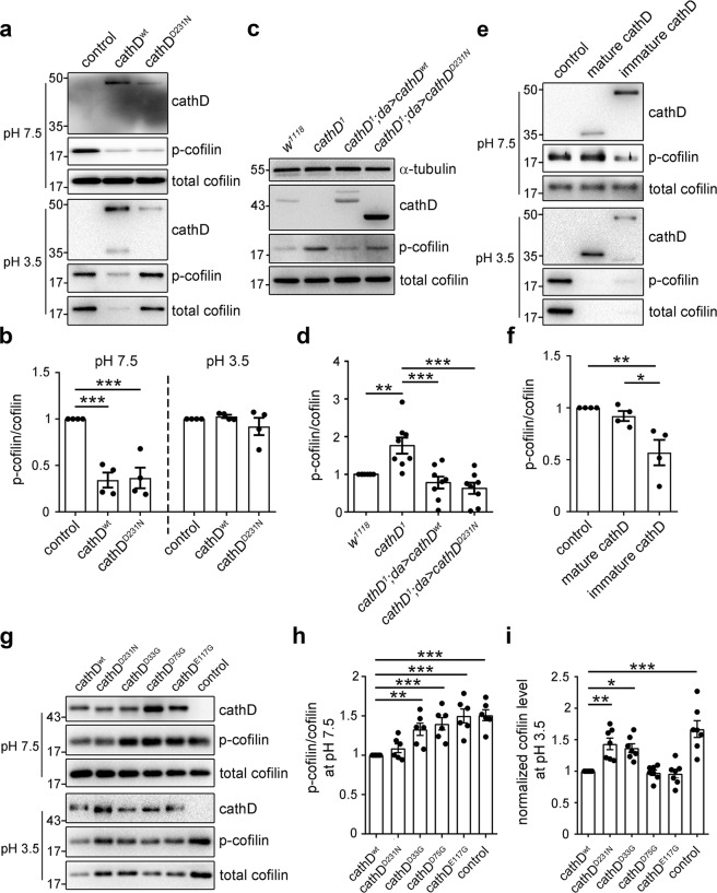
To evaluate the catalytic property of cathD phosphatase activity, we examined the effects of general phosphatase inhibitors on the phosphatase activity of purified cathD. Similar to canonical cofilin phosphatases,18,27 cathD is insensitive to classical serine/threonine phosphatase inhibitors (Supplementary information, Fig. S4a, b). In a neutral pH environment (pH 7.5), both purified cathDwt and its proteolytically inactive mutant cathDD231N decreased p-cofilin levels without altering total cofilin protein level (Fig. 4a, b). These results indicate that the cofilin phosphatase activity of cathD in neutral pH is independent of its proteolytic activity. In comparison, in an acidic condition (pH 3.5), cathDwt mainly played a hydrolytic role in cofilin degradation as shown by decreased total cofilin protein levels, whereas cathDD231N showed impaired activity in cofilin degradation (Fig. 4a, b). Similarly, overexpression of cathDwt or cathDD231N restored the elevated p-cofilin levels in cathD1 mutants, suggesting that both forms exhibit cofilin phosphatase activities in vivo (Fig. 4c, d).
Fig. 4. Functional switch of cathD activities relies on pH and its maturation.
Representative immunoblot images (a) and quantification (b) of cathD-mediated cofilin phosphorylation in different pH conditions, showing both recombinant cathDwt and cathDD231N (10 nM) dephosphorylate cofilin in neutral pH (7.5), whereas cathDwt mainly degrades cofilin in acidic pH (3.5). Note that both cathDwt or cathDD231N significantly reduce p-cofilin/cofilin ratio in neutral but not acidic pH (c). Representative immunoblot images (c) and quantification (d) of cathD-mediated cofilin phosphorylation in flies. c Elevated p-cofilin levels in cathD1 mutants are restored upon expressing either wild-type (cathD1; da > cathDwt) or proteolytically inactive cathD (cathD1; da > cathDD231N). d Quantification of p-cofilin/total cofilin levels (normalized to w1118 controls). The ratio is reduced when cathDwt or cathDD231N is expressed in cathD1 mutant background. Representative immunoblot images (e) and quantification (f) of in vitro phosphatase assay showing that immature recombinant human cathD directly dephosphorylates cofilin at pH 7.5, whereas mature cathD degrades cofilin at pH 3.5 (e). f Quantifications of p-cofilin/cofilin ratio (normalized to control) show that immature cathD dephosphorylates cofilin, whereas mature cathD shows limited phosphatase activity in neutral pH (7.5). Representative immunoblot images (g), quantification of p-cofilin/cofilin ratio (normalized to cathDwt, h) and cofilin level (normalized to cathDwt, i) from in vitro phosphatase assays using recombinant cathDwt, cathDD231N, cathDD33G, cathDD75G, and cathDE117. Note that cathDD33G, cathDD75G and cathDE117G exhibit impaired activities in cofilin dephosphorylation at pH 7.5, whereas cathDD33G and cathDD231N present suppressed proteolytic activities for cofilin degradation at pH 3.5. Data are means ± SEM, *P < 0.05, **P < 0.01, ***P < 0.001 by Mann–Whitney U test or One-way ANOVA with Tukey’s post hoc test.
Since immature cathD is unstable and tends to yield the mature form by proteolysis in acidic pH conditions,1 the effects of immature and mature cathD on cofilin dephosphorylation in neutral and acidic pH were determined, respectively. We found that immature cathD directly dephosphorylated cofilin in neutral pH (Fig. 4e, f), whereas mature cathD mainly degraded cofilin in the acidic environment (Fig. 4e). These data suggest a dual role for cathD as both a ubiquitous protease and a cofilin phosphatase in different intracellular compartments, depending on environmental pH and its maturation state.
Asp33, Asp75 and Glu117 residues are essential for cathD phosphatase activity
To elucidate the catalytic mechanism of cathD, candidate amino acid residues responsible for the phosphatase activity of cathD were screened by site-directed mutagenesis. Since charged acidic residues may activate nucleophilic attack for phosphate group transfer in catalysis,28 we speculated that aspartate (Asp) and glutamate (Glu) residues in the substrate-binding cleft might be involved. Therefore, nucleotide substitutions were made in amino acid positions 5, 12, 33, 50, 75, 117, 132, or 310 of human cathD, changing the residue Asp/Glu to Gly. Plasmids expressing cathD with different mutations were fused with a 6× His tag at the C-terminus and transfected to establish stable cell lines. Compared with the wild-type protein, cathDD33G, cathDD75G or cathDE117G mutants showed suppressed activities on cofilin dephosphorylation. In comparison, cathDwt, cathD231N and other mutants markedly reduced the p-cofilin level (Supplementary information, Fig. S4c, d).
To verify the roles of these residues in cofilin regulation, we purified cathD mutant proteins without altering their stabilities, secondary structures, or binding abilities with cofilin (Supplementary information, Fig. S4e, f), and applied these proteins to in vitro phosphatase/protease assays. In our observation, purified cathDD33G, cathDD75G and cathDE117G exhibited impaired cofilin-phosphatase activities in a neutral pH environment (pH 7.5), whereas cathDD33G and cathDD231N showed suppressed proteolytic activities in an acidic condition (pH 3.5, Fig. 4g–i). We next reintroduced different cathD mutants into a cathD-/- HeLa cell line, and examined the phosphorylation level of cofilin in these cells. We found that expression of cathDwt or proteolytically inactive cathDD231N dramatically reduced the p-cofilin level in cathD-/- cells, whereas expression of phosphatase inactive cathDD33G, cathDD75G or cathDE117G mutants showed no effect (Supplementary information, Fig. S4g, h). These results suggest that Asp33, Asp75 and Glu117 are key residues for the phosphatase activity of cathD in a neutral pH environment, and Asp33 is essential for both phosphatase and proteolytic activities of cathD in both neutral and acidic pH environments.
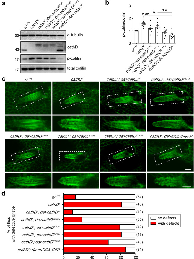
We further generated transgenic Drosophila lines ubiquitously expressing cathDD33G, cathDD75G or cathDE117G to examine the regulatory effect of these residues on cofilin activation. Compared with the wild-type cathD, mutation of Asp33, Asp75 or Glu117 suppressed dephosphorylation activity of cathD and attenuated cofilin-activation (Fig. 5a, b). Notably, cathD-mediated actin-cytoskeleton arrangement of Drosophila bristles and the percentage of flies with disordered F-actin bundles remained defective upon expressing cathD carrying any of these three mutations (Fig. 5c, d). In addition, expression of cathDwt or cathDD231N restored the elevated p-cofilin levels in LIMK1 overexpression larvae (Supplementary information, Fig. S5a, b), and reduced the lethality caused by LIMK1 overexpression, whereas cathDD33G, cathDD75G, or cathDE117G had no effect (Supplementary information, Fig. S5c). Together, these results suggest that Asp33, Asp75, and Glu117 are essential residues for the phosphatase activity of cathD.
Fig. 5. Asp33, Asp75, and Glu117 are required for the phosphatase activity of cathD.
a, b Representative Western blots indicating p-cofilin levels in extracted proteins from adult flies. Expression of wild-type cathD fully rescues the elevated p-cofilin levels caused by cathD depletion (cathD1; da > cathDwt), whereas expression of mutant cathD carrying Asp33 (cathD1; da > cathDD33G), Asp75 (cathD1; da > cathDD75G), or Glu75 (cathD1; da > cathDE117G) in cathD1 background does not restore the elevated p-cofilin levels, suggesting that mutating any of these three residues abolishes cathD phosphatase activity. Quantifications of p-cofilin/cofilin ratio (normalized to w1118) are shown in b. Note that p-cofilin levels remain elevated upon expressing cathD mutants in cathD1 background (cathD1; da > cathDD33G, cathD1; da > cathDD75G and cathD1; da > cathDE117G). c Actin-based bristle structures in pupae carrying different genotypes. Ubiquitous expression of the wild-type (cathD1; da > cathDwt) or a proteolytically inactive mutant of cathD (cathD1; da > cathDD231N) restores the over-protruded bristle phenotype upon cathD depletion (cathD1) at 34 h APF. No significant effects were detected upon expressing cathDD33G (cathD1; da > cathDD33G), cathDD75G (cathD1; da > cathDD75G) or cathDE117G (cathD1; da > cathDE117G) and bristles remain defective. Bristles were labeled by phalloidin. Dash-boxed regions are enlarged in lower panels. d The ratio of individual flies with disorganized actin bundles in bristles, showing that ubiquitous expression of cathDwt or a proteolytic inactive cathDD231N reduces the defective ratio, whereas expression of cathDD33G, cathDD75G, or cathDE117G exhibits minimal effect. Scale bar, 5 mm. Data are means ± SEM. One-way ANOVA with Tukey’s post hoc test, *P < 0.05, **P < 0.01, ***P < 0.001.
CathD modulates mitosis and cytokinesis via its cofilin phosphatase activity
Cofilin is a pivotal modulator of cell mitosis.18 Perturbations of cofilin activation induce aberrant cytokinesis,18,20 and may lead to tumorigenesis with the accumulation of aneuploid cells.29 Notably, cathD has been shown to promote tumorigenesis and cancer growth independent of its proteolytic activity.9,14,30 Considering that immature cathD is its main form in cytosol (Supplementary information, Fig. S6a), whereas disrupting the expression level or phosphatase activity of cathD caused actin aggregation (Supplementary information, Fig. S6b, c), we speculated that cathD might modulate cell division via its phosphatase activity.
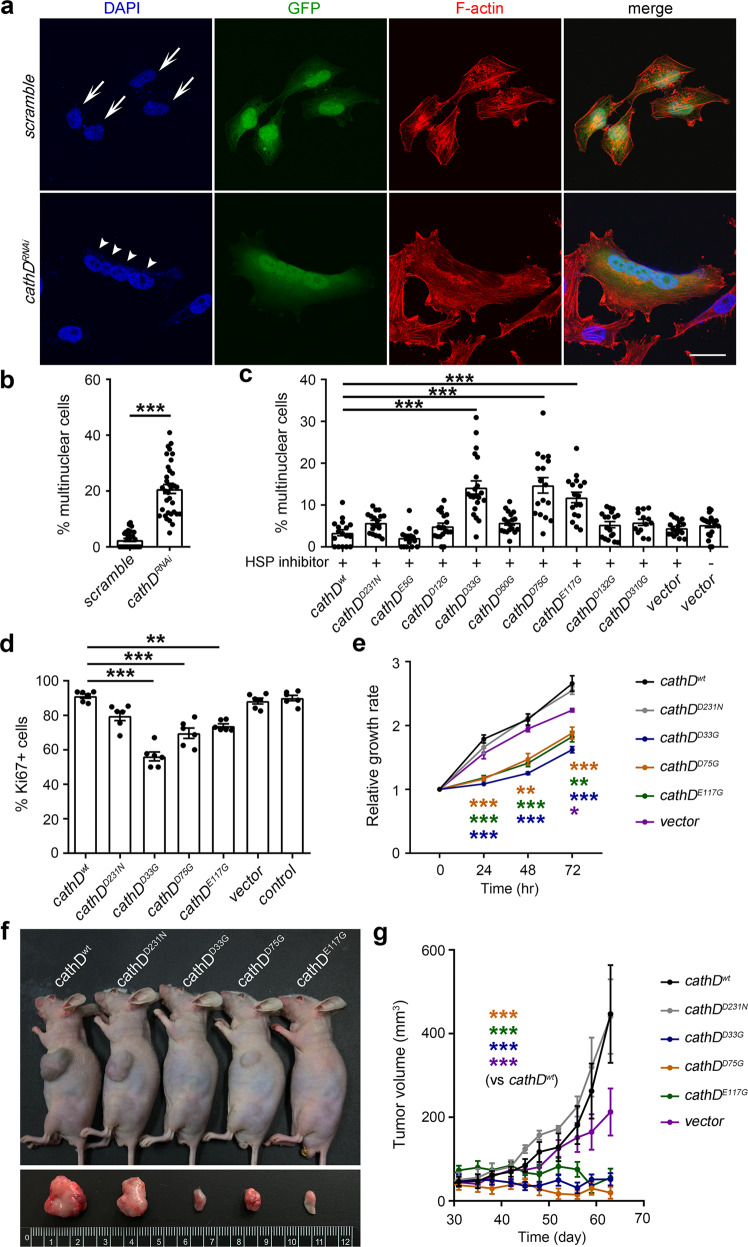
To verify our assumption, HeLa cells were transfected with plasmids expressing short hairpin RNA (shRNA) that targets cathD. Compared with the scrambled control, transfection of cathD-specific shRNA led to cytokinesis failure and the generation tetraploidy cells, indicating a dysfunction in cell mitosis and cytokinesis upon cathD depletion (Fig. 6a, b). We next generated stable cell lines expressing phosphatase-inactive mutants of cathD using the lentiviral system. Compared with controls, cathDD33G, cathDD75G and cathDE117G mutants significantly elevated the percentage of multinucleate cells with a high rate of aneuploidy (Fig. 6c; Supplementary information, Fig. S6d), suggesting that the regulatory role of cathD in cytokinesis depends on these residues.
Fig. 6. CathD regulates cell mitosis and cytokinesis via its cofilin phosphatase activity.
a, b CathD-specific (cathDRNAi) or scrambled shRNA (scramble) was co-expressed with GFP in asynchronous HeLa cells. After cultured in 15 μM Hsp inhibitor I or vehicle for 48 h, cells were fixed and stained for F-actin with rhodamine-conjugated phalloidin (red). Nuclei were counter-stained with DAPI. (a) Representative results showing that downregulation of cathD by transfection of cathD-targeting shRNA generates giant multinuclear cells. Arrows indicate nuclei in control cells, while arrowheads indicate nuclei in a tetraploidy cell induced by cathD shRNA. Scale bar, 30 μm. (b) Quantifications of multinuclear HeLa cells. Compared with controls (scramble), downregulation of cathD (cathDRNAi) increases giant multinuclear cells. c A significant increase of giant multinuclear cells was found in cathDD33G, cathDD75G, and cathDE117G mutants. Scale bar, 30 μm. d Quantifications of Ki67-positive A549 cells with indicated cathD mutants. Data are means ± SEM. One-way ANOVA followed with the Tukey’s test, **P < 0.01 and ***P < 0.001 compared with cathDwt (n = 6). e CCK-8 assay of A549 cells at indicated time points. Relative proliferation ratios were normalized to the values at 0 h. f Representative images showing A549 xenografts carrying indicated cathD mutations in nude mice after subcutaneous transplantation. g The change in tumor volume of A549 xenografts (relative to the initial volume) carrying indicated cathD mutations (n = 9). Data are means ± SEM, *P < 0.05, **P < 0.01, and ***P < 0.001 the by Mann–Whitney U test, Kruskal–Wallis test (with Dunn’s test), or ANOVA (with Tukey’s test), respectively.
Considering that cytokinesis defects lead to proliferative arrest,29 we further examined whether cathD could regulate cancer cell proliferation via its phosphatase activity. We chose lung cancer A549 cells that express low endogenous levels of cathD, and stably transfected these cells with either cathD or its mutants. Assessed by Ki67 staining and the CCK-8 proliferation assay, we found that overexpression of phosphatase-inactive cathDD33G, cathDD75G and cathDE117G mutants, rather than cathDwt or proteolytically inactive cathDD231N, dramatically impaired proliferative activities of A549 cells (Fig. 6d, e), suggesting that the cathD mitogenicity relies on its phosphatase activity. Similar results were obtained in HeLa cells carrying phosphatase-inactive cathD mutants (Supplementary information, Fig. S6e–h). Furthermore, a remarkable suppression of tumor growth was observed in cathDD33G, cathDD75G and cathDE117G xenografts (Fig. 6f, g). Taken together, cathD requires Asp33, Asp75, and Glu117 residues to regulate cofilin activity and modulate cofilin-mediated processes such as cytokinesis and mitosis.
Discussion
Emerging evidence suggests non-proteolytic functions of cathD outside acidic compartments. In the present study, we demonstrate that cathD shows a non-canonical cofilin phosphatase activity in neutral pH and regulates cofilin-mediated actin dynamics, cell mitosis and cytokinesis. Our findings identify cathD as a dual function molecule with both proteolytic and phosphatase activities, which can be switched under different pH conditions and conformational states during maturation (Fig. 7).
Fig. 7. Schematic diagram illustrating the dual role of cathD.
In acidic organelles, mature cathD hydrolyzes protein and peptide substrates as a canonical protease (left). In neutral pH environments, immature cathD precursor acts as a noncanonical phosphatase to dephosphorylate p-cofilin and modulate cofilin-mediated processes such as actin remodeling and cell mitosis (right).
Based on our recent findings that cathD balances cofilin activity in cell migration after translocation to cytosol,17 we reasoned that cytosolic cathD might non-proteolytically regulate cofilin activity. In addition to the binding interaction between cathD and cofilin, we found that recombinant cathD could directly dephosphorylate cofilin at its Ser3 residue in neutral pH in vitro. In vivo, cathD-deficient flies exhibit cofilin hyperphosphorylation, accompanied by developmental defects in actin-based structures. Similar defects have been previously reported in the absence of cofilin phosphatases,19,27 or in the presence of JAS.31 Notably, defects in actin-based structures are rescued when reintroducing cathD or the Drosophila cofilin homolog tsr into cathD1 mutants. Together with our results that cathD exhibits ssh-like phosphatase activity in cofilin activation, these observations highlight that cathD functions similarly to ssh in tuning cofilin-mediated actin remodeling.
Dual-role enzymes generally function through similar catalytic mechanisms.32–34 Strikingly, proteolytically inactive mutant cathDD231N also dephosphorylate cofilin in neutral pH, suggesting that the phosphatase property of cathD is independent on its proteolytic activity. As potent as the wild-type cathD, proteolytic inactive cathDD231N abolishes cofilin hyperphosphorylation and rescues actin-based malformations induced by cathD depletion. In addition, proteolytically inactive cathDD231N complements ssh action similarly to the wild-type cathD. Therefore, cathD exerts phosphatase and proteolytic activities through different catalytic mechanisms, and these two activities are mutually exclusive.
Enzyme precursors have been thought to be catalytically inactive. Our studies expand this notion by showing that protease precursors, as exemplified by immature cathD, may also possess catalytic activities. The proteolytically inactive immature cathD requires acidic pH and hydrolysis to change conformation and convert to the proteolytically active form.1,35,36 In various cell types, the majority of cathD and other cathepsins exist in zymogen forms,37–39 while their interaction partners are diversely distributed among different cellular compartments outside the lysosomes.40 Our findings uncover that immature cathD and possibly other immature cathepsins may exert broader functions beyond the regulation of cytoskeleton assembly and mitosis. In addition, as cofilin is diversely distributed among different cellular compartments outside the lysosomes, other potential functions of cathD merit further exploration.
In catalytic processes, polar residuals in substrate-binding groove are required for nucleophilic attacks and group transfer reactions.18,41 By site-directed mutagenesis, we identified three critical residues (Asp33, Asp75, and Glu117) mediating the cofilin phosphatase activity of cathD. CathD carrying mutation on any of these three residues does not efficiently dephosphorylate cofilin, resulting in elevated cofilin phosphorylation level. Notably, among these residues, Asp33 is also critical for the proteolytic activity of cathD. As the ionization of Asp33 does not require acidic pH environments,42 Asp33 likely activates water molecules for the subsequent substrate attack (e.g. removal of a phosphate group) in neutral pH environments. Moreover, our experiments show that cathDD33G, cathDD75G, and cathDE117G variants exhibit impaired phosphatase activities and induce the generation of giant multinucleated cells with a high rate of aneuploidy. In contrast to wild-type cathD and its proteolytically inactive cathDD231N, overexpression of phosphatase-inactive cathDD33G, cathDD75G, and cathDE117G suppresses A549 cell mitosis and proliferation. Together with previous reports,9,14 our study further provides a mechanistic insight into the mitogenicity of cathD, which relies on its cofilin phosphatase activity but not canonical proteolytic activity, in promoting tumor cell proliferation in related cancers.
Material and methods
General reagents and antibodies
Antibodies and dyes for immunofluorescence and Western blotting are listed in Supplementary information, Table S1. All other reagents were from Sigma-Aldrich and Roche. Cells were obtained from American Type Culture Collection (ATCC) and Takara Bio Inc. (Takara Bio).
Drosophila strains and genetics
All flies were reared on standard corn meal medium with 50% humidity and 12 h light/12 h dark cycles at 25 °C. UAS-cathDwt and UAS-cathDD231N were generated by Prof. Margaret S. Ho. w1118, cathD1 (gift from Dr. Mel Feany), ssh1–63 mutant flies were kindly gifts from Prof. Tadashi Uemura. FRT (42B) ubi-GFP and hs-flp; FRT (42B) were obtained from Prof. Zhao-Hui Wang. UAS-sshRNAi (107998) was supplied by the Vienna Drosophila Resource Center (VDRC). UAS-sshwt (BL9113), UAS-sshcs (BL9115), UAS-tsrwt (BL9234), UAS-tsrS3A (BL9236), UAS-cathDRNAi, and da-Gal4 (Bloomington Drosophila Stock Center) were provided by Bloomington Drosophila Stock Center. UAS-cathDwt, UAS-cathDD231N, UAS-cathDD33G, UAS-cathDD75G, UAS-cathDE117G were generated by microinjection service from the Core Facility of Drosophila Resource and Technology, Shanghai Institute of Biochemistry and Cell Biology, Chinese Academy of Sciences.
Cell culture and transfection
HEK293T, HeLa (ATCC), A549 (ATCC), and Lenti-X 293T (Takara Bio) cell lines were cultured in 10% fetal bovine serum (FBS, Thermo Fisher Scientific) in Dulbecco’s Modified Eagle’s Medium (DMEM, Thermo Fisher Scientific) with high glucose (4.5 g/L). Transient transfections were performed using TurboFect Transfection Reagent (Thermo Fisher Scientific). For Stable transfections, lentivirus was transduced into HEK293 or HeLa cell lines and selected with puromycin (Takara Bio).
Molecular cloning and plasmid constructions
Full-length human cathD and cathD carrying its proteolytically inactive mutation on Asp231 were cloned from corresponding pcDNA3.1 vectors provided by Prof. Judith Haendeler. To create cathD mutations, Asp and Glu residues in the substrate-binding cleft were altered to Gly by site-directed mutagenesis. Mutagenic primers were designed to target Glu5, Asp12, Asp33, Asp50, Asp75, Glu117, Asp132, and Asp310 residues in Supplementary information, Table S2.
Digested by restriction enzymes XhoI and EcoRI (Thermo Fisher), PCR fragments were inserted into the pcDNA 3.1/myc-His vector for transient transfection or a pLVX-Puro vector for lentiviral expression. For CRISPR/Cas9-based gene editing, sgRNA-encoding oligonucleotides were designed, synthesized and cloned into LentiCRISPRv2-GFP lentiviral vector to express Cas9, GFP, and sgRNA-targeting cathD.
Lentivirus production and stable cell line establishment
pLVX-Puro-cathDwt, LentiCRISPRv2-cathD sgRNA-GFP, or other cathD mutated constructs were transfected along with the packaging plasmid, psPax and pMD2.G envelop plasmid, into Lenti-X 293T cells at a ratio of 4:3:1. After transfection, Lenti-X 293 T cells were cultured in DMEM with high glucose (4.5 g/L) supplemented with 20% FBS for 36 h. Viral supernatants were then collected by centrifugation at 2000× g for 5 min and filtered by 0.45 μm low-protein binding filters (Merck). Harvested viral medium were added with 10 μM polybrene (Sigma-Aldrich) to improve transduction efficiency, diluted with DMEM plus 10% FBS (1:1 in volume) and added to HeLa or HEK293T cells.
After 36–48 h of lentiviral transduction, GFP-positive cells were sorted by a MoFlo Astrios EQ cell sorter (Beckman, Brea, CA) to establish stable cathD-deficient (cathD-/-) cell line. To further reintroduce cathDwt and other cathD mutants, cathD-contained lentiviral particles were added into cathD-/- HeLa cells. Transduction medium was then replaced with fresh culture medium supplemented with puromycin (Takara Bio) to a final concentration of 0.5–3 μg/mL after 48 h of lentiviral transductions to establish stable cathD-mutated cell lines.
Recombinant protein purification
Purified from the E. coli-based expression system, recombinant cathD is prone to aggregate into insoluble inclusion bodies. Therefore, we applied a mammalian expression system to produce recombinant proteins from transient or stably transfected HEK293T cells. Cells were collected and disrupted in 5 mM imdazole, 500 mM NaCl, and 20 mM Tris-HCl (pH 7.9) by sonication. His-tagged cofilin, cathD and its mutated proteins were subsequently purified under native conditions by standard affinity chromatography procedures using a purification resin (Roche Applied Science). Eluted proteins were desalted and concentrated twice by sequential centrifugation using 100-kDa and 50-kDa ultra centrifugal filters (Merck). The molecular weights of purified proteins were checked by silver staining following SDS-PAGE separation for quality control. The protein stability and the secondary structure of mutated cathD proteins were evaluated by a circular dichroism spectroscopy. Purified cathD was mainly in the 48-kDa immature form and stored at −80 °C in 150 mM NaCl, pH 7.4, 0.05% sodium azide and 25% glycerol.
Phosphatase and protease assays
Purified cathD or its mutations were mixed with cofilin or other candidate proteins in HEPES 50 mM, MgCl2 10 mM, MnCl2 2 mM, 0.2 mM DTT, and 0.1 g/mL BSA at pH 7.5 or pH 3.5, respectively for in vitro phosphatase and protease assays. Mixtures were incubated in 20 μL at 37 °C for 0.5–2 h. To evaluate the enzymatic property of cathD, classical phosphatase inhibitors okadaic acid (1 μM), calyculin A (10 μM), fenvalerate (100 nM), α-naphthyl acid phosphate (10 mM), PhosStop inhibitor cocktail (1:1000), or control vehicles were applied. Reactions were stopped by boiling in SDS-PAGE sample loading buffer (50 mM Tris-HCl, pH 6.8, 2% SDS, 0.1% bromophenol blue, 10% glycerol, 5% β-mercapto-ethanol) for 10 min.
Pull-down assay
Pull-down experiments were conducted by adding 10 μg purified his-tagged recombinant cathDwt or other cathD mutant proteins into 1 mL whole cell lysate from HEK293 cells (1 × 10⁷ cells). The mixture was incubated with His-Tag purification resin (Sigma) in a binding buffer (20 mM sodium phosphate, 300 mM sodium chloride, 10 mM imidazole, pH 7.4) at 4 °C for 6 h. After washing for three times with a wash buffer (20 mM sodium phosphate, 300 mM sodium chloride, 15 mM imidazole, pH 7.4), the binding partner of cathD and its mutant proteins were resolved with 200 μL elution buffer (20 mM sodium phosphate, 300 mM sodium chloride, 150 mM imidazole, pH 7.4) and examined by Western blotting.
Cell fractionation and cytosol isolation
The cytosolic fractions were isolated according to previously described protocols.43 Briefly, HeLa cells were collected in homogenization buffer (220 mM D-mannitol, 70 mM D-sucrose, 5 mM HEPES-KOH, 1 mM EGTA-KOH, pH 7.4) and homogenized 6 times before centrifuged at 2000× g for 10 min. The collected supernatant (S1) was centrifuged at 17,000× g for 12 min to remove major organelles (P1). The collected supernatant (S2) was then centrifuged at 100,000× g for 60 min to remove endoplasmic reticulum, Golgi and other cytosolic vesicles (P2). The resulting supernatant (S3, cytosolic fractions) was collected and further analyzed with Western blotting.
Western blot
Protein samples from adult flies were obtained through homogenization of 4 adults per sample using cold RIPA buffer (50 mM Tris, pH 7.4, 150 mM NaCl, 1% Triton X-100, 1% sodium deoxycholate, 0.1% SDS) containing Complete Protease Inhibitor Cocktail and PhosStop inhibitor cocktail (Roche Applied Science).44 Heat-shock protein 70 (Hsp70), a stress-inducible chaperone, stabilizes endosome/lysosomes by inhibiting the membrane permeability.45 To detect endogenous proteins in cathD and its mutations, stable transfected cell lines were pretreated with 5 μM HSP inhibitor I, a potent inhibitor for hsp70 which could sensitize endosomal/lysosomal membrane and enhance potential cytosolic translocation, for 24 h. Cell lysates were prepared by brief sonication in RIPA buffer on ice, followed by centrifugation at 15,000× rpm for 5 min. The concentration of samples was determined by bicinchoninic acid (BCA) assay and equal amounts of proteins were loaded onto 12% SDS-PAGE gels, followed by transferring for Western blotting. Blots were probed overnight at 4 °C with indicated antibodies in Supplementary information, Table S1.
Generation of MARCM clones and immunohistochemistry
To generate and stain MARCM clones, the cathD-deleted mutation was recombined onto the FRT (42B) chromosome for MARCM analysis by heat shock twice at 37 °C for 60 min at embryonic day 2. Heat-shocked 3rd instar larval wing discs were dissected in phosphate-buffered saline (PBS) and fixed with 4% paraformaldehyde at room temperature for 20 min. Following permeabilization with 0.3% Triton X-100 for 20 min, tissue was blocked with 4% BSA containing 0.1% Triton X-100 in PBS for 1 h at room temperature and were incubated with primary antibodies (Supplementary information, Table S1) overnight at 4 °C. Alexa Fluor 647-conjugated donkey anti-rabbit secondary antibody was applied (1:1000, Thermo Fisher Scientific).
For F-actin staining, pupal notum from cathD mutants and other genotypes at about 34 h APF were dissected, fixed with 4% paraformaldehyde, and labeled with phalloidin-conjugated with Alexa Fluor 555 or rhodamine (Thermo Fisher Scientific) for 2 h at room temperature.
Cell synchronization and immunocytochemistry
For synchronization, HeLa and A549 cells were cultured with 2 mM thymidine (Sigma) for 24 h, released for 6 h in fresh complete medium, and treated with 100 ng/mL nocodazole (Sigma) for 6 h. Mitotic cells were collected by shaking off gently, washed with HBSS twice, placed on coverslips in complete medium supplied with 15 μM HSP inhibitor I (Merck), and cultured for 48 h. Following fixation and permeabilization, cells were blocked with 10% BSA and further incubated with Ki67 antibody (1:1000, Abcam) or rhodamine-conjugated phalloidin (1:200, Thermo Fisher Scientific) for 2 h at room temperature. DAPI was applied for nucleus counterstaining. For Ki67 staining, cells were considered as positive when co-stained with DAPI in nucleoli. Images were captured by a confocal microscope (Olympus, FV1200).
Scanning electron microscopy
Actin-based structures on the adult flies with cathD1 or other mutations were determined by scanning electron microscope (Hitachi, TM-1000). Live flies were anaesthetized with 100% ether and affixed upon stubs. After completely dehydration and air dry, samples were sputter coated with tungsten-platinum by an ion sputter coater (Hitachi, MC1000) and imaged.
CCK-8 cell proliferation assay
The CCK-8 assay was applied to assess the proliferative activity of A549 cells using a CCK-8 kit (Sigma-Aldrich). Briefly, A549 cells (4 × 10³ cells/well) carrying different cathD mutations were seeded in a 96-well plate. After 0, 24, 48, or 72 h, CCK-8 solution (10 μL in 100 μL HBSS) was added and incubated for 2 h at 37 °C. Subsequently, the absorbance of the formazan at 450 nm was measured using a microplate reader (Bio-Rad, iMark) to evaluate A549 cell proliferation.
Xenograft study
Male athymic nude mice (nu/nu, 4 weeks old, obtained from Shanghai Laboratory Animal Center, Chinese Academy of Sciences) were raised under specific pathogen-free conditions and used in tumor xenograft studies. For xenograft transplantation, A549 cells overexpressing cathDwt or mutations on cathD Asp231, Asp33, Asp75G, and Glu117 residues were subcutaneously injected at the left abdomen flank with a density of 1 × 10⁷ cells/mL in 200 μL of Matrigel (Corning, 1:1 volume Matrigel/A549 cells). After injection, animals were individually housed and monitored for 95 days before sacrifice in accordance with the institutional humanitarian regulations.
Statistical analysis
Statistical analysis was performed using unpaired student t-test, Mann–Whitney U test, Kruskal–Wallis test, and one-way ANOVA followed by Tukey’s or Fisher’s LSD post hoc test when appropriate (Supplementary information, Table S3). Data were presented as means ± SEM. P < 0.05 was considered to be statistically significant.
Supplementary information
Acknowledgements
We thank Prof. Yong-Qing Zhang, Prof. Li Yu, and Prof. Wei Feng for constructive discussions. We are grateful to Prof. Chun Tang for technical support on protein structure modeling. We thank Prof. Tadashi Uemura, Prof. Zhao-Hui Wang, and Dr. Mel Feany for providing Drosophila lines; and Prof. Judith Haendeler for providing plasmid constructs. We thank the Core Facilities of Zhejiang University School of Medicine for technical assistance. This work was supported by National Key Research and Development Program of China (2016YFA0501000), National Natural Science Foundation of China (81821091, 31501128, 31490592, 31871039, 81971260, 81761138044, 81527901, 81801330, and 91832000), Key Research and Development Program of Zhejiang Province (2020C03009), Science and Technology Planning Project of Guangdong Province (2018B030331001), Key Realm R&D Program of Guangdong Province (2019B030335001), and Chinese Ministry of Education Project 111 Program (B13026).
Author contributions
Y.J.L. and T.Z. conducted experiments with assistance from S.C., D.C., C.W., X.W., D.D., L.Z., and H.L. helped with animal experiments. Y.J.L. and T.Z. analyzed data. M.S.H., Z.G. Y.J.L., and X.D.W. discussed the manuscript and contributed to experimental design. Y.J.L., X.D.W., M.S.H., and S.D. designed the study and wrote the manuscript.
Competing interests
The authors declare no competing interests.
Footnotes
These authors contributed equally: Yi-Jun Liu, Ting Zhang
Contributor Information
Yi-Jun Liu, Email: yjliu@zju.edu.cn.
Xiao-Dong Wang, Email: xiaodongwang@zju.edu.cn.
Margaret S. Ho, Email: margareth@shanghaitech.edu.cn
Supplementary information
Supplementary information accompanies this paper at 10.1038/s41422-020-00454-w.
References
- 1.Benes P, Vetvicka V, Fusek M. Cathepsin D-many functions of one aspartic protease. Crit. Rev. Oncol. Hematol. 2008;68:12–28. doi: 10.1016/j.critrevonc.2008.02.008. [DOI] [PMC free article] [PubMed] [Google Scholar]
- 2.Bach AS, et al. Nuclear cathepsin D enhances TRPS1 transcriptional repressor function to regulate cell cycle progression and transformation in human breast cancer cells. Oncotarget. 2015;6:28084–28103. doi: 10.18632/oncotarget.4394. [DOI] [PMC free article] [PubMed] [Google Scholar]
- 3.Briozzo P, Morisset M, Capony F, Rougeot C, Rochefort H. In vitro degradation of extracellular-matrix with Mr 52,000 cathepsin-D secreted by breast-cancer cells. Cancer Res. 1988;48:3688–3692. [PubMed] [Google Scholar]
- 4.Koch S, et al. Cathepsin D deficiency induces cytoskeletal changes and affects cell migration pathways in the brain. Neurobiol. Dis. 2013;50:107–119. doi: 10.1016/j.nbd.2012.10.004. [DOI] [PubMed] [Google Scholar]
- 5.Conus S, et al. Caspase-8 is activated by cathepsin D initiating neutrophil apoptosis during the resolution of inflammation. J. Exp. Med. 2008;205:685–698. doi: 10.1084/jem.20072152. [DOI] [PMC free article] [PubMed] [Google Scholar]
- 6.Ferri KF, Kroemer G. Organelle-specific initiation of cell death pathways. Nat. Cell Biol. 2001;3:E255–E263. doi: 10.1038/ncb1101-e255. [DOI] [PubMed] [Google Scholar]
- 7.Schestkowa O, Geisel D, Jacob R, Hasilik A. The catalytically inactive precursor of cathepsin D induces apoptosis in human fibroblasts and HeLa cells. J. Cell. Biochem. 2007;101:1558–1566. doi: 10.1002/jcb.21269. [DOI] [PubMed] [Google Scholar]
- 8.Laurent-Matha V, et al. Catalytically inactive human cathepsin D triggers fibroblast invasive growth. J. Cell Biol. 2005;168:489–499. doi: 10.1083/jcb.200403078. [DOI] [PMC free article] [PubMed] [Google Scholar]
- 9.Glondu M, et al. A mutated cathepsin-D devoid of its catalytic activity stimulates the growth of cancer cells. Oncogene. 2001;20:6920–6929. doi: 10.1038/sj.onc.1204843. [DOI] [PubMed] [Google Scholar]
- 10.Liaudet-Coopman E, et al. Cathepsin D: newly discovered functions of a long-standing aspartic protease in cancer and apoptosis. Cancer Lett. 2006;237:167–179. doi: 10.1016/j.canlet.2005.06.007. [DOI] [PubMed] [Google Scholar]
- 11.Zhang T, et al. Lysosomal hydrolase cathepsin D non-proteolytically modulates dendritic morphology in drosophila. Neurosci. Bull. 2020;36:1147–1157. doi: 10.1007/s12264-020-00479-6. [DOI] [PMC free article] [PubMed] [Google Scholar]
- 12.Pasqualini J. R. Breast Cancer: Prognosis, Treatment, and Prevention (CRC Press, Boca Raton, FL, 2002).
- 13.Tulone C, et al. Haematopoietic development and immunological function in the absence of cathepsin D. BMC Immunol. 2007;8:22. doi: 10.1186/1471-2172-8-22. [DOI] [PMC free article] [PubMed] [Google Scholar]
- 14.Berchem G, et al. Cathepsin-D affects multiple tumor progression steps in vivo: proliferation, angiogenesis and apoptosis. Oncogene. 2002;21:5951–5955. doi: 10.1038/sj.onc.1205745. [DOI] [PubMed] [Google Scholar]
- 15.Spyratos F, et al. Cathepsin-D—an Independent Prognostic Factor for Metastasis of Breast-Cancer. Lancet. 1989;2:1115–1118. doi: 10.1016/s0140-6736(89)91487-6. [DOI] [PubMed] [Google Scholar]
- 16.Capony F, et al. Increased secretion, altered processing, and glycosylation of pro-cathepsin D in human mammary-cancer cells. Cancer Res. 1989;49:3904–3909. [PubMed] [Google Scholar]
- 17.Liu Y. J. et al. Late endosomes promote microglia migration via cytosolic translocation of immature protease cathD. Sci. Adv. 6, eaba5783 (2020). [DOI] [PMC free article] [PubMed]
- 18.Gohla A, Birkenfeld J, Bokoch GM. Chronophin, a novel HAD-type serine protein phosphatase, regulates cofilin-dependent actin dynamics. Nat. Cell Biol. 2005;7:21–29. doi: 10.1038/ncb1201. [DOI] [PubMed] [Google Scholar]
- 19.Rogers EM, Hsiung F, Rodrigues AB, Moses K. Slingshot cofilin phosphatase localization is regulated by receptor tyrosine kinases and regulates cytoskeletal structure in the developing Drosophila eye. Mech. Dev. 2005;122:1194–1205. doi: 10.1016/j.mod.2005.07.002. [DOI] [PubMed] [Google Scholar]
- 20.Bamburg JR, Wiggan OP. ADF/cofilin and actin dynamics in disease. Trends Cell Biol. 2002;12:598–605. doi: 10.1016/s0962-8924(02)02404-2. [DOI] [PubMed] [Google Scholar]
- 21.Myllykangas L, et al. Cathepsin D-deficient drosophila recapitulate the key features of neuronal ceroid lipofuscinoses. Neurobiol. Dis. 2005;19:194–199. doi: 10.1016/j.nbd.2004.12.019. [DOI] [PubMed] [Google Scholar]
- 22.Hsiao YL, et al. Proneural proteins Achaete and Scute associate with nuclear actin to promote formation of external sensory organs. J. Cell Sci. 2014;127:182–190. doi: 10.1242/jcs.134718. [DOI] [PubMed] [Google Scholar]
- 23.Hassan BA, et al. skittles, a Drosophila phosphatidylinositol 4-phosphate 5-kinase, is required for cell viability, germline development and bristle morphology, but not for neurotransmitter release. Genetics. 1998;150:1527–1537. doi: 10.1093/genetics/150.4.1527. [DOI] [PMC free article] [PubMed] [Google Scholar]
- 24.Stephan R, Grevelhorster A, Wenderdel S, Klambt C, Bogdan S. Abi induces ectopic sensory organ formation by stimulating EGFR signaling. Mech. Dev. 2008;125:183–195. doi: 10.1016/j.mod.2007.12.002. [DOI] [PubMed] [Google Scholar]
- 25.Kraut R, Chia W, Jan LY, Jan YN, Knoblich JA. Role of inscuteable in orienting asymmetric cell divisions in Drosophila. Nature. 1996;383:50–55. doi: 10.1038/383050a0. [DOI] [PubMed] [Google Scholar]
- 26.Gohla A, Bokoch GM. 14-3-3 regulates actin dynamics by stabilizing phosphorylated cofilin. Curr. Biol. 2002;12:1704–1710. doi: 10.1016/s0960-9822(02)01184-3. [DOI] [PubMed] [Google Scholar]
- 27.Niwa R, Nagata-Ohashi K, Takeichi M, Mizuno K, Uemura T. Control of actin reorganization by Slingshot, a family of phosphatases that dephosphorylate ADF/cofilin. Cell. 2002;108:233–246. doi: 10.1016/s0092-8674(01)00638-9. [DOI] [PubMed] [Google Scholar]
- 28.Collet JF, et al. A new class of phosphotransferases phosphorylated on an aspartate residue in an amino-terminal DXDX(T/V) motif. J. Biol. Chem. 1998;273:14107–14112. doi: 10.1074/jbc.273.23.14107. [DOI] [PubMed] [Google Scholar]
- 29.Lens SMA, Medema RH. Cytokinesis defects and cancer. Nat. Rev. Cancer. 2019;19:32–45. doi: 10.1038/s41568-018-0084-6. [DOI] [PubMed] [Google Scholar]
- 30.Ohri SS, Vashishta A, Proctor M, Fusek M, Vetvicka V. The propeptide of cathepsin D increases proliferation, invasion and metastasis of breast cancer cells. Int. J. Oncol. 2008;32:491–498. [PubMed] [Google Scholar]
- 31.Tilney LG, Connelly PS, Guild GM. Microvilli appear to represent the first step in actin bundle formation in Drosophila bristles. J. Cell Sci. 2004;117:3531–3538. doi: 10.1242/jcs.01215. [DOI] [PubMed] [Google Scholar]
- 32.Kwon NH, et al. Dual role of methionyl-tRNA synthetase in the regulation of translation and tumor suppressor activity of aminoacyl-tRNA synthetase-interacting multifunctional protein-3. Proc. Natl. Acad. Sci. USA. 2011;108:19635–19640. doi: 10.1073/pnas.1103922108. [DOI] [PMC free article] [PubMed] [Google Scholar]
- 33.Latrasse D, et al. Dual function of MIPS1 as a metabolic enzyme and transcriptional regulator. Nucleic Acids Res. 2013;41:2907–2917. doi: 10.1093/nar/gks1458. [DOI] [PMC free article] [PubMed] [Google Scholar]
- 34.Coelho PA, et al. Dual role of topoisomerase II in centromere resolution and aurora B activity. PLoS Biol. 2008;6:e207. doi: 10.1371/journal.pbio.0060207. [DOI] [PMC free article] [PubMed] [Google Scholar]
- 35.Lee AY, Gulnik SV, Erickson JW. Conformational switching in an aspartic proteinase. Nat. Struct. Biol. 1998;5:866–871. doi: 10.1038/2306. [DOI] [PubMed] [Google Scholar]
- 36.Laurent-Matha V, Derocq D, Prebois C, Katunuma N, Liaudet-Coopman E. Processing of human cathepsin D is independent of its catalytic function and auto-activation: involvement of cathepsins L and B. J. Biochem. 2006;139:363–371. doi: 10.1093/jb/mvj037. [DOI] [PMC free article] [PubMed] [Google Scholar]
- 37.Erickson AH. Biosynthesis of lysosomal endopeptidases. J. Cell. Biochem. 1989;40:31–41. doi: 10.1002/jcb.240400104. [DOI] [PubMed] [Google Scholar]
- 38.Sainz JC, Garcia-Carreno F, Sierra-Beltran A, Hernandez-Cortes P. Trypsin synthesis and storage as zymogen in the midgut gland of the shrimp Litopenaeus vannamei. J. Crustac. Biol. 2004;24:266–273. [Google Scholar]
- 39.Gullberg U, Lindmark A, Nilsson E, Persson AM, Olsson I. Processing of human cathepsin-G after transfection to the rat basophilic mast-cell tumor line Rbl. J. Biol. Chem. 1994;269:25219–25225. [PubMed] [Google Scholar]
- 40.Saftig P, et al. Mice deficient for the lysosomal proteinase cathepsin-D exhibit progressive atrophy of the intestinal-mucosa and profound destruction of lymphoid-cells. EMBO J. 1995;14:3599–3608. doi: 10.1002/j.1460-2075.1995.tb00029.x. [DOI] [PMC free article] [PubMed] [Google Scholar]
- 41.Cooper JB. Aspartic proteinases in disease: a structural perspective. Curr Drug Targets. 2002;3:155–173. doi: 10.2174/1389450024605382. [DOI] [PubMed] [Google Scholar]
- 42.Gacko M, Minarowska A, Karwowska A, Minarowski L. Cathepsin D inhibitors. Folia Histochem. Cytobiol. 2007;45:291–313. [PubMed] [Google Scholar]
- 43.Sahu R, et al. Microautophagy of cytosolic proteins by late endosomes. Dev. Cell. 2011;20:131–139. doi: 10.1016/j.devcel.2010.12.003. [DOI] [PMC free article] [PubMed] [Google Scholar]
- 44.Wen X, et al. Tau accumulation via reduced autophagy mediates GGGGCC repeat expansion-induced neurodegeneration in drosophila model of ALS. Neurosci. Bull. 2020;36:1414–1428. doi: 10.1007/s12264-020-00518-2. [DOI] [PMC free article] [PubMed] [Google Scholar]
- 45.Kirkegaard T, et al. Hsp70 stabilizes lysosomes and reverts Niemann-Pick disease-associated lysosomal pathology. Nature. 2010;463:549–553. doi: 10.1038/nature08710. [DOI] [PubMed] [Google Scholar]
Associated Data
This section collects any data citations, data availability statements, or supplementary materials included in this article.


