Abstract
PURPOSE
The purpose of this in vitro study was to investigate the wear resistance and surface roughness of three interim resin materials, which were subjected to chewing simulation.
MATERIALS AND METHODS
Three interim resin materials were evaluated: (1) three-dimensional (3D) printed (digital light processing type), (2) computer-aided design and computer-aided manufacturing (CAD/CAM) milled, and (3) conventional polymethyl methacrylate interim resin materials. A total of 48 substrate specimens were prepared. The specimens were divided into two subgroups and subjected to 30,000 or 60,000 cycles of chewing simulation (n = 8). The wear volume loss and surface roughness of the materials were compared. Statistical analysis was performed using one-way analysis of variance and Tukey's post-hoc test (α=.05).
RESULTS
The mean ± standard deviation values of wear volume loss (in mm3) against the metal abrader after 60,000 cycles were 0.10 ± 0.01 for the 3D printed resin, 0.21 ± 0.02 for the milled resin, and 0.44 ± 0.01 for the conventional resin. Statistically significant differences among volume losses were found in the order of 3D printed, milled, and conventional interim materials (P<.001). After 60,000 cycles of simulated chewing, the mean surface roughness (Ra; μm) values for 3D printed, milled, and conventional materials were 0.59 ± 0.06, 1.27 ± 0.49, and 1.64 ± 0.44, respectively. A significant difference was found in the Ra value between 3D printed and conventional materials (P=.01).
CONCLUSION
The interim restorative materials for additive and subtractive manufacturing digital technologies exhibited less wear volume loss than the conventional interim resin. The 3D printed interim restorative material showed a smoother surface than the conventional interim material after simulated chewing.
Keywords: Computer-aided design, Dental restoration wear, Surface properties, Temporary dental restoration, Three-dimensional printing
INTRODUCTION
Interim fixed dental prostheses are often used for an extended period in cases of implant-supported restorations and extensive prosthetic rehabilitation.1,2 For long-term provisionalization, the interim restoration should have high wear resistance and mechanical strength, biocompatibility, and an esthetic appearance.3,4 The existing resin materials used to fabricate interim restorations can be classified according to their chemical composition: autopolymerizing polymethyl methacrylate (PMMA), polyvinyl methacrylate, polyethylene methacrylate, bis-acryl, urethane methacrylate, and microfilled resin.5 Owing to the complex environment of the oral cavity, several factors should be considered when selecting an appropriate material for a provisional restoration, including provisional timing, longevity, and ease of fabrication.6 Although conventional self-polymerizing PMMA is often selected in routine clinical practice, it exhibits a high rate of shrinkage and heat generation during polymerization3 and poor mechanical characteristics.7
With the development of computer-aided design and computer-aided manufacturing (CAD/CAM) technologies, interim prostheses can be fabricated using additive 3-dimensional (3D) printing and subtractive milling methods.8 These digital dental technologies have the advantage of lower labor costs and fewer human errors as compared with manual fabrication. Although the milling technique has been used for a longer time in dentistry and is more familiar to dentists and dental technicians, subtractive milling has several disadvantages over 3D printing, such as wastage of milling burs and restorative materials and difficulty in producing complex shapes.9 Also, a previous study showed that an interim prostheses manufactured using 3D printing had a superior fit that that of prostheses fabricated by milling or conventional methods.10 Stereolithography (SLA) and digital light processing (DLP) techniques are the most commonly used 3D printing methods to manufacture interim dental restorations. The SLA and DLP 3D printers have the advantages of high accuracy and rapid processing.11,12 These techniques use a vat of curable photopolymer resin materials, and the liquid polymer is exposed to light for polymerization.
The development of restorative materials used for digital dental technologies has significantly impacted the field of restorative dentistry in recent years.13 However, the mechanical and surface characteristics of the novel interim materials after long-term use are still unclear.14 Therefore, this in vitro study aimed to compare the wear resistance and surface roughness of 3D printed, CAD/CAM milled, and conventionally fabricated interim restorative materials. The null hypothesis of this study was that there is no difference in the wear amount and surface roughness between the tested interim materials after simulated chewing.
MATERIALS AND METHODS
Three different types of interim resin materials (Table 1) were evaluated in this study: a 3D printed resin (NextDent C&B, NextDent, Soesterberg, Netherlands), a PMMA-based CAD/CAM milled material (Yamahachi PMMA Disk, Yamahachi Dental Manufacturing, Aichi, Japan), and a conventional self-cured PMMA resin (Jet™, Lang Dental Manufacturing, Wheeling, IL, USA). To fabricate the 3D printed and milled specimens, rectangular parallelepipeds (15 × 10 × 10 mm; width × length × height) were designed using the Fusion 360 CAD software (Autodesk, Mill Valley, CA, USA), and the design files were exported in the standard tessellation language (STL) format.
Table 1. Materials used in this study.
| Group | Product | Manufacturer | Lot No. |
|---|---|---|---|
| 3D Printed resin | NextDent C&B | NextDent, Soesterberg, Netherlands | XK133N02 |
| Milled resin | Yamahachi PMMA Disk | Yamahachi Dental Manufacturing, Aichi, Japan | PA03 |
| Conventional resin | Jet™ | Lang Dental Manufacturing, Wheeling, IL, USA | Powder: 143019HC |
| Liquid: 140420AC | |||
| Abrader | EOS CobaltChrome SP2 | EOS GmbH, Krailling, Germany | H131501 |
The specimens of the 3D printed interim resin were fabricated using a DLP-type 3D printer (NextDent 5100, NextDent, Soesterberg, Netherlands) with 405-nm ultraviolet light. The specimens were 3D printed at a build angle of 0°.9 The thickness of each printing layer was set to 100 µm,15 and the support structure was attached to the bottom of the specimens. After the 3D printing process, the monomer remaining on the surface of the specimen was washed for 20 min with 90% isopropyl alcohol using a cleaning system (FH-WA-01, Formlabs, Somerville, MA, USA). Then, the specimens were subjected to a post-curing process for 30 min using a post-curing machine (LC-3DPrint Box, NextDent, Soesterberg, Netherlands). After post-curing, the support structure used for printing was removed.
To fabricate the milled specimens, CAM software program (HyperDENT® version 8.1, FOLLOW-ME! Technology GmbH, Munich, Germany) was utilized, and the PMMA resin disk (Yamahachi PMMA Disk, Yamahachi Dental Manufacturing, Aichi, Japan) was machined. A 5-axis milling machine (ARUM 5X-400, Doowon ID Co., Ltd., Daejeon, Korea) was used for the milled specimens.
For the conventional interim resin specimens, a silicon mold was fabricated and self-cured resin (Jet™, Lang Dental Manufacturing, Wheeling, IL, USA) was poured into it. The mixing ratio was 100:52, per the manufacturer's recommendations. Next, the conventional resin mixture was cured in a pot with a pressure of 0.21 MPa.
Before the wear test, all interim resin specimens were dried at 37℃ for 1 day. Subsequently, the produced specimens were polished on both sides using 600- and 1200-grit silicon carbide paper on a rotary machine (Buehler Metaserv 2000, Buehler, Germany) with water cooling. Sixteen specimens were fabricated for each interim restorative material.
The abrader was designed using CAD software (Autodesk Inventor 3D CAD, Autodesk, Mill Valley, CA, USA) with a hemisphere radius of 1.5 mm,16 because the radius of individual human cusps range between 0.6 mm and 2.4 mm.17,18 Then, the designed metal abraders were additively manufactured with Cobalt-Chrome powder (EOS CobaltChrome SP2, EOS GmbH, Krailling, Germany) using a metal 3D printer (EOSINT M270, EOS GmbH, Krailling, Germany). A brown rubber point (1200-grit Brownie® Polisher PC2, SHOFU, Kyoto, Japan) was used to polish the metal abrader surface.
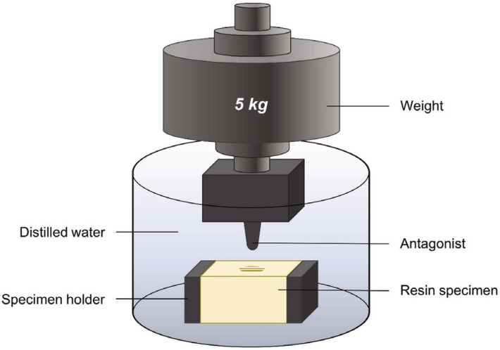
A chewing simulator (CS-4.8, SD Mechatronik, Feldkirchen-Westerham, Germany) was used to perform the wear test. The resin substrate specimens were placed in the lower specimen holders, and the metal abraders were placed in the upper holders (Fig. 1). The chambers in the machine simulated the simultaneous vertical and horizontal movements of the thermodynamic conditions. The chewing cycle was set to have a 5-mm vertical descending movement and a 2-mm horizontal movement, followed by an ascending movement with recovery of its original position. A vertical load of 5 kg was applied during the sliding motion, which is comparable to 49 N of chewing force. During the wear simulation, the specimens were subjected to thermocycling in distilled water with heat circulation at 5 – 55℃ using a heating/cooling system with a programmable logic controller. The specimens of each material were divided into two subgroups, and abraded for 30,000 or 60,000 cycles, which were considered to be equivalent to approximately 1.5 and 3 months of chewing, respectively (n = 8).19 The specimens were scanned using a multiline blue LED light scanner (D1000, 3Shape, Copenhagen, Denmark) with an accuracy of 5/8 µm (ISO 12836). The acquired images were imported into the universal reverse engineering software (Geomagic Control X 2018 version 1.2, 3D Systems, Rock Hill, SC, USA). The wear volume losses (in mm3) of the resin specimens were calculated as the difference in the volume before and after wear testing using the software.
Fig. 1. Schematic drawing of chewing simulation.
The metal antagonist with a hemisphere radius of 1.5 mm and the rectangular parallelepiped-shaped interim resin specimen are placed on the wear apparatus.
The impact of chewing simulation on the surface roughness of the materials was evaluated before and after simulated chewing. Four representative specimens were randomly selected from each group, and a confocal laser scanning microscope (LSM 800 MAT, Zeiss, Jena, Germany) was used to analyze their tested surfaces. Laser excitation at 405 nm with the C Epiplan-APOCHROMAT 209/0.7 (Zeiss, Jena, Germany) was used to obtain images. For each representative specimen, three different sites were pictured. The surface roughness of the worn area was measured using the arithmetic mean deviation of the surface roughness (Ra). Overall, 12 Ra values were collected for each group. All assessments were performed according to the ISO 4287 standards.
To evaluate the surface morphology of the specimen after chewing simulation, a representative specimen was selected for each group. A thin coating with platinum was applied to the worn surface using a sputter coater (Quorum Q150T-S, Quorum Technologies, West Sussex, UK). The wear patterns on the surface of the specimen were examined using a scanning electron microscope (SEM) (Apreo S, ThermoFisher Scientific, Waltham MA, USA) at magnifications of 200× and 1000× with 10 keV.
The mean and standard deviation (SD) of the test parameters were calculated using statistical analysis software (IBM SPSS version 25.0, IBM Corp., Chicago, IL, USA). Tests for normality and equality of variance were performed. The statistical significance of the mean difference of each parameter was evaluated at a significance level of 5% using one-way analysis of variance (ANOVA) and Tukey's post-hoc test for the three different resins. The paired t-test was used to compare the mean volume loss of each resin between the two different thermocycles.
RESULTS
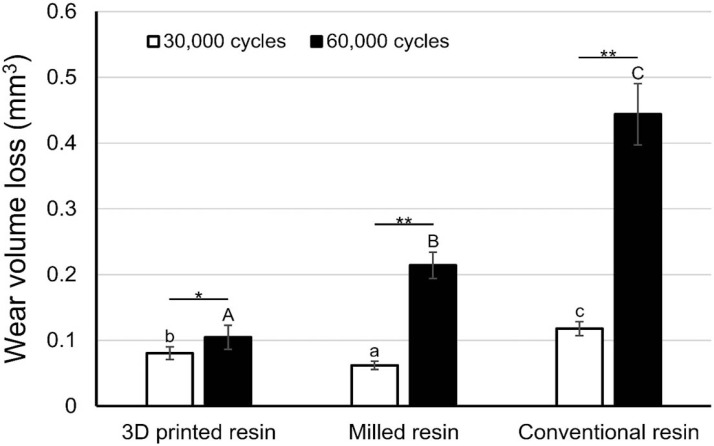
The wear volume losses of the specimens after the masticatory simulation are presented in Fig. 2. The mean ± SD volume losses (in mm3) after 30,000 and 60,000 cycles were 0.08 ± 0.09 and 0.10 ± 0.01 for the 3D printed resin, 0.06 ± 0.01 and 0.21 ± 0.02 for the milled resin, and 0.11 ± 0.01 and 0.44 ± 0.01 for the conventional resin, respectively. A significant difference in the wear volume loss was shown among the interim materials (P < .001). The wear volume loss of the 3D printed resin was lower than those of the milled and conventional resins for both cycles (P < .001). A significant difference between the loss amounts after 30,000 and 60,000 cycles was found in each resin group.
Fig. 2. Wear volume loss (mean ± standard deviation) after 30,000 and 60,000 cycles of simulated chewing. Same letters indicate no statistically significant differences.
Lowercase letters for 30,000 cycles and uppercase letters for 60,000 cycles. *P < .05, **P < .001.
The mean ± SD Ra values (µm) before (baseline) and after the wear tests at 30,000 and 60,000 cycles were 0.48 ± 0.06 and 0.58 ± 0.06 for the 3D printed resin, 0.88 ± 0.05 and 1.27 ± 0.49 for the milled resin, and 0.92 ± 0.09 and 1.63 ± 0.44 for the conventional resin, respectively. Statistically significant differences were found in the Ra of the different interim resin materials and two different cycles (Table 2).
Table 2. Mean ± standard deviation of surface roughness (Ra; µm) values for tested interim restorative materials.
| Baseline | 30,000 cycles | 60,000 cycles | |
|---|---|---|---|
| 3D printed resin | 0.13 ± 0.01Aa | 0.48 ± 0.07Ab | 0.59 ± 0.06Ac |
| Milled resin | 0.19 ± 0.03Ba | 0.88 ± 0.05Bb | 1.27 ± 0.49ABb |
| Conventional resin | 0.26 ± 0.02Ca | 0.92 ± 0.10Bb | 1.64 ± 0.44 Bc |
Same superscript letters indicate no statistically significant differences. Uppercase letters for each column, lowercase letters for each row.
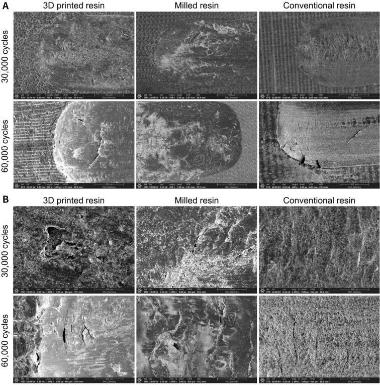
SEM images of the abraded surfaces of the specimens after the wear tests are shown in Fig. 3 (original magnification: ×200 and ×1000). All three resin types exhibited compressed and crushed features. Crack lines were observed when the metal abrader was applied for 60,000 cycles (see ×1000 images).
Fig. 3. Scanning electron microscope images of the worn resin surfaces after 30,000 and 60,000 cycles of chewing simulation. Crack lines are shown on the surfaces of specimens that simulated chewing for 60,000 cycles. (A) Original magnification: ×200, (B) Original magnification: ×1,000.
DISCUSSION
This in vitro study investigated the wear behavior and surface roughness of three different interim restorative materials at two different time intervals, and the null hypothesis of this study was rejected. The results of the study showed that there was a significant difference in wear resistance and surface roughness among the tested interim materials. The wear volume loss of the 3D printed and milled resins was less than that of the conventional resin. The 3D printed group showed a smoother surface than that of the conventional PMMA group after simulated chewing.
In the present study, interim resin materials fabricated using digital dental technologies, including 3D printing and milling, showed significantly less volume of wear than the conventional resin material, after a simulated period of 1.5 and 3 months of clinical chewing. Rayyan et al.20 have reported that CAD/CAM milled PMMA resin showed a lower percentage of weight loss due to wear than autopolymerizing conventional PMMA interim resin after subjecting the materials to 2 million cycles of a load of 40 N. Stawarczyk et al.21 have also reported that CAD/CAM milled resin materials exhibited lower wear rates than conventional manually polymerized interim resin materials. These results are in accordance with the findings of the present study.20,21 CAD/CAM milled resin materials are industrially polymerized and thus considered to exhibit better mechanical properties than conventional resin materials.20
Park et al.9 also compared the wear resistance of 3D printed interim resin and conventional PMMA resin.9 In the study,9 the 3D printed resin did not show a significantly different amount in wear volume loss compared to the conventional interim resin after 30,000 cycles of masticatory simulation.9 Similar to our study, the previous study investigated the same 3D printed resin material; however, the 3D printer and post-curing machine used in the previous study are different from those used in the current study.9 These differences in equipment might have resulted in disparate results between the previous study and the present study. Another recent study reported that 3D printed PMMA denture teeth exhibited a statistically lower depth of wear compared to the prefabricated PMMA resin denture teeth after 200,000 cycles of simulated chewing.22 Based on the results of these previous studies9,22 and our study, 3D printed resin materials are considered to have equivalent or superior wear resistance compared to the conventional PMMA materials.9,22
In this study, 3D printed interim resin showed a significantly lower Ra value than the conventional interim resin before and after simulated chewing. Previous studies have also reported that PMMA resin has a rougher surface than 3D printed resin.23,24 Furthermore, all the tested groups in this study showed increased surface roughness after masticatory simulation compared to the baseline. For both digitally and conventionally fabricated interim restorative materials, material wear leads to a rougher surface and promotes more plaque accumulation on the worn surfaces.25 The rough surface on the interim restoration could induce bacterial adhesion and dental biofilm formation, resulting in adverse effects on the periodontal health.26 In the present study, the mean Ra values of tested materials after 30,000 and 60,000 cycles were higher than the previously reported plaque accumulation threshold of 0.2 µm25 and a tongue detectable surface roughness threshold of 0.25 – 0.5 µm.27 However, since both 3D printed resin and milled resin showed similar or smoother surfaces compared to the conventionally used PMMA resin, it is considered that these 3D printed and milled materials can be used for fabricating interim restorations in clinical practice. The 3D printed resin materials have also been reported to have different surface roughness depending on the type of material28 and the printing orientation (degree);29 thus, this should also be considered when selecting the material.
The strength of this study is that it simulated chewing for a period similar to that of actual interim restoration use in clinical practice. Interim restorations are usually used for approximately 1.5 months for a simple crown restoration. However, for multiple units of prosthesis, or if the treatment also includes additional root canal treatment, periodontal surgery, or implant surgery, the interim restorations often need to be used for more than 3 months. Thus, in this study, the changes after 1.5 and 3 months were studied by subjecting the materials to 30,000 and 60,000 cycles of chewing simulation, respectively.19 To the best of our knowledge, this is the first study to evaluate the wear resistance after 3 months of using interim restorations fabricated with additive manufacturing digital technologies.
In this study, although the setting of the chewing simulator was as similar to the clinical conditions as possible, it still has the limitations of in vitro design. To facilitate the evaluation of the wear volume loss of the material itself, specimens with rectangular parallelpiped shape rather than a crown shape were used in this study. The results may be different for teeth. Furthermore, factors that influence wear include the physical properties of enamel,30,31 parafunctional habits, eating habits, and the type of antagonist material used.16,31,32,33,34,35,36 In the oral cavity, the wear process is promoted by mechanical, thermal, and chemical stimuli.37,38 Therefore, further clinical studies are needed to confirm whether the results of this study are clinically consistent.
CONCLUSION
The 3D printed resin and CAD/CAM milled resin showed greater wear resistance than the conventional interim resin after simulation of the clinical chewing period equivalent to a duration of 1.5 and 3 months. Worn 3D printed resin specimens showed a smoother surface than the conventional interim resin after chewing simulation. In terms of the wear resistance and surface roughness after chewing, the 3D printed resin material would be clinically acceptable as an interim restorative material for extended periods of clinical use.
Footnotes
This work was supported by the Korea Medical Device Development Fund grant funded by the Korean government (Ministry of Science and ICT, Ministry of Trade, Industry and Energy, Ministry of Health & Welfare, Republic of Korea, and Ministry of Food and Drug Safety) (Project Number: 202011A02).
References
- 1.Drago C. Frequency and type of prosthetic complications associated with interim, immediately loaded full-arch prostheses: a 2-year retrospective chart review. J Prosthodont. 2016;25:433–439. doi: 10.1111/jopr.12343. [DOI] [PubMed] [Google Scholar]
- 2.Drago C. Cantilever lengths and anterior-posterior spreads of interim, acrylic resin, full-arch screw-retained prostheses and their relationship to prosthetic complications. J Prosthodont. 2017;26:502–507. doi: 10.1111/jopr.12426. [DOI] [PubMed] [Google Scholar]
- 3.Patras M, Naka O, Doukoudakis S, Pissiotis A. Management of provisional restorations’ deficiencies: a literature review. J Esthet Restor Dent. 2012;24:26–38. doi: 10.1111/j.1708-8240.2011.00467.x. [DOI] [PubMed] [Google Scholar]
- 4.Perry RD, Magnuson B. Provisional materials: key components of interim fixed restorations. Compend Contin Educ Dent. 2012;33:59–60. 62. [PubMed] [Google Scholar]
- 5.Burns DR, Beck DA, Nelson SK. Committee on Research in Fixed Prosthodontics of the Academy of Fixed Prosthodontics. A review of selected dental literature on contemporary provisional fixed prosthodontic treatment: report of the Committee on Research in Fixed Prosthodontics of the Academy of Fixed Prosthodontics. J Prosthet Dent. 2003;90:474–497. doi: 10.1016/s0022-3913(03)00259-2. [DOI] [PubMed] [Google Scholar]
- 6.Priest G. Esthetic potential of single-implant provisional restorations: selection criteria of available alternatives. J Esthet Restor Dent. 2006;18:326–338. doi: 10.1111/j.1708-8240.2006.00044.x. discussion 339. [DOI] [PubMed] [Google Scholar]
- 7.Zafar MS. Prosthodontic applications of polymethyl methacrylate (PMMA): an update. Polymers (Basel) 2020;12:2299. doi: 10.3390/polym12102299. [DOI] [PMC free article] [PubMed] [Google Scholar]
- 8.Savabi O, NejatiDanesh F. A method for fabrication of temporary restoration on solid abutment of ITI implants. J Prosthet Dent. 2003;89:419. doi: 10.1067/mpr.2003.79. [DOI] [PubMed] [Google Scholar]
- 9.Park JM, Ahn JS, Cha HS, Lee JH. Wear resistance of 3D printing resin material opposing zirconia and metal antagonists. Materials (Basel) 2018;11:1043. doi: 10.3390/ma11061043. [DOI] [PMC free article] [PubMed] [Google Scholar]
- 10.Park JY, Jeong ID, Lee JJ, Bae SY, Kim JH, Kim WC. In vitro assessment of the marginal and internal fits of interim implant restorations fabricated with different methods. J Prosthet Dent. 2016;116:536–542. doi: 10.1016/j.prosdent.2016.03.012. [DOI] [PubMed] [Google Scholar]
- 11.Unkovskiy A, Bui PH, Schille C, Geis-Gerstorfer J, Huettig F, Spintzyk S. Objects build orientation, positioning, and curing influence dimensional accuracy and flexural properties of stereolithographically printed resin. Dent Mater. 2018;34:e324–e333. doi: 10.1016/j.dental.2018.09.011. [DOI] [PubMed] [Google Scholar]
- 12.Javaid M, Haleem A. Current status and applications of additive manufacturing in dentistry: a literature-based review. J Oral Biol Craniofac Res. 2019;9:179–185. doi: 10.1016/j.jobcr.2019.04.004. [DOI] [PMC free article] [PubMed] [Google Scholar]
- 13.Abduo J, Lyons K, Bennamoun M. Trends in computer-aided manufacturing in prosthodontics: a review of the available streams. Int J Dent. 2014;2014:783948. doi: 10.1155/2014/783948. [DOI] [PMC free article] [PubMed] [Google Scholar]
- 14.Alevizakos V, Mitov G, Teichert F, von See C. The color stability and wear resistance of provisional implant restorations: a prospective clinical study. Clin Exp Dent Res. 2020;6:568–575. doi: 10.1002/cre2.311. [DOI] [PMC free article] [PubMed] [Google Scholar]
- 15.Tahayeri A, Morgan M, Fugolin AP, Bompolaki D, Athirasala A, Pfeifer CS, Ferracane JL, Bertassoni LE. 3D printed versus conventionally cured provisional crown and bridge dental materials. Dent Mater. 2018;34:192–200. doi: 10.1016/j.dental.2017.10.003. [DOI] [PMC free article] [PubMed] [Google Scholar]
- 16.Preis V, Behr M, Kolbeck C, Hahnel S, Handel G, Rosentritt M. Wear performance of substructure ceramics and veneering porcelains. Dent Mater. 2011;27:796–804. doi: 10.1016/j.dental.2011.04.001. [DOI] [PubMed] [Google Scholar]
- 17.Krejci I, Albert P, Lutz F. The influence of antagonist standardization on wear. J Dent Res. 1999;78:713–719. doi: 10.1177/00220345990780021201. [DOI] [PubMed] [Google Scholar]
- 18.Jung YG, Peterson IM, Kim DK, Lawn BR. Lifetime-limiting strength degradation from contact fatigue in dental ceramics. J Dent Res. 2000;79:722–731. doi: 10.1177/00220345000790020501. [DOI] [PubMed] [Google Scholar]
- 19.DeLong R, Sakaguchi RL, Douglas WH, Pintado MR. The wear of dental amalgam in an artificial mouth: a clinical correlation. Dent Mater. 1985;1:238–242. doi: 10.1016/S0109-5641(85)80050-6. [DOI] [PubMed] [Google Scholar]
- 20.Rayyan MM, Aboushelib M, Sayed NM, Ibrahim A, Jimbo R. Comparison of interim restorations fabricated by CAD/CAM with those fabricated manually. J Prosthet Dent. 2015;114:414–419. doi: 10.1016/j.prosdent.2015.03.007. [DOI] [PubMed] [Google Scholar]
- 21.Stawarczyk B, Özcan M, Trottmann A, Schmutz F, Roos M, Hämmerle C. Two-body wear rate of CAD/CAM resin blocks and their enamel antagonists. J Prosthet Dent. 2013;109:325–332. doi: 10.1016/S0022-3913(13)60309-1. [DOI] [PubMed] [Google Scholar]
- 22.Pham DM, Gonzalez MD, Ontiveros JC, Kasper FK, Frey GN, Belles DM. Wear resistance of 3D printed and prefabricated denture teeth opposing zirconia. J Prosthodont. 2021 Jan 24; doi: 10.1111/jopr.13339. Epub ahead of print. [DOI] [PubMed] [Google Scholar]
- 23.Taşın S, Ismatullaev A, Usumez A. Comparison of surface roughness and color stainability of 3-dimensionally printed interim prosthodontic material with conventionally fabricated and CAD-CAM milled materials. J Prosthet Dent. 2021:S0022-3913(21)00075-5. doi: 10.1016/j.prosdent.2021.01.027. Epub ahead of print. [DOI] [PubMed] [Google Scholar]
- 24.Simoneti DM, Pereira-Cenci T, Dos Santos MBF. Comparison of material properties and biofilm formation in interim single crowns obtained by 3D printing and conventional methods. J Prosthet Dent. 2020:S0022-3913(20)30513-8. doi: 10.1016/j.prosdent.2020.06.026. [DOI] [PubMed] [Google Scholar]
- 25.Bollen CM, Lambrechts P, Quirynen M. Comparison of surface roughness of oral hard materials to the threshold surface roughness for bacterial plaque retention: a review of the literature. Dent Mater. 1997;13:258–269. doi: 10.1016/s0109-5641(97)80038-3. [DOI] [PubMed] [Google Scholar]
- 26.Buergers R, Rosentritt M, Handel G. Bacterial adhesion of Streptococcus mutans to provisional fixed prosthodontic material. J Prosthet Dent. 2007;98:461–469. doi: 10.1016/S0022-3913(07)60146-2. [DOI] [PubMed] [Google Scholar]
- 27.Jones CS, Billington RW, Pearson GJ. The in vivo perception of roughness of restorations. Br Dent J. 2004;196:42–45. doi: 10.1038/sj.bdj.4810881. [DOI] [PubMed] [Google Scholar]
- 28.Revilla-León M, Morillo JA, Att W, Özcan M. Chemical composition, Knoop hardness, surface roughness, and adhesion aspects of additively manufactured dental interim materials. J Prosthodont. 2020 Dec 08; doi: 10.1111/jopr.13302. Epub ahead of print. [DOI] [PubMed] [Google Scholar]
- 29.Shim JS, Kim JE, Jeong SH, Choi YJ, Ryu JJ. Printing accuracy, mechanical properties, surface characteristics, and microbial adhesion of 3D-printed resins with various printing orientations. J Prosthet Dent. 2020;124:468–475. doi: 10.1016/j.prosdent.2019.05.034. [DOI] [PubMed] [Google Scholar]
- 30.Rosentritt M, Behr M, Gebhard R, Handel G. Influence of stress simulation parameters on the fracture strength of all-ceramic fixed-partial dentures. Dent Mater. 2006;22:176–182. doi: 10.1016/j.dental.2005.04.024. [DOI] [PubMed] [Google Scholar]
- 31.Rosentritt M, Behr M, van der Zel JM, Feilzer AJ. Approach for valuating the influence of laboratory simulation. Dent Mater. 2009;25:348–352. doi: 10.1016/j.dental.2008.08.009. [DOI] [PubMed] [Google Scholar]
- 32.Hahnel S, Behr M, Handel G, Rosentritt M. Two-body wear of artificial acrylic and composite resin teeth in relation to antagonist material. J Prosthet Dent. 2009;101:269–278. doi: 10.1016/S0022-3913(09)60051-2. [DOI] [PubMed] [Google Scholar]
- 33.Johansson A, Haraldson T, Omar R, Kiliaridis S, Carlsson GE. An investigation of some factors associated with occlusal tooth wear in a selected high-wear sample. Scand J Dent Res. 1993;101:407–415. doi: 10.1111/j.1600-0722.1993.tb01140.x. [DOI] [PubMed] [Google Scholar]
- 34.Johansson A, Kiliaridis S, Haraldson T, Omar R, Carlsson GE. Covariation of some factors associated with occlusal tooth wear in a selected high-wear sample. Scand J Dent Res. 1993;101:398–406. doi: 10.1111/j.1600-0722.1993.tb01139.x. [DOI] [PubMed] [Google Scholar]
- 35.Kim SK, Kim KN, Chang IT, Heo SJ. A study of the effects of chewing patterns on occlusal wear. J Oral Rehabil. 2001;28:1048–1055. doi: 10.1046/j.1365-2842.2001.00761.x. [DOI] [PubMed] [Google Scholar]
- 36.Kadokawa A, Suzuki S, Tanaka T. Wear evaluation of porcelain opposing gold, composite resin, and enamel. J Prosthet Dent. 2006;96:258–265. doi: 10.1016/j.prosdent.2006.08.016. [DOI] [PubMed] [Google Scholar]
- 37.Mair LH, Stolarski TA, Vowles RW, Lloyd CH. Wear: mechanisms, manifestations and measurement. Report of a workshop. J Dent. 1996;24:141–148. doi: 10.1016/0300-5712(95)00043-7. [DOI] [PubMed] [Google Scholar]
- 38.Oh WS, Delong R, Anusavice KJ. Factors affecting enamel and ceramic wear: a literature review. J Prosthet Dent. 2002;87:451–459. doi: 10.1067/mpr.2002.123851. [DOI] [PubMed] [Google Scholar]


