Abstract
Biocidal agents such as formaldehyde and glutaraldehyde are able to inactivate several coronaviruses including SARS‐CoV‐2. In this article, an insight into one mechanism for the inactivation of these viruses by those two agents is presented, based on analysis of previous observations during electron microscopic examination of several members of the orthocoronavirinae subfamily, including the new virus SARS‐CoV‐2. This inactivation is proposed to occur through Schiff base reaction‐induced conformational changes in the spike glycoprotein leading to its disruption or breakage, which can prevent binding of the virus to cellular receptors. Also, a new prophylactic and therapeutic measure against SARS‐CoV‐2 using acetoacetate is proposed, suggesting that it could similarly break the viral spike through Schiff base reaction with lysines of the spike protein. This measure needs to be confirmed experimentally before consideration. In addition, a new line of research is proposed to help find a broad‐spectrum antivirus against several members of this subfamily.
Keywords: COVID‐19, cytokine storm, fasting, formaldehyde, glutaraldehyde, ketogenic diet, ketone bodies, preconditioning, Schiff base, virus inactivation
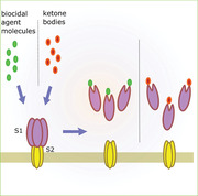
The small molecules of the biocidal agents (formaldehyde and glutaraldehyde) appear to interact with specific lysine residues in the spike protein of some coronaviruses resulting in conformational changes that can induce S1 shedding and hence spike inactivation. Ketone bodies (acetoacetate) may inactivate the spike protein in a similar way.
INTRODUCTION
Coronaviridae is a large family of enveloped single‐stranded RNA viruses. It is divided into two subfamilies: orthocoronavirinae and letovirinae.[ 1 ] Orthocoronavirinae consists of four genera named Alpha, Beta, Delta, and Gamma coronaviruses. Betacoronaviruses include several viruses that infect humans causing life‐threatening diseases such as SARS‐CoV, SARS‐CoV‐2 and Middle East respiratory syndrome coronavirus (MERS‐CoV).
Coronaviruses have a set of structural proteins, one of them is the spike “S” protein. This protein is a class I virus fusion protein that forms homotrimers giving clove‐shaped molecules protruding from the virus surface.[ 2 , 3 ] These viruses use this protein to attach to host cell receptors to allow cellular entry. Each spike monomer is composed of two subunits; S1 subunit which harbors the receptor‐binding domain (RBD) and S2 subunit which is required for the fusion of cell and virus membranes. S protein is cleaved at a site between S1 and S2 either during synthesis or during cellular entry to give the two subunits.[ 2 , 3 ] In the case of SARS‐CoV‐2, S is cleaved during synthesis at the S1/S2 site (Figure 1A) by furin[ 4 , 5 ] and the two subunits remain non‐covalently bound. This protein is further cleaved during cellular entry at another site termed S2′ by the transmembrane serine protease 2 (TMPRSS2)[ 4 ] to release the fusion peptide (FP) to initiate membrane fusion. Both cleavage events are required for successful infection by SARS‐CoV‐2. Following receptor binding, S1 subunit separates from the virus particle and the S2 subunit undergoes conformational changes ultimately bringing viral and cellular membranes closer to each other and driving their fusion to release the viral genetic material inside the cell.[ 2 ]
FIGURE 1.
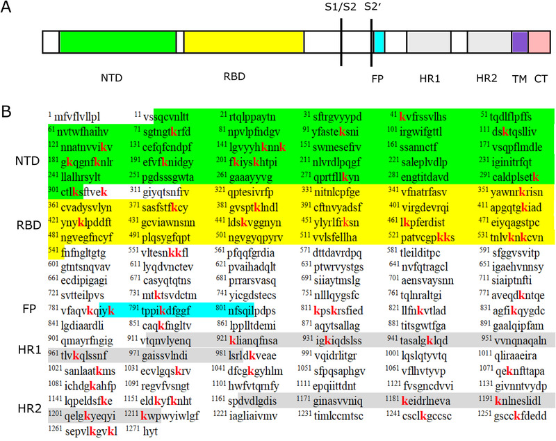
Domains (A) and amino acid sequence (B) of S protein of SARS‐CoV‐2 showing location of lysine residues (in red)
Each subunit of the spike monomers of SARS‐CoV‐2 consists of several regions (Figure 1A). The S1 subunit (residues 14–685) consists of an N‐terminal domain (NTD) and a C‐terminal domain (CTD), while the S2 subunit (residues 686–1273) includes an FP, heptapeptide repeat sequence 1,2 (HR1, HR2), transmembrane domain (TM) and cytoplasmic tail (CT).[ 6 ] The CTD acts as the receptor binding domain (RBD) for this virus,[ 7 ] in which the receptor binding motif (RBM) is the site for receptor binding. S of SARS‐CoV‐2 is a dynamic structure[ 8 ] in which the RBDs are able to transit between down and up conformations. A recent analysis of the stability of the S protein in the absence of receptors reported that spikes with RBDs in the up conformation are less stable than those with RBDs in the down conformation[ 9 ] angiotensin‐converting enzyme 2 (hACE2) receptors can only bind to RBDs in the up conformation, and this binding is suggested to help lock the RBDs in this unstable conformation until S1 monomers dissociation.[ 10 ]
S protein of SARS‐CoV‐2 is composed of 1273 amino acids, 61 of which are lysine residues (Figure 1B). These lysines are almost equally distributed between the two subunits; 30 in S1 and 31 in S2. Some of them appear to help stabilize the RBD in the down‐conformation and hence stabilize the whole S protein.[ 9 ]
Glutaraldehyde is a dialdehyde commonly used in laboratory research as a chemical fixative.[ 11 ] It has the chemical formula C5H8O2 with two carbonyl groups (one at each end). Formaldehyde is another aldehyde with only one carbonyl group and is also used in chemical fixation and virus inactivation.[ 12 ] It has the chemical formula CH2O and it can polymerize to form paraformaldehyde. Both agents have the ability to inactivate several types of coronaviruses.[ 13 ] By analyzing observations from electron microscopic examination of several coronaviruses, a mechanism for this inactivation process, via interaction with viral spikes, is proposed. Also, an intervention against SARS‐CoV‐2 is proposed, using ketone bodies (in particular acetoacetate) which circulate in the human blood and interstitial fluids, suggesting that they could inactivate the extracellular virions in a similar pattern.
FORMALDEHYDE AND GLUTARALDEHYDE APPEAR TO INDUCE THE LOSS OF VIRAL SPIKES
In one study,[ 14 ] during electron microscopic examination of Vero E6 cells infected with SARS‐CoV, Snijder et al. reported that the viral spikes of secreted extracellular virions were only visible in cryofixed samples but were rarely seen in chemically fixed samples (with 1.5% glutaraldehyde). This loss of viral spikes was also observed in the chemically‐fixed (with 2% glutaraldehyde) extracellular virions of the transmissible gastroenteritis coronavirus (genus Alphacoronavirus), which affects newborn piglets.[ 15 ] For the new virus SARS‐CoV‐2, virus particles in the supernatant of infected cell cultures that were inactivated with 2% paraformaldehyde also showed significant loss of viral spikes.[ 16 ] Also, SARS‐CoV‐2 progeny virions in glutaraldehyde‐fixed samples were reported to have few or no apparent spikes.[ 17 ]
HOW CAN THIS EFFECT BE INTERPRETED?
Chemical fixation of samples is known to cause the cessation of all biological processes inside cells, including the secretion of new virions from infected cells. This in turn eliminates the possibility of the disruption of the virus assembly process or the pre‐mature secretion of un‐assembled virions from infected cells by the action of the fixatives as an explanation for the observed effect. Therefore, this effect of chemical fixation on viral spikes most probably results from direct chemical interaction between the fixative and the spike protein. The fixatives appear to interact with the spike protein in such a way that leads to its separation from the virion. Also, since this effect is evident in several members of the orthocoronavirinae subfamily, these fixatives appear to interact with a conserved sequence of the spike protein in those members.
The next question to ask is, what is the nature of this interaction? Several studies showed that glutaraldehyde's interaction with proteins occurs predominantly with lysine amino acids on the surface of proteins.[ 18 , 19 , 20 ] The carbonyl group of glutaraldehyde reacts with the ϵ‐amino group of lysine side chain to form a Schiff base. Schiff base reaction involves the formation of a carbon‐nitrogen double bond when the carbonyl group of an aldehyde or ketone reacts with a primary amine (Figure 2).[ 21 ] Formaldehyde was also shown to form Schiff bases with lysine side chains during interaction with proteins (Figure 3).[ 22 ] Since both fixatives share in common the ability to form Schiff bases with proteins, these bases are most likely responsible for the loss of viral spikes in the mentioned studies.[ 14 , 15 , 16 , 17 ] One possible explanation is that the formation of these bases with lysine residues might alter the conformation of the spike protein leading to its separation from the virion (via separation of S1 subunit from S2 subunit) (Figure 4) or alternatively, leading to its bending. In fact, there are several observations to support this view. Firstly, glutaraldehyde was shown to induce gross conformational changes in several soluble and membrane proteins through interaction with lysine residues via Schiff base reaction[ 20 , 23 ] and thus, is expected to do the same in the spike protein. Similarly, formaldehyde was shown to be able to induce protein conformational changes as well.[ 24 , 25 ] In the majority of these tested proteins, formaldehyde and glutaraldehyde tended to reduce the α‐helix content, which in turn loosens the protein skeleton.[ 25 ] Secondly, conformational changes induced in the spike glycoprotein of the mouse hepatitis coronavirus (MHV) A59 (genus Betacoronavirus) by other means (pH changes) could dissociate the S1 subunit from the virions and abrogate virus infectivity.[ 26 ] Similarly, conformational changes induced in the spike protein of SARS‐CoV‐2 upon binding to ACE2 receptors were followed by the dissociation of S1 monomers.[ 10 ] S1 shedding following receptor binding was also observed in the cleaved S of SARS‐CoV.[ 27 ] This suggests the wide susceptibility for S1 separation following certain conformational changes induced in the cleaved spike by different means. These changes might disrupt the interactions between the two subunits ultimately driving S1 dissociation. Indeed, conformational changes induced in the spike of MHV upon binding to receptors were reported to drive the movement of S1 away from S2 and decrease the interface between them[ 28 ] which could reduce the interactions between them and facilitate S1 shedding. Similarly, in SARS‐CoV‐2, conformational changes in ACE2‐bound spikes were shown to involve a decrease in the contact area between S1 and S2 with a reported loss of interactions between them, compared with the unbound spikes.[ 10 ] It is thus conceivable to predict that conformational changes induced by the biocidal agents can do the same, namely disrupt the interactions between S1 and S2 leading to the dissociation of S1 from the virions. This may explain one mechanism by which formaldehyde and glutaraldehyde can inactivate coronaviruses (Figure 4). Based on this, other short aldehydes and ketones that are biologically‐competent and have carbonyl groups capable of forming Schiff bases with the spike protein and altering its conformation could be speculated to break the viral spikes as well and thus, have a therapeutic potential. One such compound could be the ketone bodies. Similar to glutaraldehyde and formaldehyde, ketone bodies can also alter the conformation of proteins they react with, via Schiff base reaction.[ 29 ]
FIGURE 2.
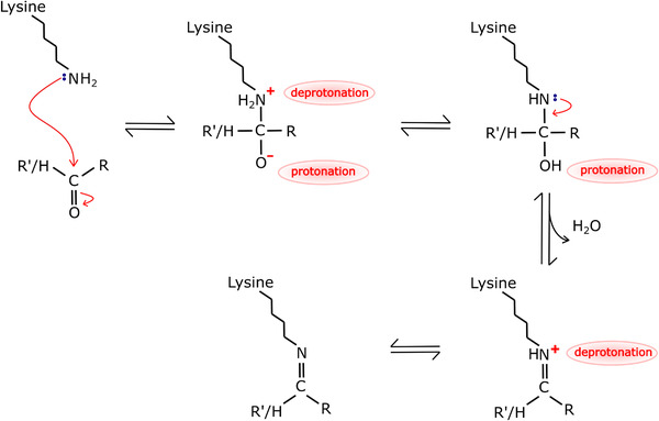
Mechanism of Schiff base reaction between ϵ‐amino group of lysine and aldehydes or ketones
FIGURE 3.
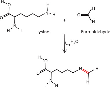
Schiff base reaction between formaldehyde and lysine
FIGURE 4.
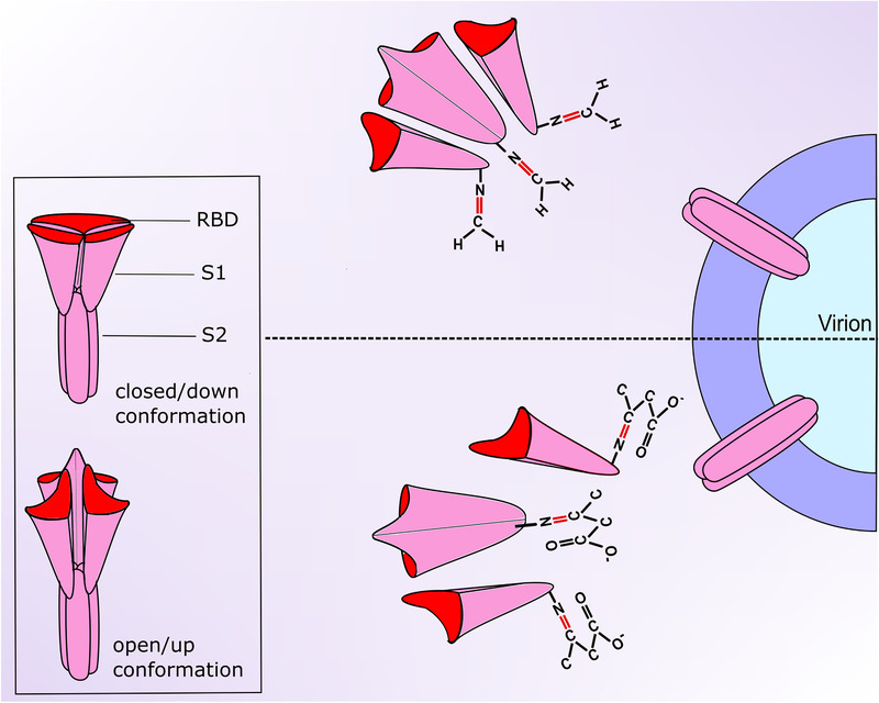
Schematic representation of the proposed SARS‐CoV‐2 spike inactivation by biocidal agents (such as formaldehyde) and by ketone bodies. Inset: representation of the spike protein with all RBDs in down or up conformation. Top: formaldehyde molecules react with lysine residues of the spike protein via Schiff base reaction and alter its conformation leading to the separation of S1 subunit from S2 subunit (which remains attached to the virion). Bottom: Ketone bodies (acetoacetate) react with the same residues of the spike protein and also alter its conformation causing the separation of S1 subunit from S2 subunit. RBDs are shown in up conformation after both reactions
CAN KETOSIS INACTIVATE THE VIRAL SPIKES?
There are three types of ketone bodies in the human blood.[ 30 ] These include beta‐hydroxybutyrate (C4H7O3 −), acetoacetate (C4H5O3 −) and the least abundant one, acetone (C3H6O). They are synthesized in the liver from fatty acids and released in the blood to act as a source of energy when there is a shortage of carbohydrate supply to the body. Thus, the level of ketone bodies increases in the plasma during periods of fasting and starvation. In fact, there are observations strongly suggesting that acetoacetate can form Schiff bases when reacting with lysine residues of proteins (Figure 5) and can alter their secondary structure,[ 29 ] similar to the effect of the mentioned fixatives. More specifically, acetoacetate reduced the α‐helix content of the tested protein, an effect similarly induced by formaldehyde and glutaraldehyde.[ 23 , 24 , 25 ] Moreover, ketone bodies are short compounds (three or four carbons long) and therefore should have accessibility to amino acids within proteins comparable to the short fixatives (formaldehyde and glutaraldehyde have one carbon and five carbons, respectively). Hence, ketone bodies are expected to effectively react with the viral spikes and break or bend them similar to the fixatives (Figure 4); however, this needs to be confirmed experimentally before consideration. The induction of ketosis would then have a therapeutic as well as a prophylactic potential. In infected patients, the loss of viral spikes after virions secretion from infected cells would inhibit or slow down the spread of infection to other cells and tissues in the body. Also, virus particles in body secretions will have low or abrogated infectivity toward other persons, thus rendering the infected patients relatively non‐infectious. In the non‐infected individuals, the presence of high titer of ketone bodies in the blood would decrease the magnitude of subsequent infection or even make them highly immune to infection.
FIGURE 5.
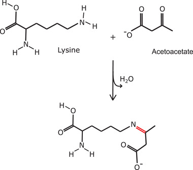
Schiff base reaction between acetoacetate and lysine
METHODS TO INDUCE KETOSIS
There are several ways to induce ketosis in the body, such as fasting, ketogenic diet or ketone supplements. Fasting has been a subject of extensive research for its desirable effects on health.[ 31 , 32 , 33 ] However, only controlled and regulated intermittent fasting should be considered, which is able to induce ketosis without weakening the immune system and the entire body. Ketogenic diet is a diet that has a high‐fat and low‐carbohydrate content and has been used regularly to treat children with refractory epilepsy and some congenital metabolic disorders.[ 34 ] This diet should only be consumed under the guidance of physicians since it is not suitable for some people.[ 34 ] There are also some ketone supplements to raise the level of ketone bodies in the blood such as ketone esters and salts.[ 35 ] These should also be used under the guidance of physicians. Ketone body infusion is another method to rapidly induce ketosis.
If such inactivating effect of acetoacetate on SARS‐CoV‐2 could be confirmed experimentally in vitro and in vivo, then some of these methods could be considered for prophylaxis such as intermittent fasting or ketogenic diet (as lifestyle regimens), while others could be considered for treatment such as acetoacetate infusion (especially in immuno‐compromised patients). It seems that infusion can rapidly raise the level of acetoacetate in the body without causing harmful or intolerable metabolic effects.[ 36 , 37 ] Further studies are needed to explore in detail other effects on metabolism and body functions. Notably, if infusion is to be used for treatment, it should be considered as early as possible during active infection before SARS‐CoV‐2 induces significant pathology, which may itself raise the level of ketone bodies in the body.[ 38 ] The dangers arising from the established damage and cytokine storm may obscure the therapeutic effects of these ketone bodies. Intermittent fasting also has been shown to have a variety of metabolic effects that are also beneficial and tolerable.[ 31 , 32 , 33 ] However, some individuals cannot tolerate fasting due to suffering from some health problems. Ketogenic diet also has several desirable positive effects on health but may have some short‐term and long‐term adverse effects[ 34 ] and thus, can only be consumed under the guidance of physicians.
DOES KETOSIS HAVE OTHER BENEFICIAL EFFECTS?
Several studies showed fasting and the consumption of ketogenic diet to have anti‐inflammatory effects as well. For example, several studies[ 39 , 40 , 41 ] reported that the levels of pro‐inflammatory cytokines such as interleukin 1β (IL‐1β), IL‐6, IL‐8, IL‐10, IL‐16, IL‐18, monocyte chemoattractant protein‐1 (MCP‐1), and tumor necrosis factor α (TNFα) decreased significantly in the plasma during fasting, many of which are involved in the cytokine storm of COVID‐19.[ 42 ] Also, the genes of several cytokines were downregulated in peripheral blood mononuclear cells during fasting.[ 43 ] Hence, intermittent fasting in non‐infected individuals may confer an additional advantage of preconditioning the body via lowering the transcript and protein levels of cytokines and thus decrease the severity of the cytokine storm if subsequent SARS‐CoV‐2 infection occurs. Ketogenic diet was also shown to inhibit the increase in the levels of IL‐1β and TNFα in the plasma of rats during inflammation and also inhibit the increase in lymphocyte counts.[ 44 ] Interestingly, ketogenic diet was shown also to increase a specific type of T cells in the lungs of mice and make them highly resistant to Influenza A virus infection.[ 45 ] There is also evidence of anti‐inflammatory effects of ketogenic diet in humans[ 46 ] (several pro‐inflammatory cytokines are reduced such as TNF‐α, IL‐6, IL‐8, and MCP‐1). Therefore, ketogenic diet consumption may also be advised as a preconditioning procedure against the cytokine storm of SARS‐CoV‐2 infection. Such preconditioning methods should be investigated in detail.
Importantly, a recent paper by Stubbs et al.[ 47 ] discussed the therapeutic potential of ketosis against several respiratory viral infections including SARS‐CoV‐2. It suggested that increased ketone bodies (beta‐hydroxybutyrate) could interfere with viral replication and reduce cell damage via several mechanisms. For example, beta‐hydroxybutyrate was proposed to help decrease oxidative stress associated with infection and reduce its damaging effects through increasing cytoplasmic NADPH, upregulating expression of antioxidant genes and directly scavenging free radicals. It was proposed also to help inhibit excessive inflammation mediated by the NLRP3 inflammasome and the proinflammatory transcription factor NF‐κB, both are activated during infection. Other proposed mechanisms include closure of mitochondrial permeability transition pore, which is used by some viruses (including the influenza virus) to dissipate the inner mitochondrial membrane potential and promote apoptosis, and inhibiting glycolysis, which is increased by some respiratory viral infection and is thought to help viral replication. Ketone bodies were also proposed to help preserve and improve organ function, tissue resistance and tolerance which may favor survival with few or no long‐term complications.[ 47 ]
CAN THE BIOCIDAL AGENTS INDUCE THE TRANSITION OF S FROM CLOSED TO OPEN STATE?
There are some questions regarding the process of spike inactivation that need to be answered. For instance, what happens to the spikes? Are they lost or just bent due to conformational changes induced by biocidal agents? Do these changes occur in S1, S2 or in both? In addition, which lysine residues are targeted by these agents and what is the nature of conformational changes induced? One possible type of conformational changes could be proposed based on a recent study investigating the stability of the spike protein of SARS‐CoV‐2.[ 9 ] This study showed the closed state, where all three RBDs are in down conformation (down‐down‐down), to be the most stable one. It also showed a progressive decrease in stability with the successive transition of the RBDs to the up conformation, with the up‐down‐down being less stable and the up‐up‐down being the least stable of the two open states examined. This was associated with an increase in the number of destabilizing residues with each up‐transition. Importantly, this study identified a set of amino acid residues that make several contacts to stabilize the dynamic RBDs in the down conformation. In the absence of receptors, the biocidal agent molecules may target some of these residues and block them causing the disruption of their contacts and inhibiting their stabilizing effect, which may either drive the release of the RBDs from the down conformation to the unstable up conformation or prevent their transition back to the down conformation, depending on whether they can reach these residues in down or up conformation. These agents not only react with lysines of proteins but also react with other amino acids such as arginine or tyrosine.[ 22 , 48 ] In addition, the small size of these molecules suggests that they could reach most of these residues easily when the RBDs are in the up conformation or even in the down conformation, which may facilitate the disruption of both intrachain and interchain stabilizing contacts. Eventually, this may allow the up transition of more than one RBD (even the three RBDs) in the absence of receptors, resulting in maximum instability of the spike protein and subsequently S1 monomers dissociation (Figure 4). This effect may also be induced by acetoacetate in vivo in analogy to the biocidal agents. An example of candidate residues that may be targeted is K386 in the RBD which makes contacts with S982, R983, and L984 in S2 of the adjacent monomer in the down conformation.[ 9 ] Such contacts are broken during transition to the up‐conformation. Noteworthy, the proposed locking of RBDs in the up conformation by acetoacetate in vivo may help prevent SARS‐CoV‐2 immune evasion and allow rapid and efficient immune detection and response, since the RBD down conformation was suggested to help this evasion.[ 5 , 49 ] Moreover, it can also increase responsiveness to antibody drug therapy. Nonetheless, this proposition does not preclude the presence of other types of conformational changes resulting from the targeting of other lysine residues at different parts of the spike protein as an explanation for the observed effect.
It is also possible that the induced conformational changes might alter the shape of the receptor binding domain or the receptor‐binding motif and thus inhibit virus–receptor interaction without or before breaking the spikes, which may represent an alternative mechanism for inactivating virus particles that retain some of their spikes after biocidal treatment. Another possibility is that biocidal agent molecules might interact with and block amino acids involved in the interaction with cellular receptors. Monitoring the binding of these treated virions to cellular receptors could be helpful.
FUTURE DIRECTIONS
There are other questions that need to be investigated in detail. For example, what is the exact nature of interaction between the fixatives and the spike protein? Does this interaction occur outside cells only or also inside cells (with unsecreted virions)? The analyzed observations showed the loss of viral spikes in extracellular virions. The effect of the fixatives on the spikes of fully‐assembled intracellular viral particles need to be assessed as well.
If the biocidal agents separate the spikes from the virions, it should be assessed whether this involves the separation of S1 subunit from the membrane‐anchored S2 subunit (which may be retained) or the dissociation of the entire spike protein from the virion. Visual inspection of the microscopic images in the analysed studies suggests the separation of the entire spike protein. However, the first possibility cannot be excluded. Supporting the first possibility, the S protein of SARS‐CoV‐2 is known to be cleaved during biosynthesis at a site between the S1 and S2 subunits and thus, S1 and S2 in extracellular virions are non‐covalently associated,[ 50 ] which may facilitate the separation of S1 subunit, presumably following conformational changes. On the other hand, S protein of SARS‐CoV is not cleaved during biosynthesis and thus, S of secreted virions remains uncleaved,[ 50 ] which support the possibility of loss of the entire S protein rather than only the S1 subunit. Importantly, a recent study[ 17 ] showed that only SARS‐CoV‐2 virions had no or few spikes in chemically fixed samples while SARS‐CoV virions had numerous spikes, which further supports the loss of only S1 subunit. Further studies are needed to solve this dilemma.
Other questions to investigate include; is Schiff base reaction solely responsible for this effect or is it a part of a sequence of reactions leading to this effect? Are there any other reactions in addition to/aside from Schiff base reaction with lysines that might have caused this effect? It should be noted that formaldehyde can form Schiff base with the side chain of the amino acid tryptophan and can also interact with the side chains of cysteine, histidine and arginine.[ 22 ] Similarly, glutaraldehyde can also react to varying degrees with the side chains of tyrosine, histidine and cysteine.[ 48 ] However, as mentioned above, its most predominant interaction, which induces conformational changes in protein substrates, is with ∊‐amino group of lysine side chain to form Schiff base. Notably, both agents are known to have a cross‐linking activity. For formaldehyde, cross links were found to form between several residues after treatment such as between lysine and argenine, between lysine and histidine and between cysteine and arginine.[ 22 ] For Glutaraldehyde, cross‐linking activity is generally believed to involve the ϵ‐amino group of lysine residues.[ 51 ] On the other hand, acetoacetate has no known cross‐linking activity. Nonetheless, both fixatives share with acetoacetate the ability to interact with lysines and to induce protein conformational changes. It should also be noted that ketones are known to be less reactive toward nucleophilic attack than aldehydes.[ 52 ] The existence of two alkyl groups attached to the carbonyl carbon of ketones reduces its partial positive charge and increases steric hindrance that diminishes accessibility of nucleophiles, compared with the carbonyl carbon of aldehydes which has only one attached alkyl group. Therefore, lysine interaction with acetoacetate is expected to progress at a slower rate than that with formaldehyde or glutaraldehde. To what extent this slower interaction can affect the S protein conformation and reactivity needs to be investigated.
The effect of various concentrations of ketone bodies (including acetoacetate and also acetone) on viral spikes should also be examined using cryofixation. In addition, their effect on virus infectivity should be assessed. Interestingly, during ketosis (either diabetic or dietary), acetoacetate can react with methylglyoxal (a metabolite produced mainly during glycolysis) to yield 3‐hydroxyhexane‐2,5‐dione, which can itself bind to lysine residues of proteins.[ 53 ] It would be interesting to examine whether this ketosis‐related compound can interact with lysines of the spike protein and alter its conformation or reactivity. Finally, research should be aimed to find or formulate biologically‐competent compounds that can function like the biocidal agents against coronaviruses (broad‐spectrum antivirus). The observed loss of viral spikes in different types of coronaviruses suggests that the biocidal agents react with a conserved sequence/s shared by all of them and thus, any similarly acting compound would inactivate most of them. This could be needed if there are future attacks by mutated or new members of this family.
CONCLUSION
Schiff base reaction between short aldehydes (the biocidal agents) and lysine residues of the spike protein is suggested to break the spikes of some coronaviruses including SARS‐CoV‐2 via conformational changes.
In analogy, acetoacetate is expected to break the viral spikes in a similar manner and thus may confer some immunity against SARS‐CoV‐2 (only after experimental confirmation).
Intermittent fasting or consumption of ketogenic diets may be considered to precondition the body to decrease the severity of the cytokine storm if subsequent SARS‐CoV‐2 infection occurs.
CONFLICT OF INTEREST
The author declares that he has no conflict of interest.
ACKNOWLEDGEMNTS
The author thanks Professor Jesse C. Hay (Division of Biological Sciences, the University of Montana), Dr. Ayano Satoh (Graduate School of Interdisciplinary Science and Engineering in Health Systems, Okayama University) and Dr. Andrew Moore for helpful comments on the manuscript.
Shaheen, A. (2021). Can ketone bodies inactivate coronavirus spike protein? The potential of biocidal agents against SARS‐CoV‐2. BioEssays, 43, e2000312. 10.1002/bies.202000312
DATA AVAILABILITY STATEMENT
Data sharing not applicable no new data generated, or the article describes entirely theoretical research.
REFERENCES
- 1. King, A. M. Q. , Lefkowitz, E. J. , Mushegian, A. R. , Adams, M. J. , Dutilh, B. E. , Gorbalenya, A. E. , Harrach, B. , Harrison, R. L. , Junglen, S. , Knowles, N. J. , Kropinski, A. M. , Krupovic, M. , Kuhn, J. H. , Nibert, M. L. , Rubino, L. , Sabanadzovic, S. , Sanfaçon, H. , Siddell, S. G. , Simmonds, P. , … Davison, A. J. (2018). Changes to taxonomy and the International Code of Virus Classification and Nomenclature ratified by the International Committee on Taxonomy of Viruses (2018). Archives of Virology, 163(9), 2601–2631. 10.1007/s00705-018-3847-1 [DOI] [PubMed] [Google Scholar]
- 2. Li, F. (2016). Structure, function, and evolution of coronavirus spike proteins. Annual Review of Virology, 3(1), 237–261. 10.1146/annurev-virology-110615-042301 [DOI] [PMC free article] [PubMed] [Google Scholar]
- 3. Millet, J. K. , & Whittaker, G. R. (2015). Host cell proteases: Critical determinants of coronavirus tropism and pathogenesis. Virus Research, 202: 120–134. 10.1016/j.virusres.2014.11.021 [DOI] [PMC free article] [PubMed] [Google Scholar]
- 4. Bestle, D. , Heindl, M. R. , Limburg, H. , van Van Lam , T. , Pilgram, O. , Moulton, H. , Stein, D. A. , Hardes, K. , Eickmann, M. , Dolnik, O. , Rohde, C. , Klenk, H. D. , Garten, W. , Steinmetzer, T. , & Böttcher‐Friebertshäuser, E. (2020). TMPRSS2 and furin are both essential for proteolytic activation of SARS‐CoV‐2 in human airway cells. Life Science Alliance, 3(9), e202000786. 10.26508/lsa.202000786. [DOI] [PMC free article] [PubMed] [Google Scholar]
- 5. Shang, J. , Wan, Y. , Luo, C. , Ye, G. , Geng, Q. , Auerbach, A. , & Li, F. (2020). Cell entry mechanisms of SARS‐CoV‐2. Proceedings of the National Academy of Sciences of the United States of America, 117(21), 11727–11734. 10.1073/pnas.2003138117 [DOI] [PMC free article] [PubMed] [Google Scholar]
- 6. Xia, S. , Zhu, Y. , Liu, M. , Lan, Q. , Xu, W. , Wu, Y. , Ying, T. , Liu, S. , Shi, Z. , Jiang, S. , & Lu, L. (2020). Fusion mechanism of 2019‐nCoV and fusion inhibitors targeting HR1 domain in spike protein. Cellular and Molecular Immunology, 17(7), 765–767. 10.1038/s41423-020-0374-2 [DOI] [PMC free article] [PubMed] [Google Scholar]
- 7. Wang, Q. , Zhang, Y. , Wu, L. , Niu, S. , Song, C. , Zhang, Z. , Lu, G. , Qiao, C. , Hu, Y. , Yuen, K. Y. , Wang, Q. , Zhou, H. , Yan, J. , & Qi, J. (2020). Structural and functional basis of SARS‐CoV‐2 entry by using human ACE2. Cell, 181(4), 894–904.e9.e9. 10.1016/j.cell.2020.03.045 [DOI] [PMC free article] [PubMed] [Google Scholar]
- 8. Lu, M. , Uchil, P. D. , Li, W. , Zheng, D. , Terry, D. S. , Gorman, J. , Shi, W. , Zhang, B. , Zhou, T. , Ding, S. , Gasser, R. , Prévost, J. , Beaudoin‐Bussières, G. , Anand, S. P. , Laumaea, A. , Grover, J. R. , Liu, L. , Ho, D. D. , Mascola, J. R. , … Mothes, W. (2020). Real‐time conformational dynamics of SARS‐CoV‐2 spikes on virus particles. Cell Host Microbe, 28(6), 880–891.e8.e8. 10.1016/j.chom.2020.11.001 [DOI] [PMC free article] [PubMed] [Google Scholar]
- 9. Moreira, R. A. , Guzman, H. V. , Boopathi, S. , Baker, J. L. , & Poma, A. B. (2020). Characterization of structural and energetic differences between conformations of the SARS‐CoV‐2 spike protein. Materials (Basel), 13(23), 5362. 10.3390/ma13235362 [DOI] [PMC free article] [PubMed] [Google Scholar]
- 10. Benton, D. J. , Wrobel, A. G. , Xu, P. , Roustan, C. , Martin, S. R. , Rosenthal, P. B. , Skehel, J. J. , & Gamblin, S. J. (2020). Receptor binding and priming of the spike protein of SARS‐CoV‐2 for membrane fusion. Nature, 588, 327–330, 10.1038/s41586-020-2772-0 [DOI] [PMC free article] [PubMed] [Google Scholar]
- 11. Dey, P. (2018). Fixation of histology samples: Principles, methods and types of fixatives. In: Basic and advanced laboratory techniques in histopathology and cytology (pp. 3–17). Singapore: Springer. 10.1007/978-981-10-8252-8_1 [DOI] [Google Scholar]
- 12. Thavarajah, R. , Mudimbaimannar, V. K. , Elizabeth, J. , Rao, U. K. , & Ranganathan, K. (2012). Chemical and physical basics of routine formaldehyde fixation. Journal of Oral and Maxillofacial Pathology, 16(3), 400–405. 10.4103/0973-029X.102496 [DOI] [PMC free article] [PubMed] [Google Scholar]
- 13. Kampf, G. , Todt, D. , Pfaender, S. , & Steinmann, E. (2020). Persistence of coronaviruses on inanimate surfaces and their inactivation with biocidal agents. Journal of Hospital Infection, 104(3), 246–251. 10.1016/j.jhi23yjn.2020.01.022 [DOI] [PMC free article] [PubMed] [Google Scholar]
- 14. Snijder, E. J. , van der Meer, Y. , Zevenhoven‐Dobbe, J. , Onderwater, J. J. , van der Meulen, J. , Koerten, H. K. , & Mommaas, A. M. (2006). Ultrastructure and origin of membrane vesicles associated with the severe acute respiratory syndrome coronavirus replication complex. Journal of Virology, 80(12), 5927–5940. 10.1128/JVI.02501-05 [DOI] [PMC free article] [PubMed] [Google Scholar]
- 15. Salanueva, I. J. , Carrascosa, J. L. , & Risco, C. (1999). Structural maturation of the transmissible gastroenteritis coronavirus. Journal of Virology, 73(10), 7952–7964. 10.1128/JVI.73.10.7952-7964.1999 [DOI] [PMC free article] [PubMed] [Google Scholar]
- 16. Zhu, N. , Zhang, D. , Wang, W. , Li, X. , Yang, B. , Song, J. , Zhao, X. , Huang, B. , Shi, W. , Lu, R. , Niu, P. , Zhan, F. , Ma, X. , Wang, D. , Xu, W. , Wu, G. , Gao, G. F. , & Tan, W. (2020). China Novel Coronavirus Investigating and Research Team . A novel coronavirus from patients with pneumonia in China, 2019. New England Journal of Medicine, 382(8), 727–733. 10.1056/NEJMoa2001017 [DOI] [PMC free article] [PubMed] [Google Scholar]
- 17. Ogando, N. S. , Dalebout, T. J. , Zevenhoven‐Dobbe, J. C. , Limpens, R. W. A. L. , van der Meer, Y. , Caly, L. , Druce, J. , de Vries, J. J. C. , Kikkert, M. , Bárcena, M. , Sidorov, I. , & Snijder, E. J. (2020). SARS‐coronavirus‐2 replication in Vero E6 cells: Replication kinetics, rapid adaptation and cytopathology. Journal of General Virology, 101(9), 925–940. 10.1099/jgv.0.001453 [DOI] [PMC free article] [PubMed] [Google Scholar]
- 18. Bowes, J. H. , & Cater, C. W. (1968). The interaction of aldehydes with collagen. Biochimica Et Biophysica Acta, 168(2), 341–352. 10.1016/0005-2795(68)90156-6 [DOI] [PubMed] [Google Scholar]
- 19. Yonath, A. , Sielecki, A. , Moult, J. , Podjarny, A. , & Traub, W. (1977). Crystallographic studies of protein denaturation and renaturation. 1. Effects of denaturants on volume and X‐ray pattern of cross‐linked triclinic lysozyme crystals. Biochemistry, 16(7), 1413–1417. 10.1021/bi00626a027 [DOI] [PubMed] [Google Scholar]
- 20. Chang, L. S. , Lin, S. R. , & Yang, C. C. (2001). Glutaraldehyde cross‐linking alters the environment around Trp(29) of cobrotoxin and the pathway for regaining its fine structure during refolding. Journal of Peptide Research, 58(2), 173–179. 10.1034/j.1399-3011.2001.00909.x [DOI] [PubMed] [Google Scholar]
- 21. Hameed, A. , Al‐Rashida, M. , Uroos, M. , Abid Ali, S. , & Khan, K. M. (2017). Schiff bases in medicinal chemistry: A patent review (2010‐2015). Expert Opinion on Therapeutic Patents, 27(1), 63–79. 10.1080/13543776.2017.1252752 [DOI] [PubMed] [Google Scholar]
- 22. Metz, B. , Kersten, G. F. , Hoogerhout, P. , Brugghe, H. F. , Timmermans, H. A. , de Jong, A. , Meiring, H. , ten Hove, J. , Hennink, W. E. , Crommelin, D. J. , & Jiskoot, W. (2004). Identification of formaldehyde‐induced modifications in proteins: Reactions with model peptides. Journal of Biological Chemistry, 279(8), 6235–6243. 10.1074/jbc.M310752200 [DOI] [PubMed] [Google Scholar]
- 23. Lenard, J. , & Singer, S. J. (1968). Alteration of the conformation of proteins in red blood cell membranes and in solution by fixatives used in electron microscopy. Journal of Cell Biology, 37(1), 117–122. 10.1083/jcb.37.1.117 [DOI] [PMC free article] [PubMed] [Google Scholar]
- 24. Fowler, C. B. , Evers, D. L. , O'Leary, T. J. , & Mason, J. T. (2011). Antigen retrieval causes protein unfolding: Evidence for a linear epitope model of recovered immunoreactivity. Journal of Histochemistry and Cytochemistry, 59(4), 366–381. 10.1369/0022155411400866 [DOI] [PMC free article] [PubMed] [Google Scholar]
- 25. Liu, Y. , Liu, R. , Mou, Y. , & Zhou, G. (2011). Spectroscopic identification of interactions of formaldehyde with bovine serum albumin. Journal of Biochemical and Molecular Toxicology, 25(2), 95–100. 10.1002/jbt.20364 [DOI] [PubMed] [Google Scholar]
- 26. Sturman, L. S. , Ricard, C. S. , & Holmes, K. V. (1990). Conformational change of the coronavirus peplomer glycoprotein at pH 8.0 and 37 degrees C correlates with virus aggregation and virus‐induced cell fusion. Journal of Virology, 64(6), 3042–3050. 10.1128/JVI.64.6.3042-3050.1990 [DOI] [PMC free article] [PubMed] [Google Scholar]
- 27. Song, W. , Gui, M. , Wang, X. , & Xiang, Y. (2018). Cryo‐EM structure of the SARS coronavirus spike glycoprotein in complex with its host cell receptor ACE2. Plos Pathogens, 14(8), e1007236. 10.1371/journal.ppat.1007236 [DOI] [PMC free article] [PubMed] [Google Scholar]
- 28. Shang, J. , Wan, Y. , Liu, C. , Yount, B. , Gully, K. , Yang, Y. , Auerbach, A. , Peng, G. , Baric, R. , & Li, F. (2020). Structure of mouse coronavirus spike protein complexed with receptor reveals mechanism for viral entry. Plos Pathogens, 16(3), e1008392. 10.1371/journal.ppat.1008392 [DOI] [PMC free article] [PubMed] [Google Scholar]
- 29. Bohlooli, M. , Ghaffari‐Moghaddam, M. , Khajeh, M. , Shahraki‐Fallah, G. , Haghighi‐Kekhaiye, B. , & Sheibani, N. (2016). The role of acetoacetate in Amadori product formation of human serum albumin. Journal of Photochemistry and Photobiology B, Biology, 163: 345–351. 10.1016/j.jphotobiol.2016.09.004 [DOI] [PMC free article] [PubMed] [Google Scholar]
- 30. Akram, M. (2013). A focused review of the role of ketone bodies in health and disease. Journal of Medicinal Food, 16(11), 965–967. 10.1089/jmf.2012.2592 [DOI] [PubMed] [Google Scholar]
- 31. Azevedo, F. R. , Ikeoka, D. , & Caramelli, B. (2013). Effects of intermittent fasting on metabolism in men. Revista Da Associacao Medica Brasileira, (1992), 59(2), 167–173. 10.1016/j.ramb.2012.09.003 [DOI] [PubMed] [Google Scholar]
- 32. Patterson, R. E. , & Sears, D. D. (2017). Metabolic effects of intermittent fasting. Annual Review of Nutrition, 37: 371–393. 10.1146/annurev-nutr-071816-064634 [DOI] [PubMed] [Google Scholar]
- 33. de Cabo, R. , & Mattson, M. P. (2019). Effects of intermittent fasting on health, aging, and disease. New England Journal of Medicine, 381(26), 2541–2551. 10.1056/NEJMra1905136 [DOI] [PubMed] [Google Scholar]
- 34. Kossoff, E. H. , Zupec‐Kania, B. A. , Amark, P. E. , Ballaban‐Gil, K. R. , Christina Bergqvist, A. G. , Blackford, R. , Buchhalter, J. R. , Caraballo, R. H. , Helen Cross, J. , Dahlin, M. G. , Donner, E. J. , Klepper, J. , Jehle, R. S. , Kim, H. D. , Christiana Liu, Y. M. , Nation, J. , Nordli, D. R. Jr. , Pfeifer, H. H. , Rho, J. M. , Stafstrom, C. E. , Thiele, E. A. , Turner, Z. , Wirrell, E. C. , Wheless, J. W. , Veggiotti, P. , & Vining, E. P. ; Charlie Foundation, Practice Committee of the Child Neurology Society; Practice Committee of the Child Neurology Society; International Ketogenic Diet Study Group . (2009). Optimal clinical management of children receiving the ketogenic diet: Recommendations of the International Ketogenic Diet Study Group. Epilepsia, 50(2), 304–317. 10.1111/j.1528-1167.2008.01765.x [DOI] [PubMed] [Google Scholar]
- 35. Stubbs, B. J. , Cox, P. J. , Evans, R. D. , Santer, P. , Miller, J. J. , Faull, O. K. , Magor‐Elliott, S. , Hiyama, S. , Stirling, M. , & Clarke, K. (2017). On the metabolism of exogenous ketones in humans. Frontiers in Physiology, 8, 848. 10.3389/fphys.2017.00848 [DOI] [PMC free article] [PubMed] [Google Scholar]
- 36. Owen, O. E. , Reichard, G. A. Jr. , Markus, H. , Boden, G. , Mozzoli, M. A. , & Shuman, C. R. (1973). Rapid intravenous sodium acetoacetate infusion in man. Metabolic and kinetic responses. Journal of Clinical Investigation, 52(10), 2606–2616. 10.1172/JCI107453 [DOI] [PMC free article] [PubMed] [Google Scholar]
- 37. Féry, F. , & Balasse, E. O. (1980). Differential effects of sodium acetoacetate and acetoacetic acid infusions on alanine and glutamine metabolism in man. Journal of Clinical Investigation, 66(2), 323–331. 10.1172/JCI109860 [DOI] [PMC free article] [PubMed] [Google Scholar]
- 38. Bruzzone, C. , Bizkarguenaga, M. , Gil‐Redondo, R. , Diercks, T. , Arana, E. , García de Vicuña, A. , Seco, M. , Bosch, A. , Palazón, A. , San Juan, I. , Laín, A. , Gil‐Martínez, J. , Bernardo‐Seisdedos, G. , Fernández‐Ramos, D. , Lopitz‐Otsoa, F. , Embade, N. , Lu, S. , Mato, J. M. , & Millet, O. (2020). SARS‐CoV‐2 infection dysregulates the metabolomic and lipidomic profiles of serum. iScience, 23(10), 101645. 10.1016/j.isci.2020.101645 [DOI] [PMC free article] [PubMed] [Google Scholar]
- 39. Aksungar, F. B. , Topkaya, A. E. , & Akyildiz, M. (2007). Interleukin‐6, C‐reactive protein and biochemical parameters during prolonged intermittent fasting. Annals of Nutrition & Metabolism, 51(1), 88–95. 10.1159/000100954 [DOI] [PubMed] [Google Scholar]
- 40. Bouwman, F. G. , de Roos, B. , Rubio‐Aliaga, I. , Crosley, L. K. , Duthie, S. J. , Mayer, C. , Horgan, G. , Polley, A. C. , Heim, C. , Coort, S. L. , Evelo, C. T. , Mulholland, F. , Johnson, I. T. , Elliott, R. M. , Daniel, H. , & Mariman, E. C. (2011). 2D‐Electrophoresis and multiplex immunoassay proteomic analysis of different body fluids and cellular components reveal known and novel markers for extended fasting. BMC Medical Genomics, 4, 24. 10.1186/1755-8794-4-24 [DOI] [PMC free article] [PubMed] [Google Scholar]
- 41. Faris, M. A. , Kacimi, S. , Al‐Kurd, R. A. , Fararjeh, M. A. , Bustanji, Y. K. , Mohammad, M. K. , & Salem, M. L. (2012). Intermittent fasting during Ramadan attenuates proinflammatory cytokines and immune cells in healthy subjects. Nutrition Research, 32(12), 947–955. 10.1016/j.nutres.2012.06.021 [DOI] [PubMed] [Google Scholar]
- 42. Mehta, P. , McAuley, D. F. , Brown, M. , Sanchez, E. , Tattersall, R. S. , & Manson, J. J. , & HLH Across Speciality Collaboration, UK . (2020). COVID‐19: Consider cytokine storm syndromes and immunosuppression. Lancet, 395(10229), 1033–1034. 10.1016/S0140-6736(20)30628-0 [DOI] [PMC free article] [PubMed] [Google Scholar]
- 43. Elliott, R. M. , de Roos, B. , Duthie, S. J. , Bouwman, F. G. , Rubio‐Aliaga, I. , Crosley, L. K. , Mayer, C. , Polley, A. C. , Heim, C. , Coort, S. L. , Evelo, C. T. , Mulholland, F. , Daniel, H. , Mariman, E. C. , & Johnson, I. T. (2014). Transcriptome analysis of peripheral blood mononuclear cells in human subjects following a 36 h fast provides evidence of effects on genes regulating inflammation, apoptosis and energy metabolism. Genes & Nutrition, 9(6), 432. 10.1007/s12263-014-0432-4 [DOI] [PMC free article] [PubMed] [Google Scholar]
- 44. Dupuis, N. , Curatolo, N. , Benoist, J. F. , & Auvin, S. (2015). Ketogenic diet exhibits anti‐inflammatory properties. Epilepsia, 56(7), e95–e98. 10.1111/epi.13038 [DOI] [PubMed] [Google Scholar]
- 45. Goldberg, E. L. , Molony, R. D. , Kudo, E. , Sidorov, S. , Kong, Y. , Dixit, V. D. , & Iwasaki, A. (2019). Ketogenic diet activates protective γδ T cell responses against influenza virus infection. Science Immunology, 4(41), eaav2026. 10.1126/sciimmunol.aav2026 [DOI] [PMC free article] [PubMed] [Google Scholar]
- 46. Forsythe, C. E. , Phinney, S. D. , Fernandez, M. L. , Quann, E. E. , Wood, R. J. , Bibus, D. M. , Kraemer, W. J. , Feinman, R. D. , & Volek, J. S. (2008). Comparison of low fat and low carbohydrate diets on circulating fatty acid composition and markers of inflammation. Lipids, 43(1), 65–77. 10.1007/s11745-007-3132-7 [DOI] [PubMed] [Google Scholar]
- 47. Stubbs, B. J. , Koutnik, A. P. , Goldberg, E. L. , Upadhyay, V. , Turnbaugh, P. J. , Verdin, E. , & Newman, J. C. (2020). Investigating ketone bodies as immunometabolic countermeasures against respiratory viral infections. Med (N Y), 1(1), 43–65. 10.1016/j.medj.2020.06.008 [DOI] [PMC free article] [PubMed] [Google Scholar]
- 48. Habeeb, A. J. , & Hiramoto, R. (1968). Reaction of proteins with glutaraldehyde. Archives of Biochemistry and Biophysics, 126(1), 16–26. 10.1016/0003-9861(68)90554-7 [DOI] [PubMed] [Google Scholar]
- 49. Taefehshokr, N. , Taefehshokr, S. , Hemmat, N. , & Heit, B. (2020). Covid‐19: Perspectives on innate immune evasion. Frontiers in Immunology, 11, 580641. 10.3389/fimmu.2020.580641 [DOI] [PMC free article] [PubMed] [Google Scholar]
- 50. Walls, A. C. , Park, Y. J. , Tortorici, M. A. , Wall, A. , McGuire, A. T. , & Veesler, D. (2020). Structure, function, and antigenicity of the SARS‐CoV‐2 spike glycoprotein. Cell, 181(2), 281–292.e6.e6. 10.1016/j.cell.2020.02.058 [DOI] [PMC free article] [PubMed] [Google Scholar]
- 51. Migneault, I. , Dartiguenave, C. , Bertrand, M. J. , & Waldron, K. C. (2004). Glutaraldehyde: Behavior in aqueous solution, reaction with proteins, and application to enzyme crosslinking. Biotechniques, 37(5), 790–802. 10.2144/04375rv01 [DOI] [PubMed] [Google Scholar]
- 52. Bruice, P. Y. (2016). Organic chemistry (8th ed.), Pearson Education. Chapter 16 [Google Scholar]
- 53. Salomón, T. , Sibbersen, C. , Hansen, J. , Britz, D. , Svart, M. V. , Voss, T. S. , Møller, N. , Gregersen, N. , Jørgensen, K. A. , Palmfeldt, J. , Poulsen, T. B. , & Johannsen, M. (2017). Ketone body acetoacetate buffers methylglyoxal via a non‐enzymatic conversion during diabetic and dietary ketosis. Cell Chemical Biology, 24(8), 935–943.e7.e7. 10.1016/j.chembiol.2017.07.012 [DOI] [PubMed] [Google Scholar]
Associated Data
This section collects any data citations, data availability statements, or supplementary materials included in this article.
Data Availability Statement
Data sharing not applicable no new data generated, or the article describes entirely theoretical research.


