Abstract
T follicular helper (Tfh) cells play an essential role in regulating the GC reaction and, consequently, the generation of high‐affinity antibodies and memory B cells. Therefore, Tfh cells are critical for potent humoral immune responses against various pathogens and their dysregulation has been linked to autoimmunity and cancer. Tfh cell differentiation is a multistep process, in which cognate interactions with different APC types, costimulatory and coinhibitory pathways, as well as cytokines are involved. However, it is still not fully understood how a subset of activated CD4+ T cells begins to express the Tfh cell‐defining chemokine receptor CXCR5 during the early stage of the immune response, how some CXCR5+ pre‐Tfh cells enter the B‐cell follicles and mature further into GC Tfh cells, and how Tfh cells are maintained in the memory compartment. In this review, we discuss recent advances on how antigen and cognate interactions are important for Tfh cell differentiation and long‐term persistence of Tfh cell memory, and how this is relevant to the current understanding of COVID‐19 pathogenesis and the development of potent SARS‐CoV‐2 vaccines.
Keywords: Antibody formation, antigen, differentiation, memory, T cells
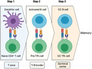
Tfh cell differentiation is a multistep process: Tfh cells first emerge after contact with DCs in the T‐cell zone (step 1), migrate to the T/B border to interact with activated B cells (step 2), and together with continued interactions with B cells seed GCs in follicles (step 3).

Introduction
Upon activation, naïve CD4+ T cells can differentiate into distinct subsets of effector Th cells with varied functions. T follicular helper (Tfh) cells form a unique subset of CD4+ T cells that provides help to B cells and is essential for GC formation and regulation [1, 2, 3, 4, 5]. The differentiation and function of Tfh cells have been shown to determine the kinetics and magnitude of GC B‐cell responses and the generation of high‐affinity antibodies [1, 2, 3, 4, 5]. Tfh cell differentiation is commonly regarded as a multistage process (Figure 1 ). During the early stages of CD4+ T‐cell responses, antigen presentation by DCs activates naïve CD4+ T cells and initiates Tfh cell differentiation including the induction of the chemokine receptor CXCR5 and the transcription factor Bcl6. Besides TCR signaling, costimulation and the local cytokine milieu are also critical for Tfh cell fate decision. After initial interactions with DCs, terminal Tfh cell differentiation requires interactions with B cells. In the past decade, great progress has been made in our understanding of Tfh cell differentiation and function. Here, we will discuss these findings and highlight some important questions regarding how cognate interactions with APCs at the different stages of Tfh cell differentiation play a central role in this process and how this knowledge can contribute to a better understanding of COVID‐19.
Figure 1.

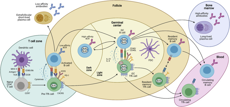
Antigen‐dependent multistep differentiation of T follicular helper cells. Naïve CD4+ T cells are primed by dendritic cells (DCs) in the T‐cell zones of secondary lymphoid organs such as the spleen or lymph nodes. This antigen‐specific interaction is mediated by presentation of processed peptides by MHC‐II molecules that are recognized by the cognate TCR. Together with the expression of costimulatory molecules and production of cytokines, these signals induce the Tfh cell differentiation program that is characterized by upregulation of the chemokine receptor CXCR5 and downregulation of CCR7, which allows these cells to migrate to the T/B border where they interact with activated B cells. This second interaction with an APC type coincides with the expression of characteristic costimulatory (e.g. ICOS) and coinhibitory receptors (e.g. PD‐1) by the Tfh cells, which may be called “pre‐Tfh” cells at this stage. After this cellular interaction, some activated B cells become extrafollicular short‐lived plasma cells (PCs) that secrete low‐affinity antibodies and some of the interacting T and B cells relocate to the follicle to form germinal centers (GCs). In these highly specialized microanatomical structures that consist of dark zones (DZ) and light zones (LZ), GC Tfh and B cells continue to interact in an antigen‐specific manner. GC Tfh cells are more polarized and express higher levels of PD‐1, Bcl6, and CXCR5 than “pre‐Tfh” cells. In addition, they express the adaptor molecule SAP that is important for interactions with GC B cells and they produce IL‐21 and IL‐4 that act on the B cells. While GC B cells mutate their immunoglobulin genes and proliferate in the DZ, GC Tfh cells provide essential signals to B cells in the LZ of the GC, where they cooperate with follicular dendritic cells (FDCs) in the selection of high‐affinity B‐cell clones and in the generation of memory B cells, which recirculate in the blood, and long‐lived plasma cells that find their niche in the bone marrow to maintain serological memory through the secretion of high‐affinity antibodies. Once the GC reaction is resolved, some memory CXCR5+ CD4+ T cells reside in close proximity with memory B cells in lymphoid organs, while others are released from the draining lymphoid organs and circulate in the blood. Both memory Tfh cell subsets express decreasing amounts of Bcl6, CXCR5, and other costimulatory/inhibitory molecules as compared to their effector counterparts. Upon antigen rechallenge, these cells rapidly acquire the effector functions of Tfh cells and strongly support secondary immune response.
DC priming for initial Tfh cell differentiation
DCs are the most important APCs for naïve CD4+ T‐cell activation and differentiation (Figure 1 ). During the initiation of the CD4+ T‐cell response, DCs are sufficient to prime naïve CD4+ T cells and generate CXCR5+ Tfh cells [6, 7]. However, further differentiation into GC Tfh cells still requires B‐cell interactions [8]. DCs can be subdivided into conventional DC (cDC) and plasmacytoid DC. cDCs can be further subdivided into cDC1 and cDC2 based on their division of labor and both subsets have been implicated in the differentiation of Tfh cells [9]. Using Batf3‐deficient mice (lacking cDC1s) and Dock8‐deficient mice (lacking cDC2s), it was shown that the migratory cDC2s, but not cDC1s, uniquely induced Tfh cell responses at the T‐B border [10]. Further, it was demonstrated that monocyte‐derived DCs were able to promote Tfh cell differentiation in specific inflammatory contexts in mice [11]. It was also demonstrated that TCR‐transgenic EBI2‐deficient CD4+ T cells after adoptive transfer and priming exhibited reduced Tfh cell differentiation in a B cell‐independent manner [12]. More precisely, most of the TCR‐transgenic EBI2‐sufficient activated CD4+ T cells colocalized with cDC2s in the outer T zone. In contrast, in the absence of EBI2, activated TCR‐transgenic CD4+ T cells were dispersed, with only some colocalizing in proximity of cDC2s. Further, the authors revealed a novel mechanism underlying DC‐T‐cell interactions at the inter‐follicular region with cDC2s expressing membrane‐bound CD25 (IL‐2R) and releasing soluble IL‐2R to absorb the surrounding IL‐2, thereby reducing IL‐2‐mediated suppression of Tfh cell differentiation as previously reported [13, 14, 15].
During CD4+ T‐cell priming, increased doses of antigen favor the differentiation of Tfh and GC Tfh cells [16]. It was also shown that in the absence of DCs, high doses of antigen were able to overcome the defective Tfh cell differentiation [17]. In the context of viral infection, it was demonstrated that mice in which DCs are constitutively ablated did not show marked alterations of Tfh cell differentiation and response after systemic low‐dose infection of mice with lymphocytic choriomeningitis virus [18]. In addition to antigen dose and nature of the antigen, it has been shown that antigen size also impacts Tfh cell differentiation. Indeed, immunization with antigen‐loaded 200 nm particles induced a stronger Tfh cell and antibody response and provided protective immunity to influenza virus infection in mice as compared to smaller (40 nm) antigen‐loaded particles, despite of inducing a similar activated T‐cell response [19]. This suggested that increasing antigen size could lead to sustained antigen presentation by DCs, resulting in enhanced Tfh cell differentiation.
TCR affinity, TCR signal strength, and TCR tonic signaling in Tfh cell differentiation
Using two distinct TCR‐transgenic mouse strains with different TCR affinities for the exact same peptide‐MHC class II (pMHCII) complex, it was shown that high‐affinity T cells developed significantly more into Tfh cells, suggesting that a high‐affinity TCR on the surface of naïve T cells preferentially induces the Tfh cell program [20]. The link between TCR affinity and Tfh cell differentiation was further studied by monitoring the progeny of naïve CD4+ T cells after pathogen infection [21]. The authors showed that naïve CD4+ T cells specific for a certain pMHCII underwent distinct effector fates based on their unique TCR, thus, each naïve T cell is poised to generate certain types of effector cells. The authors demonstrated that when the dwell time of TCR/pMHCII interaction increased, Tfh cell differentiation also increased and reached a plateau. As stated above, studies in viral infection models have shown that the IL‐2/STAT5 pathway suppresses Tfh cell differentiation [14, 15, 16]. Using IL‐2 reporter mouse strains, it was shown that newly activated CD4+ T cells could be subdivided into IL‐2+ cells enriched for Bcl6 mRNA expression and IL‐2– T cells expressing higher mRNA levels of Blimp‐1, which is encoded by Prdm1 [22]. It was further demonstrated that IL‐2 reporter expression was restricted to CXCR5+ CD4+ T cells and that specific depletion of IL‐2‐producing cells inhibited Tfh cell differentiation, suggesting that Tfh cells derived from IL‐2‐producing cells. The authors also demonstrated that IL‐2 production and Tfh cell differentiation correlated with TCR signal strength and that higher TCR signaling favored Tfh cell differentiation. Interestingly, it has been reported that low‐affinity antigen did not impact Tfh cell differentiation [23].
How TCR signaling induces downstream transcriptional regulation that influences T‐cell differentiation has recently been described. It was shown that the TCR signal‐induced transcription factor IRF4 was essential for the differentiation of Bcl6‐expressing Tfh cells [24]. It was further found that the amount of IRF4 was increased proportionately to TCR signal strength [25]. Strikingly, the authors also showed that in specific conditions of increased TCR signaling, high amounts of antigen or overexpression of IRF4, reduced Tfh cell differentiation. Mechanistically, the authors demonstrated that greater IRF4 levels allowed binding to low‐affinity binding sites that were enriched in non‐Tfh effector genes, a process that was independent of IL‐2 signaling. Another layer of difficulty was recently added to this field of investigation. It was questioned whether another TCR‐dependent factor could contribute to Tfh cell fate determination, namely peripheral TCR signaling in response to self‐pMHCII, also called tonic signaling [26]. It was revealed that tonic signaling instructed Tfh cell fate, where strong tonic signaling inhibited Tfh cell differentiation and weak tonic signaling promoted it. Overall, stronger TCR signaling favored Tfh cell differentiation either through decreased tonic signaling strength and/or through TCR activation induced by increased antigen dose.
While the above studies investigated the role of TCR signaling in Tfh cell differentiation, it remains elusive when the divergence between CXCR5– and CXCR5+ cells occurs upon activation of naïve CD4+ T cells. Using single‐cell RNA sequencing to map effector CD4+ T‐cell differentiation, the developmental trajectories of Th1 and Tfh cells were recently reconstructed during blood‐stage Plasmodium infection in mice [27]. The authors demonstrated that both cell subsets diverged after the initial cycle of cell proliferation associated with an upregulation of aerobic glycolysis and accelerated cell cycling. This is consistent with the finding that CXCR5 expression was particularly upregulated in activated CD4+ T cells that had proliferated the most and which coexpressed high levels of Bcl6 [7].
Cognate T/B interactions in Tfh cell differentiation and maintenance
After priming by APCs, activated CD4+ T cells proliferate and those cells upregulating CXCR5 and concomitantly downregulating CCR7, migrate to the T/B border where they interact with antigen‐primed B cells in a cognate fashion [28, 29] (Figure 1 ). Eventually, some of these early Tfh cells enter deeper into the B‐cell follicles and contribute to the formation of GCs, their terminal differentiation into GC Tfh cells being dependent on B cells [8, 30‐32]. Indeed, interaction with cognate B cells allows stable Bcl6 and, in turn, CXCR5 expression. Thus, efficient T‐ and B‐cell interactions are important as seen by the absence of GC Tfh cell differentiation in SAP‐deficient mice [33, 34, 35, 36]. Moreover, other costimulatory signals are critical for terminal Tfh cell differentiation. ICOS/ICOS‐L signaling was demonstrated to be crucial [37, 38, 39]. ICOS overexpression in mice that carry a mutation in the roquin gene led to extensive Tfh cell differentiation and lupus‐like syndrome [40]. Moreover, BCR stimulation inversely correlated with ICOS‐L expression, which consequently decreased Tfh cell differentiation [41]. Thus, the decision between the extra‐follicular versus the GC pathway relies on affinity for the antigen of the BCR expressed by naïve B cells [42] and the impact of BCR affinity on the quality of T/B interactions [41].
GC Tfh cells interact with GC B cells in the light zone (Figure 1 ). One important characteristic of GC reactions is affinity maturation. It was shown in mice that increased propensity to present antigen to Tfh cells and to interact with GC Tfh cells led to a decrease in the number of high‐affinity B cells since GC B cells were in less competition with each other [43]. These observations provided evidence for a model in which Tfh cells are the limiting factor that shape the GC response and affinity selection therein. Using similar technologies, another study showed that Tfh cells were able to recirculate between different GCs, thus, maximizing the diversification of T cell help during GC responses [44]. Notably, Tfh‐GC B‐cell interaction impacts the outcome of GC B cells, with stronger interaction promoting formation of plasma cell precursors versus recycling GC cell fate [45]. Continued T cell‐specific Bcl6 expression in CD4+ T cells is required for the maintenance of established Tfh cells and GCs, and prevents the trans‐differentiation of established Tfh cells into Th1 cells during acute viral infection [46]. Similarly, global miRNA expression is not only required for the induction of Tfh cells [47], but also for their maintenance [48], further highlighting the fragile nature of the Tfh cell program, which is dependent on continued presentation of antigen as well as costimulation [8, 16, 37, 38, 49].
Tfh memory
Initially, Tfh cells were thought to be effector cells that would die once the GC reaction had ended [50, 51]. Nevertheless, it was also reported that CXCR5+ CD4+ memory T cells form in mice after protein vaccination and viral infection [52, 53, 54, 55] and their existence in humans was also demonstrated [56, 57, 58, 59, 60]. While it was further shown that Bcl6 was essential for the formation of memory CD4+ T cells in mice [61], intrinsic Bcl6 expression was lower in memory cells as compared to GC Tfh cells [62, 63, 64], which correlated with a less differentiated phenotype than their effector counterparts in mice [55, 65, 66]. Finally, 30 days after immunization of Bcl6‐reporter mice, it was demonstrated that Bcl6+ CXCR5+ T cells were memory Tfh cells, since they preferentially gave rise to GC Tfh cells upon reactivation [62]. Interestingly, the Tfh cell compartment can be subdivided into resident cells, which remain in the draining LNs as well as circulating cells in nondraining secondary lymphoid organs or in the circulation (Figure 1 ). Both cell subsets have the capacity to support secondary humoral immune responses, but they reside in different anatomical locations, with resident cells being in B‐cell follicles and circulating ones in T‐cell areas or in the blood [67]. Further, this cell subdivision was demonstrated to rely on cognate interactions between resident memory B and T cells, from which the circulating T‐cell pool emerged, emphasizing the role of antigen presentation even in the memory phase. In this context, it was observed that after antigen reactivation, memory B cells induced rapid Bcl6 expression and effector function by memory Tfh cells, thereby, highlighting the close functional relationship between memory B cells and Tfh cells [68]. In mice infected with Listeria monocytogenes, it was shown that CXCR5+ memory CD4+ T cells were multipotent as they generated secondary Tfh‐ and Th1‐cell responses and these cells behaved more like central memory T cells as they expressed CCR7 and were mainly located in T‐cell areas [69]. More recently, it was reported that long‐lived Tfh cells are susceptible to cell death induced by NAD‐mediated ribosylation of P2RX7 [70]. Blocking this process during tissue isolation yielded many additional live cells that were used to confirm by single‐cell RNA sequencing that memory Tfh cells retained plasticity. Whether the memory T‐cell multipotency depends on the plasticity of these cells or whether these cells represent a heterogeneous population remains unclear.
In humans, circulating Tfh (cTfh) cells found in the blood stream represent a relatively easily accessible Tfh cell population that shares characteristics with bona fide Tfh cells found in secondary lymphoid organs [57]. It was shown that these cTfh cells originate from GCs in LNs [71] and they may serve as circulating memory cells that can be rapidly recruited to secondary immune responses. Due to their lower expression of costimulatory molecules, such as ICOS and PD‐1, which might be a consequence of missing antigenic interactions in the blood, they display a resting state, and increased activated cTfh cells can be detected after infections and vaccinations [59, 72]. Another interesting connection that still needs to be resolved is the appearance of peripheral Tfh‐like (pTfh) cells that have been described in sero‐positive rheumatoid arthritis patients [73] and in breast cancer patients [74]. These cells express many characteristics of Tfh cells, including expression of PD‐1 and ICOS and colocalization with B cells, yet they lack expression of CXCR5. In the context of Influenza infection, lung‐resident memory T cells were described to promote protective B‐cell responses in a Bcl6‐dependent fashion in nonlymphoid organs in mice [75, 76]. These T cells are CXCR5‐negative and exhibit mixed features of Tfh cells and resident memory T cells. They were named resident helper T cells (TRH). It will be important in the future to study the function of pTfh and TRH cells and their relationship to cTfh and bona fide Tfh cells.
Tfh cells in SARS‐CoV‐2 infection and vaccination
Given the importance of humoral immunity for fighting infectious pathogens, such as SARS‐CoV‐2, the current COVID‐19 pandemic has sparked great interest in the role of Tfh cells in the immune response to SARS‐CoV‐2 infection as well as their function in newly developed vaccines (Figure 2 ). Elevated frequencies of activated Tfh cells were detected in the blood of nonsevere COVID‐19 patients [77] and in the LNs of SARS‐CoV‐2‐infected rhesus macaques [78]. High frequencies of activated cTfh cells correlated with low disease severity in COVID‐19 patients [79].
Figure 2.

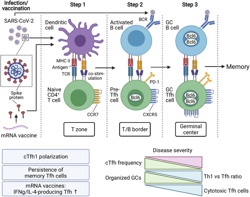
Impact of SARS‐CoV‐2 infection and vaccination on T follicular helper cell differentiation and function. Upon infection of the host with SARS‐CoV‐2, virus antigens are taken up by dendritic cells, which process and present them on MHC‐II to naïve CD4+ T cells and induce T follicular helper (Tfh) cell differentiation toward type 1 polarization. Stronger Tfh1 cell polarization inversely correlates with COVID‐19 severity. After encounter of activated B cells at the T/B border, some Tfh cells further increase their expression of Bcl6, CXCR5, and PD‐1, and form germinal centers together with cognate B cells. Absence of GC structures has been reported in symptomatic patients with severe COVID‐19 and may be connected with an increase in cytotoxic Tfh cell frequencies. Importantly, persistence of SARS‐CoV‐2‐specific memory Tfh cells has been demonstrated several months after natural infection. mRNA vaccines encoding full‐length SARS‐CoV‐2 spike protein promote efficient Tfh cell differentiation with robust coproduction of IFN‐γ and IL‐4, GC formation, and protective humoral responses against SARS‐CoV‐2.
The Th1‐polarizing conditions of a viral infection usually result in the predominant generation of type‐1 cTfh cells, such as in influenza vaccination [59, 80], following the live‐attenuated yellow fever vaccine [72], and in Hepatitis C Virus infection [81, 82]. A similar type‐1 polarization with increased ICOS expression levels has also been observed during SARS‐CoV‐2 infection in humans [83, 84, 85, 86, 87] and in rhesus macaques [78]. Importantly, functional spike‐specific CXCR5‐positive memory cTfh cells and CXCR5‐negative T cells persisted for at least 6 months after symptom onset [88, 89]. Even though Tfh cells were not analyzed, a recent report suggested antigen‐persistence in the gut driving the evolution of anti‐SARS‐CoV‐2 memory B cell and antibody responses, which might involve Tfh cells as well [90].
Single‐cell RNA‐seq analysis of COVID‐19 patients identified increased frequencies of Tfh cells with cytotoxic features and decreased frequencies of regulatory T cells among SARS‐CoV‐2‐reactive CD4+ T cells of hospitalized patients [91]. These cytotoxic Tfh cells were particularly increased early on in hospitalized versus nonhospitalized patients and their presence inversely correlated with levels of SARS‐CoV‐2 spike protein‐specific antibodies. Since cytotoxic Tfh cells have been described to directly kill B cells and their frequency correlated with disease severity in recurrent group A Streptococcus tonsillitis [92], it is plausible, that cytotoxic Tfh cells also contribute to lower humoral activity in severe COVID‐19 patients. In this regard, it is interesting to note that GCs were reported to be largely absent in postmortem spleen and LNs of acutely infected SARS‐CoV‐2 patients, with a block in Bcl6+ Tfh cells and a converse increase in Th1 cells [93]. Furthermore, reduced CXCR5 expression by B and T cells was observed in moderate and severe COVID‐19 patients, further indicating that impaired T/B crosstalk may precipitate dysregulated humoral immune responses [86, 94].
By comparing a SARS‐CoV‐2 mRNA vaccine that encoded the receptor‐binding domain (RBD) and the full‐length spike protein of SARS‐CoV‐2 with a recombinant SARS‐CoV‐2 RBD (rRBD) protein that was formulated with the MF59‐like AddaVax adjuvant, it was shown in mice, that the mRNA vaccine induced more potent Tfh cell and GC responses characterized by stronger CXCR5 and ICOS levels on Tfh cells. In addition, the mRNA vaccine led to robust coproduction of IFN‐γ and IL‐4, resembling a combined Th1/Th2 polarization, which, in contrast to the Th2‐polarized Tfh cell response elicited by the rRBD protein vaccine, translated into higher neutralizing antibody titers [95]. These data further support the efficacy of the first SARS‐CoV‐2 vaccines in humans that have been authorized by the FDA under emergency use authorization and that are also based on mRNA vaccine technology. It is anticipated that more in‐depth analyses of Tfh cells and GCs in COVID‐19 patients will provide additional insights into their critical role in anti‐SARS‐CoV‐2 immunity and vaccination.
Conclusions
Tfh cells are a critical component of potent humoral immune responses. Therefore, future studies that aim at further dissecting the identity and function of this unique CD4+ T cell population will provide additional opportunities that may be leveraged for improved vaccine design and for novel strategies in the treatment of various diseases including autoimmunity.
Conflict of interest
The authors declare no commercial or financial conflict of interest.
Abbreviations
- cDC
conventional DC
- cTfh
circulating Tfh cells
- pMHCII
peptide‐MHC class II
- pTfh
peripheral Tfh‐like cells
- RBD
receptor‐binding domain
- rRBD
recombinant SARS‐CoV‐2 RBD
- Tfh
T follicular helper cells
- TRH
resident helper T cells
Acknowledgments
This work was supported by Deutsche Forschungsgemeinschaft (DFG, German Research Foundation) under Emmy Noether Programme BA 5132/1‐2 (252623821) and Germany's Excellence Strategy EXC2151 (390873048) to D.B. and The Inspire Program from the Region Occitanie/Pyrénées‐Méditerranée (#1901175), the European Regional Development Fund (MP0022856), Institut National du Cancer (INCA‐12642), and Agence Nationale de la Recherche (ANR‐16‐CE15‐0019‐02 and ANR‐16‐CE15‐0002‐02) to N.F.; D.B. is an associated scientist of the Bundesministerium für Bildung und Forschung (BMBF, Federal Ministry of Education and Research)‐funded initiative COVIMMUNE (01KI20343). Artwork was created with BioRender.com.
[Due to a technical error the wrong file was initially displayed.]
Contributor Information
Dirk Baumjohann, Email: dirk.baumjohann@uni-bonn.de.
Nicolas Fazilleau, Email: nicolas.fazilleau@inserm.fr.
References
- 1. Crotty, S. , Helper cell biology: a decade of discovery and diseases. Immunity 2019. 50: 1132–1148. [DOI] [PMC free article] [PubMed] [Google Scholar]
- 2. Vinuesa, C. G. , Linterman, M. A. , Yu, Di , Maclennan, I. C. M. , Follicular helper T cells. Annu. Rev. Immunol. 2016. 34: 335–368. [DOI] [PubMed] [Google Scholar]
- 3. Fazilleau, N. , Mark, L. , Mcheyzer‐Williams, L. J. , Mcheyzer‐Williams, M. G. , Follicular helper T cells: lineage and location. Immunity 2009. 30: 324–335. [DOI] [PMC free article] [PubMed] [Google Scholar]
- 4. Qi, H. , T follicular helper cells in space‐time. Nat. Rev. Immunol. 2016.16: 612–625. [DOI] [PubMed] [Google Scholar]
- 5. Song, W. , Craft, J. , T follicular helper cell heterogeneity: time, space, and function. Immunol. Rev. 2019. 288: 85–96. [DOI] [PMC free article] [PubMed] [Google Scholar]
- 6. Goenka, R. , Barnett, L G. , Silver, J. S. , O'neill, P. J. , Hunter, C. A. , Cancro, M. P. , Laufer, T. M. , Cutting edge: dendritic cell‐restricted antigen presentation initiates the follicular Th cell program but cannot complete ultimate effector differentiation. J. Immunol. 2011. 187: 1091‐1095. [DOI] [PMC free article] [PubMed] [Google Scholar]
- 7. Baumjohann, D. , Okada, T. , Ansel, K. M : Distinct waves of BCL6 expression during T follicular helper cell development. J. Immunol. 2011. 187: 2089–2092. [DOI] [PubMed] [Google Scholar]
- 8. Deenick, E. K. , Chan, A. , Ma, C. S. , Gatto, D. , Schwartzberg, P. L. , Brink, R. , Tangye, S. G. , Follicular helper T cell differentiation requires continuous antigen presentation that is independent of unique B cell signaling. Immunity 2010. 33: 241–253. [DOI] [PMC free article] [PubMed] [Google Scholar]
- 9. Krishnaswamy, J. K. , Alsén, S. , Yrlid, U. , Eisenbarth, S. C. , Williams, A. , Determination of T follicular helper cell fate by dendritic cells. Front. Immunol. 2018. 9: 2169. [DOI] [PMC free article] [PubMed] [Google Scholar]
- 10. Krishnaswamy, J. K. , Gowthaman, U. , Zhang, B. , Mattsson, J. , Szeponik, L. , Liu, D. , Wu, R. et al., Migratory CD11b(+) conventional dendritic cells induce T follicular helper cell‐dependent antibody responses. Sci. Immunol. 2017. 2: eaam9169. [DOI] [PMC free article] [PubMed] [Google Scholar]
- 11. Chakarov, S. , Fazilleau, N. , Monocyte‐derived dendritic cells promote T follicular helper cell differentiation. EMBO Mol. Med. 2014. 6: 590–603. [DOI] [PMC free article] [PubMed] [Google Scholar]
- 12. Li, J. , Lu, E. , Yi, T. , Cyster, J. G. , EBI2 augments Tfh cell fate by promoting interaction with IL‐2‐quenching dendritic cells. Nature 2016. 533: 110–114. [DOI] [PMC free article] [PubMed] [Google Scholar]
- 13. Ballesteros‐Tato, A. , León, B. , Graf, B. A. , Moquin, A. , Adams, P. S. , Lund, F. E. , Randall, T. D. , Interleukin‐2 inhibits germinal center formation by limiting T follicular helper cell differentiation. Immunity 2012. 36: 847–856. [DOI] [PMC free article] [PubMed] [Google Scholar]
- 14. Johnston, R. J. , Choi, Y. S. , Diamond, J. A. , Yang, J. A. , Crotty, S. , STAT5 is a potent negative regulator of TFH cell differentiation. J. Exp. Med. 2012. 209: 243–250. [DOI] [PMC free article] [PubMed] [Google Scholar]
- 15. Nurieva, R. I. , Podd, A. , Chen, Y. , Alekseev, A. M. , Yu, M. , Qi, X. , Huang, H. et al., STAT5 protein negatively regulates T follicular helper (Tfh) cell generation and function. J. Biol. Chem 2012. 287: 11234–11239. [DOI] [PMC free article] [PubMed] [Google Scholar]
- 16. Baumjohann, D. , Preite, S. , Reboldi, A. , Ronchi, F. , Ansel, K. M , Lanzavecchia, A. , Sallusto, F. , Persistent antigen and germinal center B cells sustain T follicular helper cell responses and phenotype. Immunity 2013. 38: 596–605. [DOI] [PubMed] [Google Scholar]
- 17. Dahlgren, M. W. , Gustafsson‐Hedberg, T. , Livingston, M. , Cucak, H. , Alsén, S. , Yrlid, U. , Johansson‐Lindbom, B. , T follicular helper, but not Th1, cell differentiation in the absence of conventional dendritic cells. J. Immunol. 2015. 194: 5187–5199. [DOI] [PubMed] [Google Scholar]
- 18. Hilpert, C. , Sitte, S. , Matthies, A. , Voehringer, D. , Dendritic cells are dispensable for T cell priming and control of acute lymphocytic Choriomeningitis virus infection. J. Immunol. 2016. 197: 2780–2786. [DOI] [PubMed] [Google Scholar]
- 19. Benson, R. A. , MacLeod, M. K. , Hale, B. G. , Patakas, A. , Garside, P. and Brewer, J. M. , Antigen presentation kinetics control T cell/dendritic cell interactions and follicular helper T cell generation in vivo. Elife 2015. 4: e06994. [DOI] [PMC free article] [PubMed] [Google Scholar]
- 20. Fazilleau, N. , Mcheyzer‐Williams, L. J. , Rosen, H. , Mcheyzer‐Williams, M. G. , The function of follicular helper T cells is regulated by the strength of T cell antigen receptor binding. Nat. Immunol. 2009. 10: 375–384. [DOI] [PMC free article] [PubMed] [Google Scholar]
- 21. Tubo, N. J. , Pagán, A. J. , Taylor, J. J. , Nelson, R. W. , Linehan, J. L. , Ertelt, J. M. , Huseby, E. S. et al., Single naive CD4(+) T cells from a diverse repertoire produce different effector cell types during infection. Cell 2013. 153: 785–796. [DOI] [PMC free article] [PubMed] [Google Scholar]
- 22. DiToro, D. , Winstead, C. J. , Pham, D. , Witte, S. , Andargachew, R. , Singer, J. R. , Wilson, C. G. et al., Differential IL‐2 expression defines developmental fates of follicular versus nonfollicular helper T cells. Science 2018. 361: eaao2933. [DOI] [PMC free article] [PubMed] [Google Scholar]
- 23. Keck, S. , Schmaler, M. , Ganter, S. , Wyss, L. , Oberle, S. , Huseby, E. S. , Zehn, D. et al., Antigen affinity and antigen dose exert distinct influences on CD4 T‐cell differentiation. Proc. Natl. Acad. Sci. USA 2014. 111: 14852–14857. [DOI] [PMC free article] [PubMed] [Google Scholar]
- 24. Bollig, N. , Brustle, A. , Kellner, K. , Ackermann, W. , Abass, E. , Raifer, H. , Camara, B. et al., Transcription factor IRF4 determines germinal center formation through follicular T‐helper cell differentiation. Proc. Natl. Acad. Sci. USA 2012. 109: 8664‐8669. [DOI] [PMC free article] [PubMed] [Google Scholar]
- 25. Krishnamoorthy, V. , Kannanganat, S. , Maienschein‐Cline, M. , Cook, S. L. , Chen, J. , Bahroos, N. et al., The IRF4 gene regulatory module functions as a read‐write integrator to dynamically coordinate T helper cell fate. Immunity 2017. 47: 481–497. [DOI] [PMC free article] [PubMed] [Google Scholar]
- 26. Bartleson, J. M. , Viehmann Milam, A. A. , Donermeyer, D. L. , Horvath, S. , Xia, Y. , Egawa, T. , Allen, P. M. , Strength of tonic T cell receptor signaling instructs T follicular helper cell‐fate decisions. Nat. Immunol. 2020. 21: 1384–1396. [DOI] [PMC free article] [PubMed] [Google Scholar]
- 27. Lönnberg, T. , Svensson, V. , James, K. R. , Fernandez‐Ruiz, D. , Sebina, I. , Montandon, R. , Soon, M. S. F. et al., Single‐cell RNA‐seq and computational analysis using temporal mixture modelling resolves Th1/Tfh fate bifurcation in malaria. Sci. Immunol. 2017. 2: eaal2192. [DOI] [PMC free article] [PubMed] [Google Scholar]
- 28. Hardtke, S. , Ohl, L. , FöRster, R. , Balanced expression of CXCR5 and CCR7 on follicular T helper cells determines their transient positioning to lymph node follicles and is essential for efficient B‐cell help. Blood 2005. 106: 1924–1931. [DOI] [PubMed] [Google Scholar]
- 29. Haynes, N. M. , Allen, C. D. C. , Lesley, R. , Ansel, K. M , Killeen, N. , Cyster, J. G. , Role of CXCR5 and CCR7 in follicular Th cell positioning and appearance of a programmed cell death gene‐1high germinal center‐associated subpopulation. J. Immunol. 2007. 179: 5099–5108. [DOI] [PubMed] [Google Scholar]
- 30. Zaretsky, A. G. , Taylor, J. J. , King, I. L. , Marshall, F. A. , Mohrs, M. , Pearce, E. J. , T follicular helper cells differentiate from Th2 cells in response to helminth antigens. J. Exp. Med. 2009. 206: 991–999. [DOI] [PMC free article] [PubMed] [Google Scholar]
- 31. Johnston, R. J. , Poholek, A. C. , Ditoro, D. , Yusuf, I. , Eto, D. , Barnett, B. , Dent, A. L. et al., Bcl6 and Blimp‐1 are reciprocal and antagonistic regulators of T follicular helper cell differentiation. Science 2009. 325: 1006–1010. [DOI] [PMC free article] [PubMed] [Google Scholar]
- 32. Nurieva, R. I. , Chung, Y. , Hwang, D. , Yang, X. O. , Kang, H. S. , Ma, Li , Wang, Yi‐H et al., Generation of T follicular helper cells is mediated by interleukin‐21 but independent of T helper 1, 2, or 17 cell lineages. Immunity 2008. 29: 138–149. [DOI] [PMC free article] [PubMed] [Google Scholar]
- 33. Crotty, S. , Kersh, E. N. , Cannons, J. , Schwartzberg, P. L. , Ahmed, R. , SAP is required for generating long‐term humoral immunity. Nature 2003. 421: 282–287. [DOI] [PubMed] [Google Scholar]
- 34. Cannons, J. L. , Yu, L. J. , Jankovic, D. , Crotty, S. , Horai, R. , Kirby, M. , Anderson, S. et al., SAP regulates T cell‐mediated help for humoral immunity by a mechanism distinct from cytokine regulation. J. Exp. Med. 2006. 203: 1551–1565. [DOI] [PMC free article] [PubMed] [Google Scholar]
- 35. Qi, H. , Cannons, J. L. , Klauschen, F. , Schwartzberg, P. L. , Germain, R. N. , SAP‐controlled T‐B cell interactions underlie germinal centre formation. Nature 2008. 455: 764–769. [DOI] [PMC free article] [PubMed] [Google Scholar]
- 36. Cannons, J. L. , Qi, H. , Lu, K. T. , Dutta, M. , Gomez‐Rodriguez, J. , Cheng, J. , Wakeland, E. K. et al., Optimal germinal center responses require a multistage T cell:B cell adhesion process involving integrins, SLAM‐associated protein, and CD84. Immunity 2010. 32: 253–265. [DOI] [PMC free article] [PubMed] [Google Scholar]
- 37. Akiba, H. , Takeda, K. , Kojima, Y. , Usui, Y. , Harada, N. , Yamazaki, T. , Ma, J. et al., The role of ICOS in the CXCR5+ follicular B helper T cell maintenance in vivo. J. Immunol. 2005. 175: 2340–2348. [DOI] [PubMed] [Google Scholar]
- 38. Weber, J. P. , Fuhrmann, F. , Feist, R. K. , Lahmann, A. , Al Baz, M. S. , Gentz, L. J. , Vu Van, D. et al., ICOS maintains the T follicular helper cell phenotype by down‐regulating Kruppel‐like factor 2. J. Exp. Med. 2015. 212: 217–233. [DOI] [PMC free article] [PubMed] [Google Scholar]
- 39. Xu, H. , Li, X. , Liu, D. , Li, J. , Zhang, X. , Chen, X. , Hou, S. et al., Follicular T‐helper cell recruitment governed by bystander B cells and ICOS‐driven motility. Nature 2013. 496: 523–527. [DOI] [PubMed] [Google Scholar]
- 40. Yu, D. , Tan, A. H. , Hu, X. , Athanasopoulos, V. , Simpson, N. , Silva, D. G. , Hutloff, A. et al., Roquin represses autoimmunity by limiting inducible T‐cell co‐stimulator messenger RNA. Nature 2007. 450: 299–303. [DOI] [PubMed] [Google Scholar]
- 41. Sacquin, A. , Gador, M. , Fazilleau, N. , The strength of BCR signaling shapes terminal development of follicular helper T cells in mice. Eur. J. Immunol. 2017. 47: 1295‐1304. [DOI] [PubMed] [Google Scholar]
- 42. Paus, D. , Phan, T. G. , Chan, T. D. , Gardam, S. , Basten, A. , Brink, R. , Antigen recognition strength regulates the choice between extrafollicular plasma cell and germinal center B cell differentiation. J. Experiment. Med. 2006. 203: 1081–1091. [DOI] [PMC free article] [PubMed] [Google Scholar]
- 43. Victora, G. D. , Schwickert, T. A. , Fooksman, D. R. , Kamphorst, A. O. , Meyer‐Hermann, M. , Dustin, M. L. , Nussenzweig, M C. , Germinal center dynamics revealed by multiphoton microscopy with a photoactivatable fluorescent reporter. Cell 2010. 143: 592–605. [DOI] [PMC free article] [PubMed] [Google Scholar]
- 44. Shulman, Z. , Gitlin, A. D. , Targ, S. , Jankovic, M. , Pasqual, G. , Nussenzweig, M. C. , Victora, G. D. , T follicular helper cell dynamics in germinal centers. Science 2013. 341: 673–677. [DOI] [PMC free article] [PubMed] [Google Scholar]
- 45. Ise, W. , Fujii, K. , Shiroguchi, K. , Ito, A. , Kometani, K. , Takeda, K. , Kawakami, E. et al., T follicular helper cell‐germinal center B cell interaction strength regulates entry into plasma cell or recycling germinal center cell fate. Immunity 2018. 48: 702–715. [DOI] [PubMed] [Google Scholar]
- 46. Alterauge, D. , Bagnoli, J. W. , Dahlström, F. , Bradford, B. M. , Mabbott, N. A. , Buch, T. , Enard, W. et al., Continued Bcl6 expression prevents the transdifferentiation of established Tfh cells into Th1 cells during acute viral infection. Cell Rep. 2020. 33: 108232. [DOI] [PubMed] [Google Scholar]
- 47. Baumjohann, D. , Kageyama, R. , Clingan, J. M. , Morar, M. M. , Patel, S. , De Kouchkovsky, D. , Bannard, O. et al., The microRNA cluster miR‐17 approximately 92 promotes TFH cell differentiation and represses subset‐inappropriate gene expression. Nat. Immunol. 2013. 14: 840–848. [DOI] [PMC free article] [PubMed] [Google Scholar]
- 48. Zeiträg, J. , Dahlström, F. , Chang, Y. , Alterauge, D. , Richter, D. , Niemietz, J. and Baumjohann, D. , T cell‐expressed microRNAs critically regulate germinal center T follicular helper cell function and maintenance in acute viral infection in mice. Eur. J. Immunol. 2020. 51: 408‐413. [DOI] [PubMed] [Google Scholar]
- 49. Choi, Y. S. , Kageyama, R. , Eto, D. , Escobar, T. C. , Johnston, R. J. , Monticelli, L. , Lao, C. et al., ICOS receptor instructs T follicular helper cell versus effector cell differentiation via induction of the transcriptional repressor Bcl6. Immunity 2011. 34: P932–946. [DOI] [PMC free article] [PubMed] [Google Scholar]
- 50. Linterman, M. A. , Rigby, R. J. , Wong, R. , Silva, D. , Withers, D. , Anderson, G. , Verma, N. K. et al., Roquin differentiates the specialized functions of duplicated T cell costimulatory receptor genes CD28 and ICOS. Immunity 2009. 30: 228–241. [DOI] [PubMed] [Google Scholar]
- 51. Rasheed, A‐Ur , Rahn, H. P. , Sallusto, F. , Lipp, M. , Müller, G. , Follicular B helper T cell activity is confined to CXCR5(hi)ICOS(hi) CD4 T cells and is independent of CD57 expression. Eur. J. Immunol. 2006. 36: 1892–1903. [DOI] [PubMed] [Google Scholar]
- 52. Fazilleau, N. , Eisenbraun, M. D. , Malherbe, L. , Ebright, J. N. , Pogue‐Caley, R. R. , Mcheyzer‐Williams, L. J. , Mcheyzer‐Williams, M. G. , Lymphoid reservoirs of antigen‐specific memory T helper cells. Nat. Immunol. 2007. 8: 753–761. [DOI] [PubMed] [Google Scholar]
- 53. Weber, J. P. , Fuhrmann, F. , Hutloff, A. , T follicular helper cells survive as long‐term memory cells. Eur. J. Immunol. 2012. 42: 1981–1988. [DOI] [PubMed] [Google Scholar]
- 54. Lüthje, K. , Kallies, A. , Shimohakamada, Y. , Belz, G. T. , Light, A. , Tarlinton, D. M. , Nutt, S. L. , The development and fate of follicular helper T cells defined by an IL‐21 reporter mouse. Nat. Immunol. 2012. 13: 491–498. [DOI] [PubMed] [Google Scholar]
- 55. Hale, J. S , Youngblood, B. , Latner, D. R. , Mohammed, A. Ur R. , Ye, L. , Akondy, R. S. , Wu, T. et al., Distinct memory CD4 T cells with commitment to T follicular helper‐ and T helper 1‐cell lineages are generated after acute viral infection. Immunity 2013. 38: 805–817. [DOI] [PMC free article] [PubMed] [Google Scholar]
- 56. Chevalier, N. , Jarrossay, D. , Ho, E. , Avery, D. T. , Ma, C. S. , Yu, Di , Sallusto, F. et al., CXCR5 expressing human central memory CD4 T cells and their relevance for humoral immune responses. J. Immunol. 2011. 186: 5556–5568. [DOI] [PubMed] [Google Scholar]
- 57. Morita, R. , Schmitt, N. , Bentebibel, S. E. , Ranganathan, R. , Bourdery, L. , Zurawski, G. , Foucat, E. et al., Human blood CXCR5(+)CD4(+) T cells are counterparts of T follicular cells and contain specific subsets that differentially support antibody secretion. Immunity 2011. 34: 108–121. [DOI] [PMC free article] [PubMed] [Google Scholar]
- 58. Locci, M. , Havenar‐Daughton, C. , Landais, E. , Wu, J. , Kroenke, M. A. , Arlehamn, C. L. , Su, L. F. et al., Human circulating PD‐1CXCR3CXCR5 memory Tfh cells are highly functional and correlate with broadly neutralizing HIV antibody responses. Immunity 2013. 39: 758–769. [DOI] [PMC free article] [PubMed] [Google Scholar]
- 59. Bentebibel, S. E. , Lopez, S. , Obermoser, G. , Schmitt, N. , Mueller, C. , Harrod, C. , Flano, E. et al., Induction of ICOS+CXCR3+CXCR5+ TH cells correlates with antibody responses to influenza vaccination. Sci. Transl. Med. 2013. 5: 176ra132. [DOI] [PMC free article] [PubMed] [Google Scholar]
- 60. He, J. , Tsai, L. M. , Leong, Y. A. , Hu, X. , Ma, C. S. , Chevalier, N. , Sun, X. et al., Circulating precursor CCR7(lo)PD‐1(hi) CXCR5(+) CD4(+) T cells indicate Tfh cell activity and promote antibody responses upon antigen reexposure. Immunity 2013. 39: 770–781. [DOI] [PubMed] [Google Scholar]
- 61. Ichii, H. , Sakamoto, A. , Arima, M. , Hatano, M. , Kuroda, Y. , Tokuhisa, T. , Bcl6 is essential for the generation of long‐term memory CD4+ T cells. Int. Immunol. 2007. 19: 427–433. [DOI] [PubMed] [Google Scholar]
- 62. Liu, X. , Yan, X. , Zhong, B. , Nurieva, R. I. , Wang, A. , Wang, X. , Martin‐Orozco, N. , Wang, Y. et al., Bcl6 expression specifies the T follicular helper cell program in vivo. J. Exp. Med. 2012. 209: 1841–1852. [DOI] [PMC free article] [PubMed] [Google Scholar]
- 63. Kitano, M. , Moriyama, S. , Ando, Y. , Hikida, M. , Mori, Y. , Kurosaki, T. , Okada, T. , Bcl6 protein expression shapes pre‐germinal center B cell dynamics and follicular helper T cell heterogeneity. Immunity 2011. 34: 961–972. [DOI] [PubMed] [Google Scholar]
- 64. Choi, Y. S. , Yang, J. A. , Yusuf, I. , Johnston, R. J. , Greenbaum, J. , Peters, B. , Crotty, S. , Bcl6 expressing follicular helper CD4 T cells are fate committed early and have the capacity to form memory. J. Immunol. 2013. 190: 4014–4026. [DOI] [PMC free article] [PubMed] [Google Scholar]
- 65. Yusuf, I. , Kageyama, R. , Monticelli, L. , Johnston, R. J. , Ditoro, D. , Hansen, K. , Barnett, B. et al., Germinal center T follicular helper cell IL‐4 production is dependent on signaling lymphocytic activation molecule receptor (CD150). J. Immunol. 2010. 185: 190–202. [DOI] [PMC free article] [PubMed] [Google Scholar]
- 66. Iyer, S. S. , Latner, D. R. , Zilliox, M. J. , Mccausland, M. , Akondy, R. S. , Penaloza‐Macmaster, P. , Hale, J. S. et al., Identification of novel markers for mouse CD4 T follicular helper cells. Eur. J. Immunol. 2013. 43: 3219–3232. [DOI] [PMC free article] [PubMed] [Google Scholar]
- 67. Asrir, A. , Aloulou, M. , Gador, M. , Pérals, C. , Fazilleau, N. , Interconnected subsets of memory follicular helper T cells have different effector functions. Nat. Commun. 2017. 8: 847. [DOI] [PMC free article] [PubMed] [Google Scholar]
- 68. Ise, W. , Inoue, T. , Mclachlan, J. B. , Kometani, K. , Kubo, M. , Okada, T. , Kurosaki, T. , Memory B cells contribute to rapid Bcl6 expression by memory follicular helper T cells. Proc. Natl. Acad. Sci. U.S.A. 2014. 111: 11792–11797. [DOI] [PMC free article] [PubMed] [Google Scholar]
- 69. Pepper, M. , Pagán, A. J. , Igyártó, B. Z. , Taylor, J. J. , Jenkins, M. K. , Opposing signals from the bcl6 transcription factor and the interleukin‐2 receptor generate T helper 1 central and effector memory cells. Immunity 2011. 35: 583–595. [DOI] [PMC free article] [PubMed] [Google Scholar]
- 70. Künzli, M. , Schreiner, D. , Pereboom, T. C. , Swarnalekha, N. , Litzler, L. C. , Lötscher, J. , Ertuna, Y. I. et al., Long‐lived T follicular helper cells retain plasticity and help sustain humoral immunity. Sci. Immunol. 2020. 5: eaay5552. [DOI] [PubMed] [Google Scholar]
- 71. Vella, L. A. , Buggert, M. , Manne, S. , Herati, R. S. , Sayin, I. , Kuri‐Cervantes, L. , Bukh Brody, I. et al., T follicular helper cells in human efferent lymph retain lymphoid characteristics. J. Clin. Invest. 2019. 129: 3185–3200. [DOI] [PMC free article] [PubMed] [Google Scholar]
- 72. Huber, J. E. , Ahlfeld, J. , Scheck, M. K. , Zaucha, M. , Witter, K. , Lehmann, L. , Karimzadeh, H. et al., Dynamic changes in circulating T follicular helper cell composition predict neutralising antibody responses after yellow fever vaccination. Clin. Transl. Immunol. 2020. 9: e1129. [DOI] [PMC free article] [PubMed] [Google Scholar]
- 73. Rao, D. A. , Gurish, M. F. , Marshall, J L. , Slowikowski, K. , Fonseka, C Y. , Liu, Y. , Donlin, L T. et al., Pathologically expanded peripheral T helper cell subset drives B cells in rheumatoid arthritis. Nature 2017. 542: 110–114. [DOI] [PMC free article] [PubMed] [Google Scholar]
- 74. Gu‐Trantien, C. , Loi, S. , Garaud, S. , Equeter, C. , Libin, M. , De Wind, A. , Ravoet, M. et al., CD4+ follicular helper T cell infiltration predicts breast cancer survival. J Clin Invest 2013. 213:2873–2892. [DOI] [PMC free article] [PubMed] [Google Scholar]
- 75. Swarnalekha, N. , Schreiner, D. , Litzler, L C. , Iftikhar, S. , Kirchmeier, D. , Künzli, M. , Son, Y. M. et al., T resident helper cells promote humoral responses in the lung. Sci Immunol 2021. 6: eabb6808. [DOI] [PMC free article] [PubMed] [Google Scholar]
- 76. Son, Y. M. , Cheon, I. S. , Wu, Y. , Li, C. , Wang, Z. , Gao, X. , Chen, Y. et al., Tissue‐resident CD4(+) T helper cells assist the development of protective respiratory B and CD8(+) T cell memory responses. Sci. Immunol. 2021. 6: eabb6852. [DOI] [PMC free article] [PubMed] [Google Scholar]
- 77. Thevarajan, I. , Nguyen, T H. O. , Koutsakos, M. , Druce, J. , Caly, L. , Van De Sandt, C E. , Jia, X. et al., Breadth of concomitant immune responses prior to patient recovery: a case report of non‐severe COVID‐19. Nat Med 2020. 26: 453–455. [DOI] [PMC free article] [PubMed] [Google Scholar]
- 78. Shaan Lakshmanappa, Y. , Elizaldi, S R. , Roh, J W. , Schmidt, B A. , Carroll, T D. , Weaver, K D. , Smith, J C. et al., SARS‐CoV‐2 induces robust germinal center CD4 T follicular helper cell responses in rhesus macaques. Nat Commun 2021. 12: 541. [DOI] [PMC free article] [PubMed] [Google Scholar]
- 79. Rydyznski Moderbacher, C. , Ramirez, S I. , Dan, J M. , Grifoni, A. , Hastie, K M. , Weiskopf, D. , Belanger, S. et al., Antigen‐specific adaptive immunity to SARS‐CoV‐2 in acute COVID‐19 and associations with age and disease severity. Cell 2020. 183: 996–1012. [DOI] [PMC free article] [PubMed] [Google Scholar]
- 80. Koutsakos, M. , Wheatley, A K. , Loh, L. , Clemens, E. B , Sant, S. , Nüssing, S. , Fox, A. et al., Circulating T(FH) cells, serological memory, and tissue compartmentalization shape human influenza‐specific B cell immunity. Sci Transl Med 2018. 10: eabb6852. [DOI] [PubMed] [Google Scholar]
- 81. Zhang, J. , Liu, W. , Wen, Bo , Xie, T. , Tang, P. , Hu, Y. , Huang, L. et al., Circulating CXCR3(+) Tfh cells positively correlate with neutralizing antibody responses in HCV‐infected patients. Sci Rep 2019. 9: 10090. [DOI] [PMC free article] [PubMed] [Google Scholar]
- 82. Smits, M. , Zoldan, K. , Ishaque, N. , Gu, Z. , Jechow, K. , Wieland, D. , Conrad, C. et al., Follicular T helper cells shape the HCV‐specific CD4+ T cell repertoire after virus elimination. J Clin Invest 2020. 130: 998–1009. [DOI] [PMC free article] [PubMed] [Google Scholar]
- 83. Juno, J A. , Tan, H. ‐. X. , Lee, W. S. , Reynaldi, A. , Kelly, H G. , Wragg, K. , Esterbauer, R. et al., Humoral and circulating follicular helper T cell responses in recovered patients with COVID‐19. Nat Med 2020.26: 1428–1434. [DOI] [PubMed] [Google Scholar]
- 84. Gong, F. , Dai, Y. , Zheng, T. , Cheng, L. , Zhao, D. , Wang, H. , Liu, M. et al., Peripheral CD4+ T cell subsets and antibody response in COVID‐19 convalescent individuals. J Clin Invest 2020. 130: 6588–6599. [DOI] [PMC free article] [PubMed] [Google Scholar]
- 85. Neidleman, J. , Luo, X. , Frouard, J. , Xie, G. , Gill, G. , Stein, E S. , Mcgregor, M. et al., SARS‐CoV‐2‐specific T cells exhibit phenotypic features of helper function, lack of terminal differentiation, and high proliferation potential. Cell Rep Med 2020. 1: 100081. [DOI] [PMC free article] [PubMed] [Google Scholar]
- 86. Mathew, D. , Giles, J. R. , Baxter, A. E. , Oldridge, D. A. , Greenplate, A. R. , Wu, J. E. , Alanio, C. et al., Deep immune profiling of COVID‐19 patients reveals distinct immunotypes with therapeutic implications. Science 2020. 369: eabc8511. [DOI] [PMC free article] [PubMed] [Google Scholar]
- 87. Zhang, J. , Wu, Q. , Liu, Z. , Wang, Q. , Wu, J. , Hu, Y. , Bai, T. et al., Spike‐specific circulating T follicular helper cell and cross‐neutralizing antibody responses in COVID‐19‐convalescent individuals. Nat Microbiol 2021. 6: 51–58. [DOI] [PubMed] [Google Scholar]
- 88. Rodda, L. B. , Netland, J. , Shehata, L. , Pruner, K. B. , Morawski, P. A. , Thouvenel, C. D. , Takehara, K. K. et al., Functional SARS‐CoV‐2‐specific immune memory persists after mild COVID‐19. Cell 2020. 184: 169‐183. [DOI] [PMC free article] [PubMed] [Google Scholar]
- 89. Dan, J M. , Mateus, J. , Kato, Yu , Hastie, K M. , Yu, E. D. , Faliti, C E. , Grifoni, A. et al., Immunological memory to SARS‐CoV‐2 assessed for up to 8 months after infection. Science 2021. 371: eabf4063. [DOI] [PMC free article] [PubMed] [Google Scholar]
- 90. Gaebler, C. , Wang, Z. , Lorenzi, J C. C. , Muecksch, F. , Finkin, S. , Tokuyama, M. , Cho, A. et al., Evolution of antibody immunity to SARS‐CoV‐2. Nature 2021. 591: [DOI] [PMC free article] [PubMed] [Google Scholar]; 639–644.
- 91. Meckiff, B J. , Ramírez‐Suástegui, C. , Fajardo, V. , Chee, S J. , Kusnadi, A. , Simon, H. , Eschweiler, S. et al., Imbalance of regulatory and cytotoxic SARS‐CoV‐2‐reactive CD4(+) T cells in COVID‐19. Cell 2020. 183: 1340–1353. [DOI] [PMC free article] [PubMed] [Google Scholar]
- 92. Dan, J. M. , Havenar‐Daughton, C. , Kendric, K. , Al‐Kolla, R. , Kaushik, K. , Rosales, S. L. , Anderson, E. L. et al., Recurrent group A Streptococcus tonsillitis is an immunosusceptibility disease involving antibody deficiency and aberrant TFH cells. Sci Transl Med 2019. 11. [DOI] [PMC free article] [PubMed] [Google Scholar]
- 93. Kaneko, N. , Kuo, H. ‐. H. , Boucau, J. , Farmer, J R. , Allard‐Chamard, H. , Mahajan, V S. , Piechocka‐Trocha, A. et al., Loss of Bcl‐6‐expressing T follicular helper cells and germinal centers in COVID‐19. Cell 2020. 183: 143–157. [DOI] [PMC free article] [PubMed] [Google Scholar]
- 94. Su, Y. , Chen, D. , Yuan, D. , Lausted, C. , Choi, J. , Dai, C L. , Voillet, V. et al., Multi‐omics resolves a sharp disease‐state shift between mild and moderate COVID‐19. Cell 2020. 183: 1479–1495. [DOI] [PMC free article] [PubMed] [Google Scholar]
- 95. Lederer, K. , Castaño, D. , Gómez Atria, D. , Oguin, T H. , Wang, S. , Manzoni, T B. , Muramatsu, H. et al., SARS‐CoV‐2 mRNA vaccines foster potent antigen‐specific germinal center responses associated with neutralizing antibody generation. Immunity 2020. 53: 1281–1295. [DOI] [PMC free article] [PubMed] [Google Scholar]


