Originally published in:
Radiology: Cardiothoracic Imaging 2020;2(6):e200219
Erratum in:
Radiology: Cardiothoracic Imaging 2021;3(3):e219001
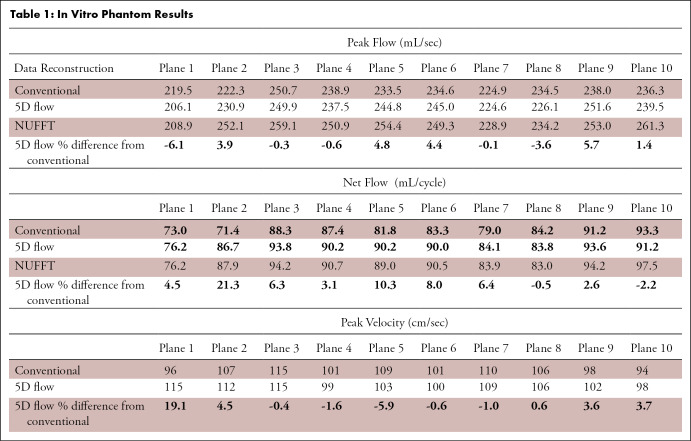
This erratum corrects an error in Table 1. The original table wrongly duplicated the peak flow values for Conventional and 5D flow into the Net Flow category and a corresponding incorrect percent difference for both Net Flow and Peak Flow. As a result, the original article incorrectly stated net flow was within 7% of conventional 4D flow at all planes. Using the corrected values, 5D flow peak flow, peak velocity, and net flow still showed good-to-excellent agreement with values within 7% of conventional at most planes. Higher variability (8.0% to 21.3% difference) was demonstrated in planes at the edge of the acquired FOV (1, 2, 5, 6) for net flow and peak velocity. In addition, the original peak flow values were reported correctly; however, the percent difference was incorrectly represented as 5D flow having up to a 6.5% difference compared to conventional 4D flow. The updated calculations depict up to a 6.1% difference in peak flow between techniques.
Table 1:
In Vitro Phantom Results
While the demonstrated variability in net flow was higher than originally suggested, 5D flow-derived parameters nonetheless showed good-to-excellent agreement and none of the original conclusions were altered by these new values.
The table has been amended online and should appear as follows:


