

Dear Editor,
A 56‐year‐old woman, with personal history of lichen planus 7 years prior that had been successfully treated with topical therapy, was referred to our dermatology outpatient unit from a primary care centre. She complained of pruritic lesions that had appeared 48 h after the second dose of the COVID‐19 vaccine (Comirnaty, Pfizer, New York, NY, USA; BioNTech, Mainz, Germany). The lesions had developed in the ankles and subsequently extended to the flexural wrist and forearms, periumbilical area, mammary and axillary folds.
When asked about possible alternative triggers, the patient denied any signs of infection, changes in medication or stress in the weeks prior. Upon physical examination, she showed polygonal, well‐delimited, erythematous papules in the ankles (Fig. 1), periumbilical area (Fig. 2), flexural wrist and forearms and mammary and axillary folds. Dermoscopy examination revealed slight desquamation and Wickham's striae. No mucosal or nail involvement was appreciated.
Figure 1.
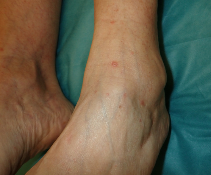
Flat‐topped, erythematous papules on the ankle.
Figure 2.
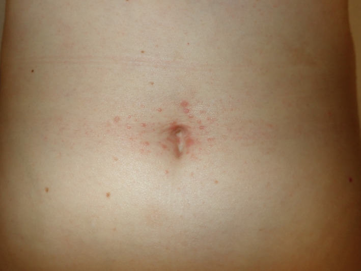
Similar lesions located in the periumbilical area.
A punch biopsy of one of the papules on the arm was performed, showing typical findings of lichen planus with epidermal hyperplasia forming a characteristic saw‐tooth appearance with wedge‐shaped hypergranulosis, vacuolar degeneration of basal layer and dense lymphocytic infiltrate in the superficial dermis. The patient started treatment with high‐potency topical corticosteroids.
Lichen planus is a T cell‐mediated inflammatory disease of unknown origin. Its clinical presentation varies depending on the location in which it appears, affecting mostly the skin, where it shows as violaceous, shiny, polygonal, flat‐topped papules that can be intensely pruritic. This condition has been related to previous exposure to certain agents such as drugs, vaccines and viruses. 1 Lately, it has also been related to the COVID‐19 infection. 1 Hepatitis B virus (HBV), influenza, rabies, Diphtheria, Tetanus, Pertussis (DTaP) and measles, mumps and rubella (MMR) are some of the vaccines that have been previously associated with flares of lichen planus, with HBV being the most common agent implicated. The exact mechanism and the component responsible for this event are yet to be uncovered. 2
There is not little uncertainty surrounding vaccination against COVID‐19. Due to the pressing need, vaccines have been developed and approved at an extraordinarily quick pace. 3 Currently approved vaccines base their action on administering the host with sequences that encode the viral spike protein. This essentially signifies the first‐time gene therapy‐based vaccines, the long‐term effects of which are still unknown, are going to be administered globally. 4 So, despite their safety and efficacy having been demonstrated in respective clinical trials, it is reasonable to be cautious and conduct a more intensive postmarketing vigilance. 5
What has been shown in clinical trials is that the leading vaccines elicit a Th1 response, rising the serum levels of IL‐2, TNFα and IFNγ. 6 , 7 These precise cytokines are involved in the appearance of lichen planus. Although we still do not completely understand its pathogenesis, the up‐regulation of Th1 and increase of pro‐apoptotic cytokines such as TNFα and IFNγ have been described as key agents responsible for basal keratinocyte apoptosis seen in this skin condition. 1
Consequently, these vaccines could imply a surge of certain skin diseases mediated by the previously mentioned factors. Not only could we see flares of lichen planus, but also psoriasis, atopic dermatitis, vitiligo, acne vulgaris, pemphigus vulgaris, neutrophilic dermatoses and certain connective tissue diseases. 4
Given the experience with different vaccines and the mechanism of action of the novel COVID‐19 vaccines, it is plausible that the latter could be responsible for flares of certain skin dermatoses. Although there is still no definite evidence, as dermatologists, we should be aware of the possibility and keep an eye out for worsening or debut of these diseases after the COVID‐19 vaccine.
Funding source
No funding sources supported this work.
Conflicts of interest
None of the authors report any conflicts of interest.
Acknowledgement
The patient in this manuscript has given written informed consent to the publication of her case details.
References
- 1. Lehman J, Tollefson M, Gibson L. Lichen planus. Int J Dermatol 2009; 48: 682–694. [DOI] [PubMed] [Google Scholar]
- 2. Diaz‐Guimaraens B, Dominguez‐Santas M, Suarez‐Valle A, Fernandez‐Nieto D, Jimenez‐Cauhe J, Ballester A. Annular lichen planus associated with coronavirus SARS‐CoV‐2 disease (COVID‐19). Int J Dermatol 2021; 60: 246–247. [DOI] [PubMed] [Google Scholar]
- 3. Rosengard HC, Wheat CM, Tilson MP, Cuda JD. Lichen planus following tetanus–diphtheria–acellular pertussis vaccination: a case report and review of the literature. SAGE Open Med Case Rep 2018; 6: 2050313X1775033. [DOI] [PMC free article] [PubMed] [Google Scholar]
- 4. Koven S. Developing Covid‐19 vaccines at pandemic speed. N Engl J Med 2020; 28: 1–2. [DOI] [PubMed] [Google Scholar]
- 5. Sinha A, Kumar R, Singh AR. Implication of mass COVID‐19 vaccination on dermatology practice in 2021. Dermatol Ther 2021; e14765. 10.1111/dth.14765 [DOI] [PMC free article] [PubMed] [Google Scholar]
- 6. Polack FP, Thomas SJ, Kitchin N et al. Safety and efficacy of the BNT162b2 mRNA Covid‐19 vaccine. N Engl J Med 2020; 383: 2603–2615. [DOI] [PMC free article] [PubMed] [Google Scholar]
- 7. Sahin U, Muik A, Derhovanessian E et al. COVID‐19 vaccine BNT162b1 elicits human antibody and TH1 T cell responses. Nature 2020; 586: 594–599. [DOI] [PubMed] [Google Scholar]