Abstract
Background
Infection with Salmonella enterica usually results in diarrhea, fever, and abdominal cramps, but some people become asymptomatic or chronic carrier as a source of infection for others. This study aimed to analyze the difference in serotype, antimicrobial resistance, and genetic profiles between Salmonella strains isolated from patients and those from asymptomatic people in Nantong city, China.
Methods
A total of 88 Salmonella strains were collected from patients and asymptomatic people from 2017 to 2018. Serotyping, antimicrobial susceptibility testing, and PFGE analysis were performed to analyze the characteristics of these strains.
Results
Twenty serotypes belonging to 8 serogroups were identified in the 88 Salmonella strains. S. Typhimurium remained to be the predominant serotype in strains from both patients and asymptomatic people. Among the 27 strains from patients, S. Enteritidis and S. Rissen were shown as the other two major serotypes, while S. London, S. Derby, and S. Meleagridis were demonstrated as the other significant serotypes among the 61 strains from asymptomatic people. Antimicrobial resistance testing revealed that 84.1% of strains from both resources were multi-drug resistant. PFGE displayed a highly discriminative ability to differentiate strains belonging to S. Derby, S. Typhimurium, etc., but could not efficiently differentiate serotypes like S. Enteritidis.
Conclusions
This study’s results demonstrated that S. Typhimurium could cause human infection in both symptomatic and asymptomatic state; S. London, S. Derby, and S. Meleagridis usually cause asymptomatic infection, while S. Enteritidis infection mainly results in human diseases. The high multi-drug resistance rate detected in the antimicrobial resistance and diverse PFGE profiles of these strains implied that the strains were isolated from different sources, and the increased surveillance of Salmonella from both patients and asymptomatic people should be taken to control the disease.
Supplementary Information
The online version contains supplementary material available at 10.1186/s12879-021-06340-z.
Keywords: Salmonella, Asymptomatic infection, PFGE, Antimicrobial susceptibility
Background
Salmonellosis is an infection with bacteria called Salmonella. Human salmonellosis generally manifests two kinds of disorders: typhoid fever caused by typhoidal Salmonella enterica serotypes S. Typhi, S. Paratyphi A, S. Paratyphi B, and S. Paratyphi C, and another is gastroenteritis caused by nontyphoidal Salmonella (NTS) serotypes such as S. Enteritidis and S. Typhimurium [1]. Salmonella can cause symptomatic infections, which is defined as the occurrence of any number of watery stools during 24 h period accompanied by fever, vomiting or abdominal cramps [2]. Besides, some people can get a Salmonella infection without any symptoms and clear the infection within a few days, or become persistent carrier of the bacteria, which are described as asymptomatic or chronic infection, respectively [3]. A bacterial load of 106–108 CFU of NTS organisms is needed to cause symptomatic disease in healthy adults [4]. It is estimated that approximately 93.8 million gastroenteritis cases and 155,000 deaths are attributed to NTS worldwide annually [5]. According to the United States and European Food Safety Authority (EFSA) data, S. Enteritidis and S. Typhimurium have remained the top serotypes causing clinical human salmonellosis [6, 7]. Both serotypes were predominantly contributed to human gastroenteritis due to Salmonella infection in hospitals from different provinces or cities of China [8–11]. With difference to the serotypes identified in patients, S. Derby, S. London, and S. Senftenberg have been demonstrated to be the major Salmonella serotypes in asymptomatic food handlers as well as S. Typhimurium and S. Enteritidis [12]. Therefore, it is necessary to characterize the difference in Salmonella serotypes between patients and asymptomatic people.
Phenotypic and genotypic methods allow the identification and characterization of bacterial strains with different sources [13]. Salmonella serovars’ appearance with multidrug-resistant (MDR) patterns has increased rapidly and become a heavy burden on the clinical treatment of salmonellosis [4, 14]. A report of 1826 NTS isolates from human patients in Guangdong province of China revealed that 46% of the isolates were MDR, and 72% showed resistance to at least one antimicrobial [8]. Among 109 Salmonella isolates from diarrheagenic children in Beijing, 50% of the strains showed resistance to at least three antimicrobials, and 12.8% were resistant to six [15]. In Malaysia, seven Salmonella strains were isolated from a total of 317 asymptomatic food handlers, and these strains showed multidrug-resistance to ampicillin, chloramphenicol, trimethoprim-sulfamethoxazole, sulfonamides, streptomycin, and tetracycline [16]. Since the bacteria have evolved to be resistant to various antimicrobials, constant antibiotic surveillance is warranted, and the genotyping techniques are essential to tracing the source of the bacteria. The pulsed-field gel electrophoresis (PFGE) was considered as the golden standard genotyping technique for differentiation of Salmonella isolates [17, 18]. Furthermore, it can also be used to trace the source of infections and the transmission route of the strains [18].
This study investigated the serotypes distribution, antimicrobial resistance, and PFGE profiles of S. enterica isolated from patients and asymptomatic people in Nantong, China. By comparing the characteristics of Salmonella strains from two different kinds of sources, we could develop effective strategies to control Salmonella infection in humans.
Methods
Serotyping of Salmonella isolates from human
A total of 88 Salmonella isolates were obtained from patients and asymptomatic people by the Center for Disease Control and Prevention, Nantong, China. Among the 88 strains, 61 was isolated from people on physical examination without any symptoms, while 27 was isolated from patients with diarrhea. This study received ethical approval from the Ethics Committees of Center for Disease Control and Prevention of Nantong city. Serotyping of the isolates was performed using slide agglutination test according to the instructions of Salmonella antisera kit (Tianrun Bio-Pharmaceutical Co. Ltd., Ningbo, China) based on somatic O, as well as phase 1 and phase 2 flagella antigens. The serotype identification of each strain was based on the Kauffmann-White scheme [19].
Antimicrobial susceptibility testing
Susceptibility testing of the 88 Salmonella isolates with the Sensititre National Antimicrobial Resistance Monitoring System Gram-negative susceptibility plates (Customized version, Sensititre; Trek Diagnostic Systems, Inc., Westlake, OH) was performed according to the manufacturer’s instructions. Twenty-six antimicrobial agents (antimicrobial abbreviations and dilution concentration ranges are given in parentheses, in micrograms per milliliter) were used as follows: ampicillin (AMP, 2–64); ampicillin-sulbactam (AMS, 2–64 and 1–32); amoxicillin-clavulanic acid (AMC, 2–64 and 1–32); aztreonam (AZM, 1–32); cefazolin (CFZ, 0.5–16); cefotaxime (CTX, 0.25–8); ceftazidime (CAZ, 0.5–16); cefoxitin (CFX, 2–64); cefepine (FEP, 0.25–16); gentamicin (GEN, 1–32); amikacin (AMI, 1–32); imipenem (IMI, 0.25–8); meropenem (MEM, 0.06–4); chloramphenicol (CHL, 2–64); trimethoprim/sulfamethoxazole (SXT, 0.25–8 and 4.75–152); sulfisoxazole (SUL, 32–512); tetracycline (TET, 1–32); minocycline (MIN, 1–32); nalidixic acid (NAL, 4–64); ciprofloxacin (CIP, 0.03–32); levofloxacin (LEV, 0.125–8); doxycycline (DOX, 0.5–16); kanamycin (KAN, 8–64); streptomycin (STR, 4–32); colistin sulphate (CT, 0.5–16); polymyxin B (PB, 0.5–16); The Clinical and Laboratory Standards Institute (CLSI, 2018) on Antimicrobial Susceptibility Testing breakpoints were used to assess the results [20]. Escherichia coli ATCC25922 with known antimicrobial resistance profiles was used as a quality control organism.
PFGE
PFGE was conducted to reveal the clonal-relatedness of Salmonella strains following the standardized laboratory protocol for molecular subtyping of Salmonella by PFGE [17]. The S. Braenderup H9812 was used as a marker strain. Briefly, agarose-embedded genomic DNA samples were digested with XbaI (Takara, Japan) at 37 °C for 2 h. The Chef Mapper electrophoresis system was then used to separate restriction fragments in 0.5X Tris-borate- ethylenediaminetetraacetic acid (EDTA; TBE) extended-range buffer (Bio-Rad, United States) with recirculation at 14 °C for 18–20 h. The gel was stained with GelRed and visualized under UV light to record the gel results with TIFF images. The genetic patterns for each of the strains were compared and analyzed using the BioNumericus version 7.5 software (Applied Maths, Belgium). A dendrogram was produced using the Dice coefficient correlation and unweighted pair group method using the arithmetic mean algorithm (UPGMA) with 1.5% optimization and a band position tolerance.
Results
Prevalence and distribution of serotypes for Salmonella isolates from humans
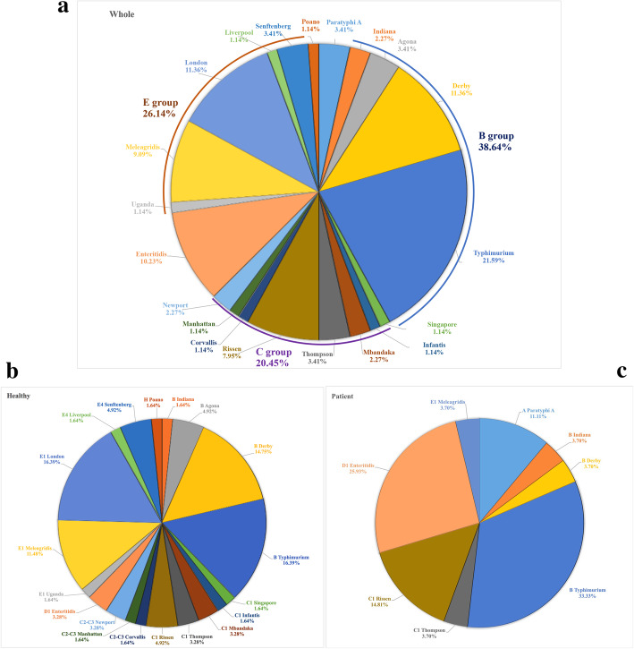
A total of 88 Salmonella strains were isolated, 49 (55.7%) from males and 39 (44.7%) from females in Nantong city (Table 1). Nearly 69.3% (61/88) of strains were isolated from asymptomatic people, while 30.7% (27/88) were from patients (Table 1). Among the 27 strains from patients, 8 (29.6%) were isolated from children. All of the strains with their information, including the age, sex, and samples, were listed in Table S1. Among the identified 88 Salmonella isolates, 20 serotypes distributed in 8 serogroups were identified, with B (38.6%) and E (26.1%) as the top 2 predominant serogroups (Table 2). The S. Typhimurium serotype was detected in 21.6% of the 88 isolates, followed by 12.5% of S. London, 11.4% of S. Derby, 10.2% of S. Enteritidis, and 7.5% of both S. Rissen and S. Meleagridis (Table 2, Fig. 1a). However, compared to 19 serotypes identified in 61 isolates from asymptomatic people, 8 serotypes were detected in 27 isolates from patients (Table 2, Fig. 1b). Only 1 out of 11 S. London isolates was obtained from a patient, while 77.8% (7/9) of S. Enteritidis can cause human disease, and all of the 3 S. Paratyphi A strains were isolated from patients (Table 2, Fig. 1c). S Typhimurium (16.4%) and S. London (16.4%) was the predominant serotype in the isolates from asymptomatic people, followed by S. Derby (14.8%) and S. Meleagridis (11.5%) (Fig. 1b). In contrast, S. Typhimurium (33.3%) and S. Enteritidis (25.9%) were the top 2 serotypes causing symptoms in patients, followed by S. Rissen (14.8%) (Fig. 1c).
Table 1.
The sampling information of Salmonella from humans
| Variable | Value |
|---|---|
| Sex | na (%) |
| Male | 49 (55.7%) |
| Female | 39 (44.3%) |
| Age | median (range) |
| 37 (1–77) | |
| Place | n (%) |
| Nantong CDC | 61 (69.3%) |
| Chongchuan district | 23 (26.1%) |
| Other counties | 4 (4.6%) |
| Source | n (%) |
| Asymptomatic people | 61 (69.3%) |
| Patient | 27 (30.7%) |
an represents the number of strains
Table 2.
Distribution of serotypes for 88 Salmonella isolates from patients and asymptomatic people
| O group (no) | Serotype (no, percentagea) | NO. | Percentage (%) |
|---|---|---|---|
| A (3) | Paratyphi A (3, 100%) | 3 | 3.4 |
| B (34) | Indiana (1, 50%) | 2 | 2.3 |
| Agona | 3 | 3.4 | |
| Derby (1, 10%) | 10 | 11.4 | |
| Typhimurium (9, 47.4%) | 19 | 21.6 | |
| C1 (14) | Singapore | 1 | 1.1 |
| Infantis | 1 | 1.1 | |
| Mbandaka | 2 | 2.3 | |
| Thompson (1, 33.3%) | 3 | 3.4 | |
| Rissen (4, 57.1%) | 7 | 8.0 | |
| C2-C3 (4) | Corvallis | 1 | 1.1 |
| Manhattan | 1 | 1.1 | |
| Newport | 2 | 2.3 | |
| D1 (9) | Enteritidis (7, 77.8%) | 9 | 10.2 |
| E1 (19) | Uganda | 1 | 1.1 |
| Meleagridis (1, 12.5%) | 8 | 9.1 | |
| London | 10 | 11.4 | |
| E4 (4) | Liverpool | 1 | 1.1 |
| Senftenberg | 3 | 3.4 | |
| H (1) | Poano | 1 | 1.1 |
arepresents the number of strains isolated from patients with diarrhea, fever, or abdominal cramps in hospitals and the percentage of these strains among the same serotype
Fig. 1.
Prevalence of Salmonella serotypes in humans. The distribution of Salmonella serotypes among 88 isolates from humans (a), including 61 isolates from asymptomatic people (b) and 27 isolates from patients (c). The B group, C group, and E group represents Salmonella serogroup B (O:4), C [C1(O:7); C2-C3(O:8)], E [E1(O:3,10); E4 (O:1,3,19)], respectively
Antimicrobial resistance
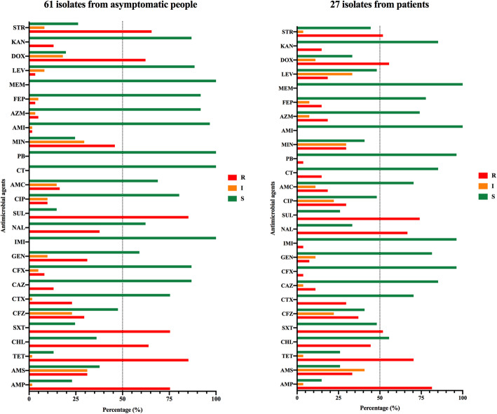
A total of 8 (8/88, 9.1%) strains were susceptible to all the 26 antimicrobials, and all of the 88 isolates showed susceptibility to meropenem, a type of carbapenems used to treat symptomatic Salmonella infection (Fig. 2). S. Derby showed a high rate of multi-drug resistance to these antimicrobials (90%, 9/10), followed by S. Enteritidis (88.9%, 8/9), S. Meleagridis (85.7%, 6/7), S. Typhimurium (84.2%, 16/19), S. London (81.8%, 9/11), and S. Rissen (71.4%, 5/7). Besides, 85.2% (52/61) of the isolates from asymptomatic humans and 85.2% (23/27) of those from patients were identified to be MDR (Fig. 2).
Fig. 2.
Antimicrobial resistance of human Salmonella isolates. The percentage of strains with resistance to each antimicrobial agent. The “R” displayed as red column represents resistant, the “I” displayed as orange column represents intermediate, and the “S” displayed as green column represents susceptible
Among the 26 antimicrobial agents belonging to 8 classes (β-Lactamases, aminoglycosides, carbapenems, polymyxins, phenicols, sulfonamides, tetracyclines, fluoroquinolones), resistance to sulfisoxazole, tetracycline, and ampicillin was found in 81.8, 80.7, 77.3% of isolates, respectively (Fig. 2). These top three antimicrobials were also seen in the 61 isolates from asymptomatic people. Besides, 73.8% (45/61) of these isolates showed resistance to the three antimicrobials (Fig. 2). However, ampicillin (81.5%), tetracycline (70.4%), and nalidixic acid (66.8%) were the top three antimicrobials found in 27 isolates from patients. Resistance to all of the three antimicrobials was seen in 51.9% (14/27) of these isolates.
Among the 88 isolates, the S. Meleagridis F2–6 and S. Rissen F5–5 strain showed resistance to amikacin and imipenem, respectively (Fig. 2). Four isolates belonging to three serotypes (S. Parytyphi A, S. Enteritidis, S. Typhimurium) were resistant against polymyxin E, an antibiotic medication used as a last-resort treatment for MDR gram-negative infections, and all four strains were isolated from patients. The S. Enteritidis F6–1 strain displayed resistance to both polymyxin E and polymyxin B.
PFGE analysis
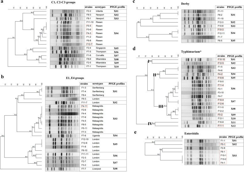
The 88 strains were then subjected to the PFGE analysis with the restriction enzyme XbaI for inter-serotype differentiation to reveal their genetic relationship. Among the 16 strains belonging to 8 serotypes in C1 and C2-C3 groups, 10 profiles (X01 to X10) were identified with X04 shared by all of the 8 S. Rissen strains isolated from both patients and asymptomatic people (Fig. 3a). Among the 23 strains belonging to E1 and E4 groups, 8 genetic profiles (designated X01 to X08) were presented to show the genetic difference of 6 serotypes (Fig. 3b). The X01 and X03 represented 3 S. Senftenberg and 8 S. Meleagridis strains, respectively (Fig. 3b). Except for one strain of S. London without clear DNA bands, the other 9 strains were distributed in 4 different profiles, which were X02, X05, X06, and X07 (Fig. 3b).
Fig. 3.
PFGE analysis of Salmonella strains. PFGE profiles and phylogenic relationship of strains belonging to C1 and C2-C3 groups (a), E1 and E4 groups (b), S. Derby (c), S. Typhimurium (d), and S. Enteritidis (e). The strains in red box were isolated from patients. The “I”, “II”, “III”, and “IV” represents the four clusters of S. Typhimurium (including S. Typhimurium monophasic variants) divided by PFGE analysis
Among the 34 strains belonging to the B group, S. Typhimurium and S. Derby took up 85.3% with diverse PFGE profiles (Fig. 3c, d). Twelve profiles (X01 to X12) were identified in 19 S. Typhimurium strains (Fig. 3d). The X01, X02, X04, X05, X07, X019, and X12 profiles were detected in strains isolated from patients, while the X03, X08, X10, and X11 profiles were found in strains isolated from asymptomatic people (Fig. 3d). The X06 profile consists of five strains from both sources, but 80% (4/5) of them in this profile were from asymptomatic people (Fig. 3d). Except for one strain of S. Derby with smear patterns, the other 9 strains displayed 5 PFGE profiles (X01 to X05) with X01 and X04 as predominant profiles, each of which are shared by 3 strains (Fig. 3c). The only one strain F10–3 isolated from the patient belonged to the X04 genetic profile (Fig. 3c). In our study, S. Enteritidis is the only detected serotype in the D group. Three profiles (X01, X02, and X03) were presented for 8 out of 9 S. Enteritidis strains. Four strains (F6–1, F10–1, F10–2, and F4–1) belonging to the X03 were obtained from patients, and X03 was identified as the predominant PFGE profile (6/8, 75%) for the 8 strains (Fig. 3e).
Discussion
Distribution of Salmonella spp. in patients and asymptomatic people
Salmonella is one of the major pathogens causing human diarrhea, which is closely related to the consumption of bacterially contaminated foods. Therefore, Salmonella reports have been published every year by the US National Enteric Disease Surveillance system, the European Food Safety Authority (EFSA), and the European Centre for Disease Prevention and Control (ECDC). According to the US and EU reports, S. Enteritidis and S. Typhimurium (including S. Typhimurium monophasic variants) have been the top 2 serotypes causing human salmonellosis [6, 7]. Our study showed that S. Typhimurium is the predominant serotype from diarrhea patients in Nantong city, followed by S. Enteritidis, correspondent to the previous reports in China [8]. However, in asymptomatic people, S. London became the predominant serotype as well as S. Typhimurium, followed by S. Derby and S. Meleagridis. The difference in serotype distribution in patients and asymptomatic people reflected that many NTS serotypes could infect humans without any symptom, but these serotypes were underestimated in the existing surveillance system for mostly patients. Additionally, these NTS serotypes have frequently been isolated from pig, chicken, and their associated meat products [14, 21], implying the potential transmission of Salmonella from animal foods to humans. Except for S. Typhimurium causing disease or no symptoms, S. Rissen displayed a similar characteristic to S. Typhimurium in human infection (Table 2). In 2009, S. Rissen caused > 80 people infection in over 4 different states of the USA, and the cases for human infection by S. Rissen were also reported in Denmark, Thailand, UK, and China [22–24]. Among 208 S. Rissen isolates from human samples, 108 out of the isolates were from patients, 100 isolates were from the asymptomatic carriers [22], reflecting that nearly 50% of the people infected with S. Rissen were in the asymptomatic state as well as in our study (Table 2). However, some serotypes have been mainly isolated from patients, such as the typhoidal S. Paratyphi A (100%) obtained from blood samples, and nontyphoidal S. Enteritidis (77.8%) collected from diarrheagenic patients.
Multidrug-resistance of Salmonella spp.
Among the used 26 antimicrobial agents belonging to 8 different types, 84.1% (74/88) of Salmonella isolates showed resistance to at least three types of antimicrobials, which is dramatically higher than the reported 46 and 50% of Salmonella isolates from patients were MDR in Guangdong and Beijing, respectively [8, 15]. Among the 61 Salmonella isolates from asymptomatic people, 49.2% (30/61) of the strains showed resistance to 6 out of 8 types of antimicrobial agents, while 73.8% showed resistance to 5 types of antimicrobials. This demonstrated that the NTS organisms from asymptomatic people showed strong resistance to antimicrobials as well as the human diarrheal or bloodborne isolates, which is similar to the report that 81.8% of Salmonella isolates from asymptomatic food handlers were MDR [25]. However, the result was different from the recently reported fewer MDR NTS isolates in asymptomatic children than in symptomatic individuals in Vietnamese [26]. Twenty-three out of 88 Salmonella isolates showed resistance to one or more antimicrobial agents belonging to extended-spectrum β-lactamases (ESBLs). One ESBL S. Thompson strain displayed resistance to all of the detected β-lactamases, including aztreonam, cefepime, cefazolin, ceftazidime, and amoxicillin/clavulanic acid. The emergence of ESBL-producing Salmonella may cause a substantial increase in treatment costs and prolonged treatment periodicity [27].
PFGE differentiation of Salmonella strains
Although the whole-genome sequencing (WGS)-based typing methods have been considered highly discriminative epidemiological tools, PFGE has a relatively high concordance with epidemiological relatedness [28]. The PFGE profiles not only showed perfect correspondence to serotypes belonging to C1, C2–3 or E1, E4 serogroups (Fig. 3a, b), but also displayed a highly discriminative ability to different strains of serotypes like S. London, S. Newport, and S. Mabandaka, which is potentially caused by the integration of new genetic elements for adaption to adverse conditions [29].
S. Derby is another frequently reported serotype isolated from both human and animal or animal foods. Most isolates were obtained from asymptomatic people, revealing that it is not the predominant serotype causing severe infections in humans. In this study, the 9 S. Derby strains were divided into 5 profiles by PFGE analysis, which has been confirmed as a molecular subtyping method used to differentiate S. Derby strains (Fig. 3c). Ten PFGE profiles were obtained in 16 S. Derby strains of human origin with 9 antimicrobial resistance patterns [30]. With difference to S. Derby, S. Typhimurium is the predominant serotype causing either human diarrhea or asymptomatic infection [30]. Four clusters were identified in 19 S. Typhimurium strains, most of which are distributed in cluster III, including 7 and 4 strains from asymptomatic people and patients, respectively (Fig. 3d). This is the reason for considering S. Typhimurium as one of the most important serotypes in the National Salmonella surveillance system [6]. Another serotype, S. Enteritidis has been confirmed as the predominant serotype causing human diarrhea, and it could not be efficiently differentiated by PFGE (Fig. 3e). Other molecular typing methods, such as CRISPR typing and whole-genome sequencing (WGS) based typing, can be further used to study the evolutionary relationship of these isolates [31, 32].
Although PFGE has been considered as the “gold standard” for bacterial typing, the WGS has superior resolution to PFGE, and it can differentiate isolates which were indistinguishable by PFGE. WGS can distinguish stains with difference at only a single nucleotide and provide higher resolution than the other molecular typing methods [33]. In this study, the PFGE showed high discriminatory power in some serotypes, such as S. Typhimurium, but it could not efficiently distinguish S. Enteritidis isolates. Further analysis will be performed to reveal the relationship of the isolates belonging to the same serotype from different sources.
Conclusion
This study compared the serotypes, antimicrobial resistance phenotypes, and genetic profiles of Salmonella strains between asymptomatic people and patients. The results revealed that S. Typhimurium is the predominant serotype causing human infection in both symptomatic and asymptomatic state. The other NTS including S. London, S. Derby, and S. Meleagridis mainly cause asymptomatic infection, while S. Enteritidis infection commonly results in human diseases. The high multi-drug resistance rate detected in these strains and diverse PFGE profiles showed no significant difference in strains between symptomatic and asymptomatic individuals, implying that all human-related Salmonella strains can induce both human salmonellosis and asymptomatic infection. Therefore, increased surveillance of Salmonella from both patients and asymptomatic people should be taken to control the transmission of the pathogen.
Supplementary Information
Additional file 1: Supplementary Table 1. The information and antimicrobial resistance phenotype of human Salmonella isolates.
Acknowledgements
Not applicable.
Abbreviations
- NTS
Nontyphoidal Salmonella
- MDR
Multidrug-resistant
- EFSA
European Food Safety Authority
- PFGE
Pulsed-field gel electrophoresis
- CFU
Colony-forming unit
- AMP
Ampicillin
- AMS
Ampicillin-sulbactam
- AMC
Amoxicillin-clavulanic acid
- AZM
Aztreonam
- CFZ
Cefazolin
- CTX
Cefotaxime
- CAZ
Ceftazidime
- CFX
Cefoxitin
- FEP
Cefepine
- GEN
Gentamicin
- AMI
Amikacin
- IMI
Imipenem
- MEM
Meropenem
- CHL
Chloramphenicol
- SXT
Trimethoprim/sulfamethoxazole
- SUL
Sulfisoxazole
- TET
Tetracycline
- MIN
Minocycline
- NAL
Nalidixic acid
- CIP
Ciprofloxacin
- LEV
Levofloxacin
- Dox
Doxycyclin
- KAN
Kanamycin
- STR
Streptomycin
- CT
Colistin sulphate
- PB
Polymyxin B
- CLSI
Clinical and Laboratory Standards Institute
- ATCC
American Type Culture Collection
- ECDC
The European Centre for Disease Prevention and Control
- CRISPR
Clustered regularly interspaced short palindromic repeats
- WGS
The Whole-genome sequencing
- ESBLs
Extended-spectrum β-lactamases
- UPGMA
The arithmetic mean algorithm
- UV
Ultraviolet
Authors’ contributions
HX and QL conceived the design of this study. HX, WbZ, and WZ collected the bacteria strains and performed the experiments. KZ, YZ, and ZW facilitated the data collection and performed the analysis. HX drafted the manuscript. KZ, YL and QL revised the manuscript. All authors read and approved the final manuscript.
Funding
This research was financially supported by Jiangsu Key Laboratory of Zoonosis (R1703 to HX), Qing Lan Project of Yangzhou University (QL), and National Natural Science Foundation of China (32072821 to QL). The funding body played no role in the design of the study, in the collection, analysis, and interpretation of data, and in the writing of the manuscript.
Availability of data and materials
The datasets used and/or analysed during the current study are available from the corresponding author on reasonable request.
Declarations
Ethics approval and consent to participate
This study received ethical approval by the Human Research Ethics Committee of Nantong Center for Disease Control and Prevention (38–2017/1701). All the bacterial isolates were provided with written consents for the research in this study.
Consent for publication
Not applicable.
Competing interests
The authors declare that they have no conflict of interest.
Footnotes
Publisher’s Note
Springer Nature remains neutral with regard to jurisdictional claims in published maps and institutional affiliations.
Haiyan Xu, Weibing Zhang, and Kai Zhang contributed equally to this work.
References
- 1.Harrois D, Breurec S, Seck A, Delaune A, Le Hello S, Pardos de la Gandara M, et al. Prevalence and characterization of extended-spectrum β-lactamase-producing clinical Salmonella enterica isolates in Dakar, Senegal, from 1999 to 2009. Clin Microbiol Infect. 2014;20(2):109–116. doi: 10.1111/1469-0691.12339. [DOI] [PubMed] [Google Scholar]
- 2.Jertborn M, Haglind P, Iwarson S, Svennerholm AM. Estimation of symptomatic and asymptomatic Salmonella infections. Scand J Infect Dis. 1990;22(4):451–455. doi: 10.3109/00365549009027077. [DOI] [PubMed] [Google Scholar]
- 3.Paudyal N, Pan H, Wu B, Zhou X, Zhou X, Chai W, et al. Persistent asymptomatic human infections by Salmonella enterica serovar Newport in China. mSphere. 2020;5(3):e00163–e00120. doi: 10.1128/mSphere.00163-20. [DOI] [PMC free article] [PubMed] [Google Scholar]
- 4.Chen HM, Wang Y, Su LH, Chiu CH. Nontyphoid Salmonella infection: microbiology, clinical features, and antimicrobial therapy. Pediatr Neonatol. 2013;54(3):147–152. doi: 10.1016/j.pedneo.2013.01.010. [DOI] [PubMed] [Google Scholar]
- 5.Majowicz SE, Musto J, Scallan E, Angulo FJ, Kirk M, O'Brien SJ, Jones TF, Fazil A, Hoekstra RM, International Collaboration on Enteric Disease 'Burden of Illness' Studies The global burden of nontyphoidal Salmonella gastroenteritis. Clin Infect Dis. 2010;50(6):882–889. doi: 10.1086/650733. [DOI] [PubMed] [Google Scholar]
- 6.Centers for Disease Control and Prevention . National Salmonella surveillance annual report, 2016. Atlanta: US Department of Health and Human Services, CDC; 2018. [Google Scholar]
- 7.European Food Safety Authority The European Union summary report on trends and sources of zoonoses, zoonotic agents and food-borne outbreaks in 2017. EFSA J. 2018;16(12):e5500. doi: 10.2903/j.efsa.2018.5500. [DOI] [PMC free article] [PubMed] [Google Scholar]
- 8.Liang Z, Ke B, Deng X, Liang J, Ran L, Lu L, He D, Huang Q, Ke C, Li Z, Yu H, Klena JD, Wu S. Serotypes, seasonal trends, and antibiotic resistance of non-typhoidal Salmonella from human patients in Guangdong province, China, 2009-2012. BMC Infect Dis. 2015;15(1):53. doi: 10.1186/s12879-015-0784-4. [DOI] [PMC free article] [PubMed] [Google Scholar]
- 9.Ke Y, Lu W, Liu W, Zhu P, Chen Q, Zhu Z. Non-typhoidal Salmonella infections among children in a tertiary hospital in Ningbo, Zhejiang, China, 2012-2019. PLoS Negl Trop Dis. 2020;14(10):e0008732. doi: 10.1371/journal.pntd.0008732. [DOI] [PMC free article] [PubMed] [Google Scholar]
- 10.Li Y, Xie X, Xu X, Wang X, Chang H, Wang C, Wang A, He Y, Yu H, Wang X, Zeng M. Nontyphoidal Salmonella infection in children with acute gastroenteritis: prevalence, serotypes, and antimicrobial resistance in Shanghai, China. Foodborne Pathog Dis. 2014;11(3):200–206. doi: 10.1089/fpd.2013.1629. [DOI] [PubMed] [Google Scholar]
- 11.Ran L, Wu S, Gao Y, Zhang X, Feng Z, Wang Z, Kan B, Klena JD, Lo Fo Wong DMA, Angulo FJ, Varma JK. Laboratory-based surveillance of nontyphoidal Salmonella infections in China. Foodborne Pathog Dis. 2011;8(8):921–927. doi: 10.1089/fpd.2010.0827. [DOI] [PubMed] [Google Scholar]
- 12.Xu H, Zhang W, Guo C, Xiong H, Chen X, Jiao X, Su J, Mao L, Zhao Z, Li Q. Prevalence, serotypes, and antimicrobial resistance profiles among Salmonella isolated from food catering workers in Nantong, China. Foodborne Pathog Dis. 2019;16(5):346–351. doi: 10.1089/fpd.2018.2584. [DOI] [PubMed] [Google Scholar]
- 13.Scott TM, Rose JB, Jenkins TM, Farrah SR, Lukasik J. Microbial source tracking: current methodology and future directions. Appl Environ Microbiol. 2002;68(12):5796–5803. doi: 10.1128/aem.68.12.5796-5803.2002. [DOI] [PMC free article] [PubMed] [Google Scholar]
- 14.Xu C, Ren X, Feng Z, Fu Y, Hong Y, Shen Z, Zhang L, Liao M, Xu X, Zhang J. Phenotypic characteristics and genetic diversity of Salmonella enterica serotype Derby isolated from human patients and foods of animal origin. Foodborne Pathog Dis. 2017;14(10):593–599. doi: 10.1089/fpd.2017.2278. [DOI] [PubMed] [Google Scholar]
- 15.Qu M, Lv B, Zhang X, Yan H, Huang Y, Qian H, Pang B, Jia L, Kan B, Wang Q. Prevalence and antibiotic resistance of bacterial pathogens isolated from childhood diarrhea in Beijing, China (2010-2014) Gut Pathog. 2016;8(1):31. doi: 10.1186/s13099-016-0116-2. [DOI] [PMC free article] [PubMed] [Google Scholar]
- 16.Woh PY, Thong KL, Behnke JM, Lewis JW, Zain SNM. Characterization of nontyphoidal Salmonella isolates from asymptomatic migrant food handlers in peninsular Malaysia. J Food Prot. 2017;80(8):1378–1383. doi: 10.4315/0362-028X.JFP-16-342. [DOI] [PubMed] [Google Scholar]
- 17.Centers for Disease Control and Prevention . The CDC PulseNet one-day (24–28 h) standardized laboratory protocol for molecular subtyping of Escherichia coli O157:H7, non-typhoidal Salmonella serotypes, and Shigella sonnei by Pulsed Field Gel Electrophoresis (PFGE) 2017. [Google Scholar]
- 18.Diaz-Torres O, Lugo-Melchor OY, de Anda J, Gradilla-Hernandez MS, Amezquita-Lopez BA, Meza-Rodriguez D. Prevalence, distribution, and diversity of Salmonella strains isolated from a subtropical lake. Front Microbiol. 2020;11:521146. doi: 10.3389/fmicb.2020.521146. [DOI] [PMC free article] [PubMed] [Google Scholar]
- 19.Grimont PA, Weill FX. Antigenic formulae of the Salmonella serovars. 9. Paris: WHO Collaborating Center for Reference and Research on Salmonella, Institut Pasteur; 2007. [Google Scholar]
- 20.Clinical and Laboratory Standards Institute . Performance standards for antimicrobial susceptibility testing. 28. Wayne: CLSI supplement M100; 2018. [Google Scholar]
- 21.Ma S, Lei C, Kong L, Jiang W, Liu B, Men S, Yang Y, Cheng G, Chen Y, Wang H. Prevalence, antimicrobial resistance, and relatedness of Salmonella isolated from chickens and pigs on farms, abattoirs, and markets in Sichuan province, China. Foodborne Pathog Dis. 2017;14(11):667–677. doi: 10.1089/fpd.2016.2264. [DOI] [PubMed] [Google Scholar]
- 22.Xu X, Biswas S, Gu G, Elbediwi M, Li Y, Yue M. Characterization of multidrug resistance patterns of emerging Salmonella enterica serovar Rissen along the food chain in China. Antibiotics (Basel) 2020;9:660. doi: 10.3390/antibiotics9100660. [DOI] [PMC free article] [PubMed] [Google Scholar]
- 23.Hendriksen RS, Bangtrakulnonth A, Pulsrikarn C, Pornreongwong S, Hasman H, Song SW, Aarestrup FM. Antimicrobial resistance and molecular epidemiology of Salmonella Rissen from animals, food products, and patients in Thailand and Denmark. Foodborne Pathog Dis. 2008;5(5):605–619. doi: 10.1089/fpd.2007.0075. [DOI] [PubMed] [Google Scholar]
- 24.Irvine N. Communicable diseases monthly report, Northern Ireland edition: The Health Protection Agency; 2009. Available online: http://www.publichealth.hscni.net/directorate-public-health/health-protection/surveillance-data. Accessed 15 Oct 2020.
- 25.Solomon FB, Wada FW, Anjulo AA, Koyra HC, Tufa EG. Burden of intestinal pathogens and associated factors among asymptomatic food handlers in South Ethiopia: emphasis on salmonellosis. BMC Res Notes. 2018;11(1):502. doi: 10.1186/s13104-018-3610-4. [DOI] [PMC free article] [PubMed] [Google Scholar]
- 26.Parisi A, Le Thi Phuong T, Mather AE, Jombart T, Thanh Tuyen H, Phu Huong Lan N, et al. Differential antimicrobial susceptibility profiles between symptomatic and asymptomatic non-typhoidal Salmonella infections in Vietnamese children. Epidemiol Infect. 2020;148:e144. doi: 10.1017/S0950268820001168. [DOI] [PMC free article] [PubMed] [Google Scholar]
- 27.Zhang SX, Zhou YM, Tian LG, Chen JX, Tinoco-Torres R, Serrano E, Li SZ, Chen SH, Ai L, Chen JH, Xia S, Lu Y, Lv S, Teng XJ, Xu W, Gu WP, Gong ST, Zhou XN, Geng LL, Hu W. Antibiotic resistance and molecular characterization of diarrheagenic Escherichia coli and non-typhoidal Salmonella strains isolated from infections in Southwest China. Infect Dis Poverty. 2018;7(1):53. doi: 10.1186/s40249-018-0427-2. [DOI] [PMC free article] [PubMed] [Google Scholar]
- 28.Tang S, Orsi RH, Luo H, Ge C, Zhang G, Baker RC, Stevenson A, Wiedmann M. Assessment and comparison of molecular subtyping and characterization methods for Salmonella. Front Microbiol. 2019;10:1591. doi: 10.3389/fmicb.2019.01591. [DOI] [PMC free article] [PubMed] [Google Scholar]
- 29.Prosser JI, Bohannan BJ, Curtis TP, Ellis RJ, Firestone MK, Freckleton RP, et al. The role of ecological theory in microbial ecology. Nat Rev Microbiol. 2007;5(5):384–392. doi: 10.1038/nrmicro1643. [DOI] [PubMed] [Google Scholar]
- 30.Valdezate S, Vidal A, Herrera-Leon S, Pozo J, Rubio P, Usera MA, et al. Salmonella Derby clonal spread from pork. Emerg Infect Dis. 2005;11(5):694–698. doi: 10.3201/eid1105.041042. [DOI] [PMC free article] [PubMed] [Google Scholar]
- 31.Li Q, Wang X, Yin K, Hu Y, Xu H, Xie X, Xu L, Fei X, Chen X, Jiao X. Genetic analysis and CRISPR typing of Salmonella enterica serovar Enteritidis from different sources revealed potential transmission from poultry and pig to human. Int J Food Microbiol. 2018;266:119–125. doi: 10.1016/j.ijfoodmicro.2017.11.025. [DOI] [PubMed] [Google Scholar]
- 32.Ktari S, Ksibi B, Ghedira K, Fabre L, Bertrand S, Maalej S, et al. Genetic diversity of clinical Salmonella enterica serovar Typhimurium in a university hospital of south Tunisia, 2000–2013. Infect Genet Evol. 2020;85:104436. doi: 10.1016/j.meegid.2020.104436. [DOI] [PubMed] [Google Scholar]
- 33.Salipante SJ, SenGupta DJ, Cummings LA, Land TA, Hoogestraat DR, Cookson BT. Application of whole-genome sequencing for bacterial strain typing in molecular epidemiology. J Clin Microbiol. 2015;53(4):1072–1079. doi: 10.1128/JCM.03385-14. [DOI] [PMC free article] [PubMed] [Google Scholar]
Associated Data
This section collects any data citations, data availability statements, or supplementary materials included in this article.
Supplementary Materials
Additional file 1: Supplementary Table 1. The information and antimicrobial resistance phenotype of human Salmonella isolates.
Data Availability Statement
The datasets used and/or analysed during the current study are available from the corresponding author on reasonable request.


