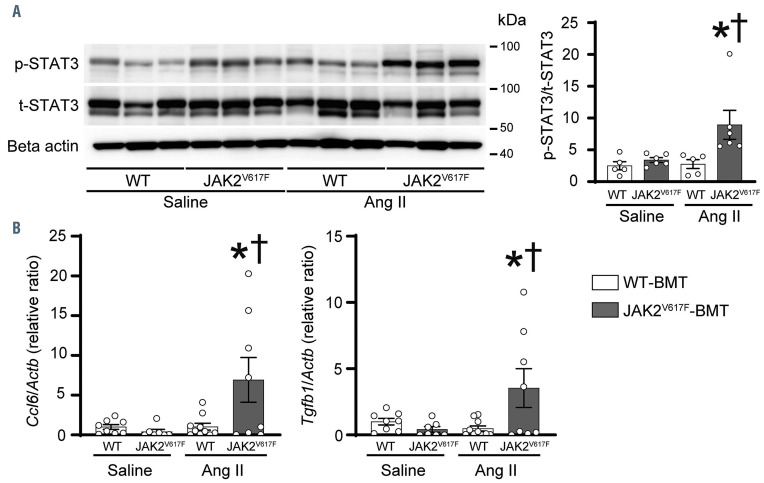
Figure 4.
Hematopoietic JAK2 V617F increases STAT3 phosphorylation and cytokine expression in response to angiotensin II infusion in the abdominal aorta. (A) Western blot analysis on the STAT3 in the abdominal aorta. Aorta extracts from the WT-BMT mice or JAK2V617F-BMT mice were immunoblotted with the indicated antibodies. Phosphorylated STAT3 (p-STAT3) to total STAT3 (t-STAT3) ratios are shown in the bar graphs. The average value for the saline-infused WT-BMT mice was set to 1 (n=5–6). actin was used as the loading control. (B) Relative mRNA expression levels of Ccl6 and Tgfb1 in the aorta. Actb was used for normalization. The average value for saline-infused WT-BMT mice was set to 1 (n=7–10). All data are presented as mean ± standard error of the mean. *P<0.05 vs. the corresponding saline-infused mice and †P<0.05 vs. the Ang II-infused WT-BMT mice by one-way ANOVA with Tukey post-hoc analysis. WT, ApoE−/−: mice transplanted with bone marrow (BM) cells from wild-type (WT) mice; JAK2V617F, ApoE−/−: mice transplanted with BM cells from JAK2V617F mice; BMT: BM transplantation; Ang II: angiotensin II.

