PLOS ONE logoLink to PLOS ONE
. 2021 Jul 2;16(7):e0253618. doi: 10.1371/journal.pone.0253618

Delftia acidovorans secretes substances that inhibit the growth of Staphylococcus epidermidis through TCA cycle-triggered ROS production

Tomotaka Ohkubo 1,2, Yasuhiko Matsumoto 1, Otomi Cho 1, Yuki Ogasawara 2, Takashi Sugita 1,*
Editor: Rashid Nazir3
PMCID: PMC8253425  PMID: 34214099

Abstract

The proportion of Staphylococcus aureus in the skin microbiome is associated with the severity of inflammation in the skin disease atopic dermatitis. Staphylococcus epidermidis, a commensal skin bacterium, inhibits the growth of S. aureus in the skin. Therefore, the balance between S. epidermidis and S. aureus in the skin microbiome is important for maintaining healthy skin. In the present study, we demonstrated that the heat-treated culture supernatant of Delftia acidovorans, a member of the skin microbiome, inhibits the growth of S. epidermidis, but not that of S. aureus. Comprehensive gene expression analysis by RNA sequencing revealed that culture supernatant of D. acidovorans increased the expression of genes related to glycolysis and the tricarboxylic acid cycle (TCA) cycle in S. epidermidis. Malonate, an inhibitor of succinate dehydrogenase in the TCA cycle, suppressed the inhibitory effect of the heat-treated culture supernatant of D. acidovorans on the growth of S. epidermidis. Reactive oxygen species production in S. epidermidis was induced by the heat-treated culture supernatant of D. acidovorans and suppressed by malonate. Further, the inhibitory effect of the heat-treated culture supernatant of D. acidovorans on the growth of S. epidermidis was suppressed by N-acetyl-L-cysteine, a free radical scavenger. These findings suggest that heat-resistant substances secreted by D. acidovorans inhibit the growth of S. epidermidis by inducing the production of reactive oxygen species via the TCA cycle.

Introduction

Atopic dermatitis (AD) is a chronic inflammatory skin disease characterized by variable clinical features, such as relapsing pruritus and eczema [1,2]. Genetic and environmental factors are associated with the onset and exacerbation of AD [35]. The skin of AD patients exhibits an elevated pH and barrier disruption. Moreover, the skin microbial composition is altered in AD patients compared with healthy humans. Microbial diversity on the human skin contributes to maintain healthy skin by modulating immune responses [68]. In the skin of AD patients, the microbial diversity is reduced due to an increased proportion of Staphylococcus aureus in the skin microbiome [4]. S. aureus exacerbates AD and induces Th2 cytokines and proteases in human skin [4,9,10]. On the other hand, Staphylococcus epidermidis, a coagulase-negative Staphylococcus, inhibits S. aureus growth by producing antimicrobial peptides and short-chain fatty acids [11,12]. The proportion of S. aureus in the skin of AD patients was decreased after transplantation of coagulase-negative Staphylococcus strains having antimicrobial activity [11]. Disrupting the balance between S. aureus and coagulase-negative Staphylococcus with antimicrobial activity against S. aureus in the skin microbiome is associated with exacerbation of AD [11,12]. The relative abundance of S. aureus is increased in the skin of patients with psoriasis (PS) as well as in those with AD [13,14]. S. epidermidis also inhibits the growth of Cutibacterium acnes, which is associated with acne vulgaris (AV) [1517]. Therefore, S. epidermidis might play a key role in various skin disorders.

Delftia acidovorans, a gram-negative bacterium, is detected in the skin of AD patients by microbiome analysis [1822]. D. acidovorans secretes a compound with antimicrobial activity against S. aureus, Enterococcus faecalis, Acinetobacter baumannii, Klebsiella pneumoniae, and Pseudomonas aeruginosa [23]. Thus, D. acidovorans may affect bacterial growth in the skin microbiome, but the effect of D. acidovorans on the growth of S. epidermidis remains unclear.

In the present study, we investigated the effect of D. acidovorans on the growth of S. epidermidis and elucidated its mechanisms. We found that the heat-treated culture supernatant (CS) of D. acidovorans inhibited S. epidermidis growth. Furthermore, we demonstrated that D. acidovorans heat-treated CS induced the production of reactive oxygen species (ROS) via the tricarboxylic acid cycle (TCA) cycle in S. epidermidis. Our findings provide important insight into how D. acidovorans affects the skin microbiome by inhibiting the growth of S. epidermidis, resulting in a skin microbiome imbalance related to various skin disorders.

Materials & methods

Reagents

Malonic acid, N-acetyl-L-cysteine (NAC), and gentamicin sulfate were purchased from Wako Pure Chemical Corporation (Osaka, Japan). Menadione was purchased from MilliporeSigma (St. Louis, MO, USA). 2,7-Dichlorodihydrofluorescein diacetate was purchased from Cayman Chemical (Ann Arbor, MI, USA).

Strain and growth conditions

The NBRC100911 strain of Staphylococcus epidermidis (S. epidermidis) and the NBRC100910 strain of Staphylococcus aureus (S. aureus) were obtained from the National Institute of Technology and Evaluation (Tokyo, Japan). S. epidermidis and S. aureus strains were spread on nutrient agar (5 g/L sodium chloride [Wako Pure Chemical Corporation], 5 g/L beef extract [Becton Dickinson, Franklin Lakes, NJ, USA], 10 g/L hipolypepton [Wako Pure Chemical Corporation], and 15 g/L agar powder [Wako Pure Chemical Corporation] before autoclaving) and grown overnight at 37°C. The JCM6218 strain of D. acidovorans was obtained from the Japan Collection of Microorganisms (Ibaraki, Japan). The D. acidovorans JCM6218 strain was spread on nutrient agar and grown overnight at 27°C.

D. acidovorans culture supernatant preparation

A colony of the D. acidovorans JCM6218 strain was inoculated in 3 mL nutrient broth (NB) and incubated overnight at 32°C. The bacterial culture (3 mL) was inoculated into 300 mL NB and incubated overnight at 32°C with shaking (200 rpm, ZWY-240 Incubator Shaker, LABWIT Scientific, VIC, Australia). Bacterial cells were removed by centrifugation at 6300 rpm for 5 min (TOMY-MX305, TOMY Digital Biology Co. Ltd, Tokyo, Japan). The D. acidovorans CS was filtered using the Vacuum Filtration 500 rapid filter MAX (TPP, Schaffhausen, Switzerland). D. acidovorans CS incubated for 30 min at 100°C was used as D. acidovorans heat-treated CS in this study.

Bacterial growth inhibition assay

Bacterial growth inhibition assays were performed according to a previous report [24]. Samples including D. acidovorans CS and inhibitors were diluted with NB to the appropriate concentrations and dispensed in 50-μL aliquots into a 96-well plate (TPP). S. epidermidis and S. aureus suspensions (2 x 10⁴ cells/ml) were prepared with NB and 50 μL was added to each well. After incubating at 37°C for 12 h or 32°C for 18 h, absorbance at 630 nm was measured using a microplate reader (iMark microplate reader; Bio-Rad Laboratories Inc., Hercules, CA, USA).

RNA sequencing analysis

RNA-sequencing (RNA-seq) analysis was performed according to a previous report [24]. An S. epidermidis suspension was prepared with NB to an absorbance of 1 at 630 nm. The suspension was diluted in the same volume of D. acidovorans CS and incubated at 37°C for 4 h. After incubation, RNA was extracted from the cells using EZ-Beads (AMR, Inc., Gifu, Japan) and a High Pure RNA Isolation kit (Roche, Basel, Switzerland) according to the manufacturer’s instructions. The RNA-seq library was prepared from the RNA using an NEBNext rRNA Depletion Kit and NEBNext Ultra II RNA Library Prep Kit for Illumina (New England Biolabs Japan Inc., Tokyo, Japan) and subjected to RNA-seq analysis using MiSeq (Illumina Inc., San Diego, CA, USA). Gene function information was examined using the GenBank (https://www.ncbi.nlm.nih.gov/genbank/) and pathway information was analyzed using the Kyoto Encyclopedia of Genes and Genomes (KEGG; https://www.genome.jp/kegg/kegg_ja.html).

Time-kill assay

The S. epidermidis suspension (1×10⁵ cells/ml) was prepared with phosphate-buffered saline (PBS) and treated with 20% heat-treated NB, 20% D. acidovorans heat-treated CS, or gentamicin (final concentration 10 μg/mL). Each sample was incubated at 37°C or 32°C. Aliquots were serially diluted in PBS and 100 μL of the diluted samples was spread on a nutrient agar plate at 0, 3, and 6 h. The viable cell number was determined by counting the colonies on the plate after incubation for 24 h at 37°C.

Measuring ROS production

Quantification of ROS production in S. epidermidis with 2’,7’-dichlorodihydrofluorescein diacetate) was performed according to previous reports [25,26]. S. epidermidis was suspended in 3 mL NB and incubated overnight at 37°C with shaking (150 rpm, ZWY-240 incubator shaker, LABWIT Scientific). The culture was suspended in NB to an absorbance of 0.001 at 630 nm and exposed to 100 μM 2’,7’-dichlorodihydrofluorescein diacetate (final concentration) for 1 h at 37°C. After incubation, the samples were centrifuged at 14,000 rpm for 5 min (TOMY-MX105) and the supernatants were removed. The bacterial pellets were suspended in fresh NB and treated with D. acidovorans heat-treated CS or menadione, an ROS inducer. Fluorescence (λ excitation = 485 nm, λ emission = 538 nm) was measured using the Fluoroskan Ascent (Thermo Fisher Scientific, Waltham, MA, USA). Fluorescence was determined by subtracting the background fluorescence.

Statistical analysis

Statistical differences between groups were analyzed by the Student t-test, Tukey-Kramer test, Dunnett test, and Williams test. All experiments were performed at least twice. Each experiment was performed in triplicate and error bars indicate the standard deviations of the means. A P value of less than 0.05 was considered statistically significant.

Results

Growth inhibitory effect of substances secreted by D. acidovorans against S. epidermidis

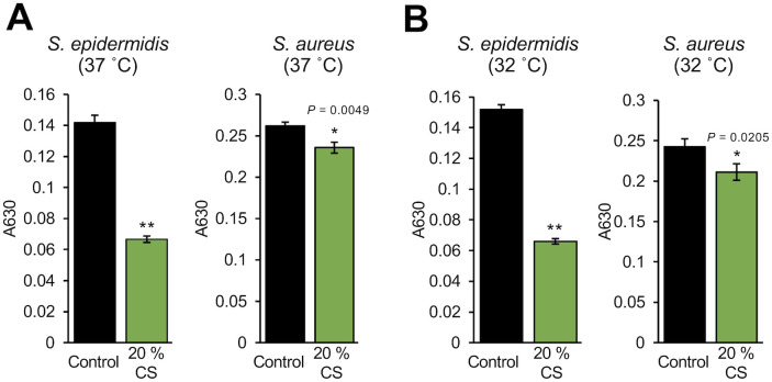
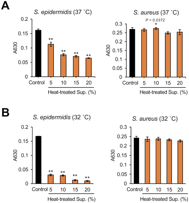
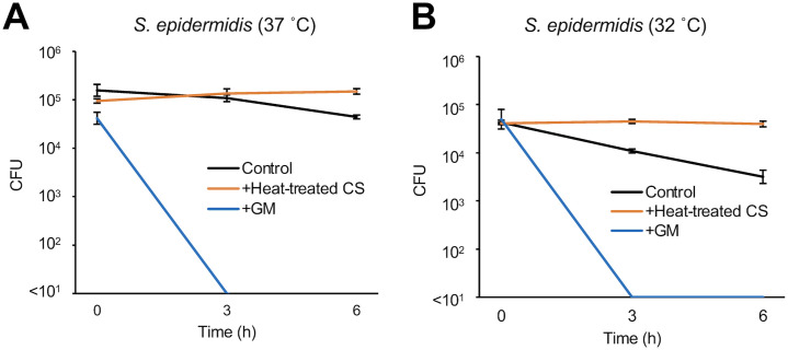
We investigated whether D. acidovorans produces substances that affect the growth of S. epidermidis. Compared with S. aureus, the growth of S. epidermidis at 37°C or 32°C was significantly inhibited by D. acidovorans CS (Fig 1). D. acidovorans heat-treated CS also inhibited S. epidermidis growth in a dose-dependent manner (Fig 2). We next examined whether the inhibitory activity of D. acidovorans heat-treated CS against S. epidermidis is bactericidal or bacteriostatic. D. acidovorans heat-treated CS did not decrease the viable number of S. epidermis in PBS (Fig 3). On the other hand, gentamicin, which has bactericidal activity, decreased the number of viable bacteria within 3 h (Fig 3). These results suggest that heat-stable substances secreted by D. acidovorans exhibit bacteriostatic activity, but not bactericidal activity, against S. epidermidis.

Fig 1. Inhibitory activity of the culture supernatant of D. acidovorans against S. epidermidis growth.

Fig 1

S. epidermidis (1×10⁴ cells/mL) and S. aureus (1×10⁴ cells/mL) in NB were mixed with 20% D. acidovorans CS. After incubating at 37°C for 12 h (A) or 32°C for 18 h (B), absorbance at 630 nm was measured using a microplate reader. Error bars indicate the standard deviations (SD) of the means (n = 3). Statistical differences between groups were analyzed by the Student t-test. (*: P < 0.05, **: P < 0.001).

Fig 2. Dose dependency of the inhibitory effect of heat-treated culture supernatant of D. acidovorans against S. epidermidis growth.

Fig 2

S. epidermidis (1×10⁴ cells/mL) and S. aureus (1×10⁴ cells/mL) in NB were mixed with 5% to 20% D. acidovorans heat-treated CS (Heat-treated CS). After incubating at 37°C for 12 h (A) or 32°C for 18 h (B), absorbance at 630 nm was measured using a microplate reader. Error bars indicate the SD of the means (n = 3). Statistical differences between groups were analyzed by the Williams test. (*: P < 0.05, **: P < 0.001).

Fig 3. Time-kill assay of heat-treated culture supernatant of D. acidovorans against S. epidermidis.

Fig 3

S. epidermidis (1×10⁵ cells/mL) in PBS was mixed with 20% NB (Control), 20% D. acidovorans heat-treated CS (Heat-treated CS), and 10 μg/mL gentamicin (GM) at 37°C (A) or 32°C (B). Each line shows the number of viable cells of S. epidermidis at 0, 3, and 6 h. Error bars indicate the SD of the means (n = 3).

Role of the S. epidermidis TCA cycle in the inhibitory effect of heat-stable substances secreted by D. acidovorans

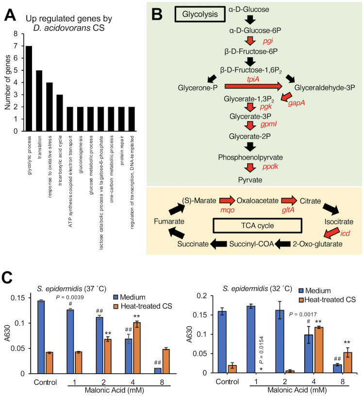
To understand the mechanism of action of the substances secreted by D. acidovorans, we analyzed gene expression in S. epidermidis in response to the D. acidovorans culture supernatant (CS). Expression of 100 genes in S. epidermidis was increased more than 2-fold by the D. acidovorans CS and expression of 7 genes was decreased to less than half (S1 and S2 Tables in S1 File). Gene ontology term analysis revealed that S. epidermidis genes related to glycolysis, the TCA cycle, and the response to oxidative stress were upregulated by the D. acidovorans CS (Fig 4A). Pathway analysis showed that expression of the 6 genes related to glycolysis and 3 genes related to the TCA cycle in S. epidermidis was increased more than 2-fold by the D. acidovorans CS (Fig 4B). Because pyruvate produced by glycolysis triggers TCA cycle activation, we focused on the relationship between activation of the TCA cycle, which is downstream of glycolysis, and the inhibitory activity of D. acidovorans heat-treated CS against S. epidermidis. Malonic acid (4 mM), a TCA cycle inhibitor, suppressed the inhibitory effect of the D. acidovorans heat-treated CS against S. epidermidis growth (Fig 4C). These results suggest that substances secreted by D. acidovorans inhibit the growth of S. epidermidis via TCA cycle regulation.

Fig 4. Suppressive effect of a TCA cycle inhibitor on the inhibitory effect of heat-treated culture supernatant of D. acidovorans against S. epidermidis growth.

Fig 4

(A) Gene expression analysis of S. epidermidis (1×10⁹ cells/mL) mixed with 50% D. acidovorans CS at 37°C for 4 h. The graphs show the number of upregulated genes in S. epidermidis after D. acidovorans CS treatment. (B) Upregulated genes related to glycolysis and the TCA cycle. Red letters represent genes that increased more than 2-fold after treatment with D. acidovorans CS in S. epidermidis. (C) S. epidermidis (1×10⁴ cells/mL) in NB was mixed with 1–4 mM malonic acid and 15% D. acidovorans heat-treated CS (Heat-treated CS). After incubating at 37°C for 12 h or 32°C for 18 h, absorbance at 630 nm was measured using a microplate reader. Error bars indicate the SD of the means (n = 3). Statistical differences of each group were analyzed by the Dunnett test. (#: P < 0.05 (Medium), # #: P < 0.001 (Medium), *: P < 0.05 (Heat-treated CS), **: P < 0.001 (Heat-treated CS)).

Heat-stable substances secreted by D. acidovorans induce ROS production in S. epidermidis

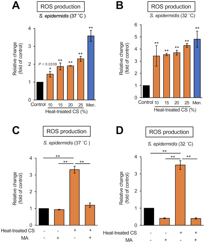
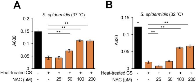
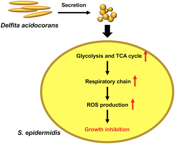
The TCA cycle is linked to ROS production in S. epidermidis [27]. ROS production in S. epidermidis was induced by exposure to the D. acidovorans heat-treated CS (Fig 5A and 5B). The addition of malonic acid inhibited the ROS production in S. epidermidis induced by the D. acidovorans heat-treated CS (Fig 5C and 5D). Addition of a free radical scavenger, NAC, suppressed the S. epidermidis growth inhibition in a dose-dependent manner (Fig 6). These results suggest that substances secreted by D. acidovorans inhibit the growth of S. epidermidis via TCA cycle-triggered ROS production (Fig 7).

Fig 5. ROS production induced in S. epidermidis by heat-treated culture supernatant of D. acidovorans.

Fig 5

(A, B) ROS production in S. epidermidis (1×10⁶ cells/mL) mixed with NB (Control), 10–25% D. acidovorans heat-treated CS (Heat-treated CS), or 70 μg/mL menadione (Men.) at 37°C (A) or 32°C (B) for 24 h. Menadione was used as a positive control. Fluorescence (λ excitation = 485 nm, λ emission = 538 nm) was measured using the Fluoroskan Ascent. Error bars indicate the SD of the means (n = 3). Statistical differences between groups were analyzed by the Dunnett test. (*: P < 0.05, **: P < 0.001). (C, D) ROS production in S. epidermidis (1×10⁶ cells/mL) mixed with NB (Control), 4 mM malonic acid (MA), 25% D. acidovorans heat-treated CS (Heat-treated CS), or 4 mM MA + 25% heat-treated CS at 37°C (C) or 32°C (D) for 24 h. Fluorescence (λ excitation = 485 nm, λ emission = 538 nm) was measured using the Fluoroskan Ascent. Error bars indicate the SD of the means (n = 3). Statistical differences between groups were analyzed by the Tukey-Kramer test. (*: P < 0.05, **: P < 0.001).

Fig 6. Suppressive effect of N-acetyl-L-cysteine on the inhibitory activity of heat-treated culture supernatant of D. acidovorans against S. epidermidis growth.

Fig 6

S. epidermidis (1×10⁴ cells/mL) in NB was mixed simultaneously with 15% D. acidovorans heat-treated CS (Heat-treated CS) and 25–200 μM N-acetyl-cysteine (NAC). After incubating at 37°C for 12 h (A) or 32°C for 18 h (B), absorbance at 630 nm was measured using a microplate reader. Error bars indicate the SD of the means (n = 3). Statistical differences between groups were analyzed by the Tukey-Kramer test. (*: P < 0.05, **: P < 0.001).

Fig 7. Model of the action of heat-stable substances secreted by D. acidovorans against S. epidermidis.

Fig 7

Heat-stable substances secreted by D. acidovorans induce the expression of the genes related to glycolysis and the TCA cycle and subsequently increase ROS production in S. epidermidis. The induced ROS production leads to growth inhibition in S. epidermidis.

Discussion

The findings of the present study demonstrated that heat-stable substances secreted by D. acidovorans inhibited the growth of S. epidermidis, but not that of S. aureus. The heat-stable substances induced ROS production through the TCA cycle in S. epidermidis. Moreover, the inhibitory effect against S. epidermidis growth by the heat-stable substances was suppressed by NAC, a radical scavenger. Our findings suggest that D. acidovorans secretes heat-stable substances that inhibit S. epidermidis growth by TCA cycle-triggered ROS production.

A previous study demonstrated that D. acidovorans secreted heat-stable antimicrobial substances that inhibit the growth of S. aureus [23]. Therefore, we assessed the inhibitory activity of D. acidovorans heat-treated CS against S. epidermidis. We demonstrated that heat-stable substances secreted by D. acidovorans induced the expression of genes related to glycolysis and the TCA cycle, and the inhibitory effect against growth of S. epidermidis was suppressed by malonic acid, an inhibitor of succinate dehydrogenase in the TCA cycle. On the other hand, 4 mM malonic acid alone inhibited S. epidermidis growth, but this inhibitory activity was suppressed by the heat-stable substances of D. acidovorans. Therefore, we assumed that the inhibitory effect of malonic acid on the TCA cycle competed with the increased gene expression in the TCA cycle induced by the heat-stable substances secreted by D. acidovorans. The finding also suggests that the heat-stable substances of D. acidovorans affect the TCA cycle in S. epidermidis. We also assumed that the effect heat-stable substances of D. acidovorans on the TCA cycle of S. epidermidis led to an increase in the ROS production because the ROS production induced by the heat-stable substances of D. acidovorans was inhibited by malonic acid. Furthermore, the suppressive effect of NAC suggested that the induced ROS production in S. epidermidis caused the inhibitory effect of the heat-stable substances of D. acidovorans against the growth of S. epidermidis. S. aureus, on the other hand, produces staphyloxanthin, an antioxidant that confers resistance to ROS [28,29]. We speculated that the heat-stable substances of D. acidovorans exhibit no inhibitory effects against S. aureus growth because of differences in the resistance to ROS.

D. acidovorans is often isolated and detected by microbiome analysis from clinical samples, including from the skin of patients with AD [18,19,3034]. We found that heat-stable substances secreted by D. acidovorans inhibited the growth of S. epidermidis, but not the growth of S. aureus. In the skin of patients with AD, the relative abundance of S. aureus is significantly increased, and enterotoxin or protease derived from S. aureus exacerbates inflammation of AD [4,9,10]. The relative abundance of S. aureus is also increased in the skin of patients with PS and Th17 cytokines are induced by S. aureus [13,14]. S. epidermidis inhibits the growth of S. aureus, which might be associated with AD and PS [11,12]. Moreover, S. epidermidis inhibits the growth of C. acnes, which is associated with AV [1517]. We therefore hypothesized that the inhibitory activity of substances secreted by D. acidovorans against S. epidermidis is indirectly associated with exacerbation of AD, PS, and AV. D. acidovorans might be related to various skin disorders. Additional studies are needed to elucidate the relationship between D. acidovorans and AD, PS, and AV.

Previous studies reported that D. acidovorans secretes delftibactin, which has antimicrobial activity against S. aureus [23,35]. Delftibactin reacts in a solution containing AuCl3 and forms a gold precipitation, resulting in detoxification by chelating Au3+ [35]. We demonstrated that the D. acidovorans heat-treated CS used in this study did not form a gold precipitation by mixing it with a solution containing AuCl3 (S1 Fig in S1 File). Furthermore, gold toxicity against S. epidermidis was not inhibited by D. acidovorans heat-treated CS (S2 Fig in S1 File). Therefore, we assumed that the D. acidovorans heat-treated CS used in this study may not contain a sufficient amount of delftibactin to inhibit the growth of S. aureus. We considered that differences in both the strain and culture conditions between previous studies and the present study could affect the composition of delftibactin. Tejman-Yarden et al. indicated the existence of a fraction other than the delftibactin fraction that exerts antimicrobial activity in the culture supernatant of D. acidovorans. In addition, a substance characterized as C39H68N14O17, which is similar to the composition of delftibactin is present in the delftibactin fraction [23]. It is possible that the active substances focused on in this study are in these fractions. Identifying the active substances in the D. acidovorans heat-treated CS is an important topic for future studies.

Conclusion

D. acidovorans secretes heat-stable substances that have inhibitory activity against S. epidermidis growth through TCA cycle-triggered-ROS production. How D. acidovorans affects the proportion of S. aureus and S. epidermidis in the human skin microbiome is an important topic for future studies.

Supporting information

S1 File

(DOCX)

S1 Dataset

(XLSX)

Data Availability

All data identified in the present study are provided in the paper and its Supplementary information (S1 Dataset). The RNA seq data have been deposited at DDBJ/ENA/GenBank: Submission ID = mpu_microbiology-0001, BioProject = PRJDB11464, BioSample = SAMD00294166, SAMD00294165, Accession number = DRA011812.

Funding Statement

This study was supported by the Japan Society for the Promotion of Science, JSPS, https://www.jsps.go.jp/english/index.html, (grant number JP20K07208 to O.C., JP16K08384, and JP19K07176 to T.S.). The funders did not play any role in the study design, data collection and analysis, decision to publish, or preparation of the manuscript.

References

  • 1.Katayama I, Aihara M, Ohya Y, Saeki H, Shimojo N, Shoji S, et al. Japanese guidelines for atopic dermatitis 2017. Allergol Int. 2017;66: 230–247. doi: 10.1016/j.alit.2016.12.003 [DOI] [PubMed] [Google Scholar]
  • 2.Eckert L, Gupta S, Amand C, Gadkari A, Mahajan P, Gelfand JM. The burden of atopic dermatitis in US adults: Health care resource utilization data from the 2013 National Health and Wellness Survey. J Am Acad Dermatol. 2018;78: 54–61.e1. doi: 10.1016/j.jaad.2017.08.002 [DOI] [PubMed] [Google Scholar]
  • 3.Meylan P, Lang C, Mermoud S, Johannsen A, Norrenberg S, Hohl D, et al. Skin Colonization by Staphylococcus aureus Precedes the Clinical Diagnosis of Atopic Dermatitis in Infancy. J Invest Dermatol. 2017;137: 2497–2504. doi: 10.1016/j.jid.2017.07.834 [DOI] [PubMed] [Google Scholar]
  • 4.Kong HH, Oh J, Deming C, Conlan S, Grice EA, Beatson MA, et al. Temporal shifts in the skin microbiome associated with disease flares and treatment in children with atopic dermatitis. Genome Res. Cold Spring Harbor Lab; 2012;22: 850–859. doi: 10.1101/gr.131029.111 [DOI] [PMC free article] [PubMed] [Google Scholar]
  • 5.Kennedy EA, Connolly J, Hourihane JO, Fallon PG, McLean WHI, Murray D, et al. Skin microbiome before development of atopic dermatitis: Early colonization with commensal staphylococci at 2 months is associated with a lower risk of atopic dermatitis at 1 year. J Allergy Clin Immunol. 2017;139: 166–172. doi: 10.1016/j.jaci.2016.07.029 [DOI] [PMC free article] [PubMed] [Google Scholar]
  • 6.Naik S, Bouladoux N, Linehan JL, Han S-J, Harrison OJ, Wilhelm C, et al. Commensal-dendritic-cell interaction specifies a unique protective skin immune signature. Nature. Nature Publishing Group; 2015;520: 104–108. doi: 10.1038/nature14052 [DOI] [PMC free article] [PubMed] [Google Scholar]
  • 7.Linehan JL, Harrison OJ, Han S-J, Byrd AL, Vujkovic-Cvijin I, Villarino AV, et al. Non-classical Immunity Controls Microbiota Impact on Skin Immunity and Tissue Repair. Cell. 2018;172: 784–796.e18. doi: 10.1016/j.cell.2017.12.033 [DOI] [PMC free article] [PubMed] [Google Scholar]
  • 8.Lai Y, Cogen AL, Radek KA, Park HJ, Macleod DT, Leichtle A, et al. Activation of TLR2 by a small molecule produced by Staphylococcus epidermidis increases antimicrobial defense against bacterial skin infections. J Invest Dermatol. 2010;130: 2211–2221. doi: 10.1038/jid.2010.123 [DOI] [PMC free article] [PubMed] [Google Scholar]
  • 9.Williams MR, Nakatsuji T, Sanford JA, Vrbanac AF, Gallo RL. Staphylococcus aureus Induces Increased Serine Protease Activity in Keratinocytes. J Invest Dermatol. 2017;137: 377–384. doi: 10.1016/j.jid.2016.10.008 [DOI] [PMC free article] [PubMed] [Google Scholar]
  • 10.Syed AK, Reed TJ, Clark KL, Boles BR, Kahlenberg JM. Staphlyococcus aureus phenol-soluble modulins stimulate the release of proinflammatory cytokines from keratinocytes and are required for induction of skin inflammation. Camilli A, editor. Infect Immun. 2015;83: 3428–3437. doi: 10.1128/IAI.00401-15 [DOI] [PMC free article] [PubMed] [Google Scholar]
  • 11.Nakatsuji T, Chen TH, Narala S, Chun KA, Two AM, Yun T, et al. Antimicrobials from human skin commensal bacteria protect against Staphylococcus aureus and are deficient in atopic dermatitis. Sci Transl Med. 2017;9: eaah4680. doi: 10.1126/scitranslmed.aah4680 [DOI] [PMC free article] [PubMed] [Google Scholar]
  • 12.Traisaeng S, Herr DR, Kao H-J, Chuang T-H, Huang C-M. A Derivative of Butyric Acid, the Fermentation Metabolite of Staphylococcus epidermidis, Inhibits the Growth of a Staphylococcus aureus Strain Isolated from Atopic Dermatitis Patients. Toxins (Basel). Multidisciplinary Digital Publishing Institute; 2019;11: 311. doi: 10.3390/toxins11060311 [DOI] [PMC free article] [PubMed] [Google Scholar]
  • 13.Totté JEE, van der Feltz WT, Bode LGM, van Belkum A, van Zuuren EJ, Pasmans SGMA. A systematic review and meta-analysis on Staphylococcus aureus carriage in psoriasis, acne and rosacea. Eur J Clin Microbiol Infect Dis. 2016;35: 1069–1077. doi: 10.1007/s10096-016-2647-3 [DOI] [PMC free article] [PubMed] [Google Scholar]
  • 14.Chang H-W, Yan D, Singh R, Liu J, Lu X, Ucmak D, et al. Alteration of the cutaneous microbiome in psoriasis and potential role in Th17 polarization. Microbiome. BioMed Central; 2018;6: 154–27. doi: 10.1186/s40168-018-0533-1 [DOI] [PMC free article] [PubMed] [Google Scholar]
  • 15.Wang Y, Kuo S, Shu M, Yu J, Huang S, Dai A, et al. Staphylococcus epidermidis in the human skin microbiome mediates fermentation to inhibit the growth of Propionibacterium acnes: implications of probiotics in acne vulgaris. Appl Microbiol Biotechnol. Springer Berlin Heidelberg; 2014;98: 411–424. doi: 10.1007/s00253-013-5394-8 [DOI] [PMC free article] [PubMed] [Google Scholar]
  • 16.Christensen GJM, Scholz CFP, Enghild J, Rohde H, Kilian M, Thürmer A, et al. Antagonism between Staphylococcus epidermidis and Propionibacterium acnes and its genomic basis. BMC Genomics. BioMed Central; 2016;17: 152–14. doi: 10.1186/s12864-016-2489-5 [DOI] [PMC free article] [PubMed] [Google Scholar]
  • 17.Nakamura K, O’Neill AM, Williams MR, Cau L, Nakatsuji T, Horswill AR, et al. Short chain fatty acids produced by Cutibacterium acnes inhibit biofilm formation by Staphylococcus epidermidis. Sci Rep. Nature Publishing Group; 2020;10: 21237–12. doi: 10.1038/s41598-020-77790-9 [DOI] [PMC free article] [PubMed] [Google Scholar]
  • 18.Brandwein M, Fuks G, Israel A, Sabbah F, Hodak E, Szitenberg A, et al. Skin Microbiome Compositional Changes in Atopic Dermatitis Accompany Dead Sea Climatotherapy. Photochem Photobiol. 2019;95: 1446–1453. doi: 10.1111/php.13119 [DOI] [PubMed] [Google Scholar]
  • 19.Li W, Xu X, Wen H, Wang Z, Ding C, Liu X, et al. Inverse Association Between the Skin and Oral Microbiota in Atopic Dermatitis. J Invest Dermatol. 2019;139: 1779–1787.e12. doi: 10.1016/j.jid.2019.02.009 [DOI] [PubMed] [Google Scholar]
  • 20.Leung DYM, Calatroni A, Zaramela LS, LeBeau PK, Dyjack N, Brar K, et al. The nonlesional skin surface distinguishes atopic dermatitis with food allergy as a unique endotype. Sci Transl Med. 2019;11: eaav2685. doi: 10.1126/scitranslmed.aav2685 [DOI] [PMC free article] [PubMed] [Google Scholar]
  • 21.Tay ASL, Li C, Nandi T, Chng KR, Andiappan AK, Mettu VS, et al. Atopic dermatitis microbiomes stratify into ecological dermotypes enabling microbial virulence and disease severity. J Allergy Clin Immunol. 2020. doi: 10.1016/j.jaci.2020.09.031 [DOI] [PubMed] [Google Scholar]
  • 22.Byrd AL, Deming C, Cassidy SKB, Harrison OJ, Ng W-I, Conlan S, et al. Staphylococcus aureus and Staphylococcus epidermidis strain diversity underlying pediatric atopic dermatitis. Sci Transl Med. 2017;9: eaal4651. doi: 10.1126/scitranslmed.aal4651 [DOI] [PMC free article] [PubMed] [Google Scholar]
  • 23.Tejman-Yarden N, Robinson A, Davidov Y, Shulman A, Varvak A, Reyes F, et al. Delftibactin-A, a Non-ribosomal Peptide With Broad Antimicrobial Activity. Front Microbiol. Frontiers; 2019;10: 2377. doi: 10.3389/fmicb.2019.02377 [DOI] [PMC free article] [PubMed] [Google Scholar]
  • 24.Matsumoto Y, Nakashima T, Cho O, Ohkubo T, Kato J, Sugita T. Pyruvate-triggered TCA cycle regulation in Staphylococcus aureus promotes tolerance to betamethasone valerate. Biochem Biophys Res Commun. 2020;528: 318–321. doi: 10.1016/j.bbrc.2020.05.035 [DOI] [PubMed] [Google Scholar]
  • 25.Van Acker H, Gielis J, Acke M, Cools F, Cos P, Coenye T. The Role of Reactive Oxygen Species in Antibiotic-Induced Cell Death in Burkholderia cepacia Complex Bacteria. Kaufmann GF, editor. PLoS ONE. Public Library of Science; 2016;11: e0159837. doi: 10.1371/journal.pone.0159837 [DOI] [PMC free article] [PubMed] [Google Scholar]
  • 26.Langlois J-P, Millette G, Guay I, Dubé-Duquette A, Chamberland S, Jacques P-É, et al. Bactericidal Activity of the Bacterial ATP Synthase Inhibitor Tomatidine and the Combination of Tomatidine and Aminoglycoside Against Persistent and Virulent Forms of Staphylococcus aureus. Front Microbiol. Frontiers; 2020;11: 805. doi: 10.3389/fmicb.2020.00805 [DOI] [PMC free article] [PubMed] [Google Scholar]
  • 27.Thomas VC, Chittezham Thomas V, Kinkead LC, Janssen A, Schaeffer CR, Woods KM, et al. A dysfunctional tricarboxylic acid cycle enhances fitness of Staphylococcus epidermidis during β-lactam stress. Gilmore MS, editor. mBio. American Society for Microbiology; 2013;4: 233. doi: 10.1128/mBio.00437-13 [DOI] [PMC free article] [PubMed] [Google Scholar]
  • 28.Nasser A, Moradi M, Jazireian P, Safari H, Alizadeh-Sani M, Pourmand MR, et al. Staphylococcus aureus versus neutrophil: Scrutiny of ancient combat. Microb Pathog. 2019;131: 259–269. doi: 10.1016/j.micpath.2019.04.026 [DOI] [PubMed] [Google Scholar]
  • 29.Clauditz A, Resch A, Wieland K-P, Peschel A, Götz F. Staphyloxanthin plays a role in the fitness of Staphylococcus aureus and its ability to cope with oxidative stress. Infect Immun. American Society for Microbiology Journals; 2006;74: 4950–4953. doi: 10.1128/IAI.00204-06 [DOI] [PMC free article] [PubMed] [Google Scholar]
  • 30.Yildiz H, Sünnetçioğlu A, Ekin S, Baran Aİ, Özgökçe M, Aşker S, et al. Delftia acidovorans pneumonia with lung cavities formation. Turgut E, editor. Colomb Med. 2019;50: 215–221. doi: 10.25100/cm.v50i3.4025 [DOI] [PMC free article] [PubMed] [Google Scholar]
  • 31.Kawamura I, Yagi T, Hatakeyama K, Ohkura T, Ohkusu K, Takahashi Y, et al. Recurrent vascular catheter-related bacteremia caused by Delftia acidovorans with different antimicrobial susceptibility profiles. J Infect Chemother. 2011;17: 111–113. doi: 10.1007/s10156-010-0089-x [DOI] [PubMed] [Google Scholar]
  • 32.Mahmood S, Taylor KE, Overman TL, McCormick MI. Acute infective endocarditis caused by Delftia acidovorans, a rare pathogen complicating intravenous drug use. J Clin Microbiol. 2012;50: 3799–3800. doi: 10.1128/JCM.00553-12 [DOI] [PMC free article] [PubMed] [Google Scholar]
  • 33.Bilgin H, Sarmis A, Tigen E, Soyletir G, Mulazimoglu L. Delftia acidovorans: A rare pathogen in immunocompetent and immunocompromised patients. Can J Infect Dis Med Microbiol. Hindawi; 2015;26: 277–279. doi: 10.1155/2015/973284 [DOI] [PMC free article] [PubMed] [Google Scholar]
  • 34.Wen A, Fegan M, Hayward C, Chakraborty S, Sly LI. Phylogenetic relationships among members of the Comamonadaceae, and description of Delftia acidovorans (den Dooren de Jong 1926 and Tamaoka et al. 1987) gen. nov., comb. nov. Int J Syst Bacteriol. Microbiology Society; 1999;49 Pt 2: 567–576. doi: 10.1099/00207713-49-2-567 [DOI] [PubMed] [Google Scholar]
  • 35.Johnston CW, Wyatt MA, Li X, Ibrahim A, Shuster J, Southam G, et al. Gold biomineralization by a metallophore from a gold-associated microbe. Nat Chem Biol. Nature Publishing Group; 2013;9: 241–243. doi: 10.1038/nchembio.1179 [DOI] [PubMed] [Google Scholar]

Decision Letter 0

Rashid Nazir

25 Mar 2021

PONE-D-21-01227

Delftia acidovorans secretes substances that inhibit the growth of Staphylococcus epidermidis through TCA cycle-triggered ROS production

PLOS ONE

Dear Dr. Sugita,

Thank you for submitting your manuscript to PLOS ONE. After careful consideration, we feel that it has merit but does not fully meet PLOS ONE’s publication criteria as it currently stands. Therefore, we invite you to submit a revised version of the manuscript that addresses the points raised during the review process.

Please submit your revised manuscript by May 09 2021 11:59PM. If you will need more time than this to complete your revisions, please reply to this message or contact the journal office at plosone@plos.org. When you're ready to submit your revision, log on to https://www.editorialmanager.com/pone/ and select the 'Submissions Needing Revision' folder to locate your manuscript file.

Please include the following items when submitting your revised manuscript:

  • A rebuttal letter that responds to each point raised by the academic editor and reviewer(s). You should upload this letter as a separate file labeled 'Response to Reviewers'.

  • A marked-up copy of your manuscript that highlights changes made to the original version. You should upload this as a separate file labeled 'Revised Manuscript with Track Changes'.

  • An unmarked version of your revised paper without tracked changes. You should upload this as a separate file labeled 'Manuscript'.

If you would like to make changes to your financial disclosure, please include your updated statement in your cover letter. Guidelines for resubmitting your figure files are available below the reviewer comments at the end of this letter.

If applicable, we recommend that you deposit your laboratory protocols in protocols.io to enhance the reproducibility of your results. Protocols.io assigns your protocol its own identifier (DOI) so that it can be cited independently in the future. For instructions see: http://journals.plos.org/plosone/s/submission-guidelines#loc-laboratory-protocols

We look forward to receiving your revised manuscript.

Kind regards,

Rashid Nazir

Academic Editor

PLOS ONE

Journal Requirements:

When submitting your revision, we need you to address these additional requirements.

1. Please ensure that your manuscript meets PLOS ONE's style requirements, including those for file naming. The PLOS ONE style templates can be found at

https://journals.plos.org/plosone/s/file?id=wjVg/PLOSOne_formatting_sample_main_body.pdf and

https://journals.plos.org/plosone/s/file?id=ba62/PLOSOne_formatting_sample_title_authors_affiliations.pdf

2. PLOS requires an ORCID iD for the corresponding author in Editorial Manager on papers submitted after December 6th, 2016. Please ensure that you have an ORCID iD and that it is validated in Editorial Manager. To do this, go to ‘Update my Information’ (in the upper left-hand corner of the main menu), and click on the Fetch/Validate link next to the ORCID field. This will take you to the ORCID site and allow you to create a new iD or authenticate a pre-existing iD in Editorial Manager. Please see the following video for instructions on linking an ORCID iD to your Editorial Manager account: https://www.youtube.com/watch?v=_xcclfuvtxQ

Additional Editor Comments (if provided):

Please revise your manuscript according to the reviewers feedback.

[Note: HTML markup is below. Please do not edit.]

Reviewers' comments:

Reviewer's Responses to Questions

Comments to the Author

1. Is the manuscript technically sound, and do the data support the conclusions?

The manuscript must describe a technically sound piece of scientific research with data that supports the conclusions. Experiments must have been conducted rigorously, with appropriate controls, replication, and sample sizes. The conclusions must be drawn appropriately based on the data presented.

Reviewer #1: Partly

Reviewer #2: Yes

**********

2. Has the statistical analysis been performed appropriately and rigorously?

Reviewer #1: Yes

Reviewer #2: Yes

**********

3. Have the authors made all data underlying the findings in their manuscript fully available?

The PLOS Data policy requires authors to make all data underlying the findings described in their manuscript fully available without restriction, with rare exception (please refer to the Data Availability Statement in the manuscript PDF file). The data should be provided as part of the manuscript or its supporting information, or deposited to a public repository. For example, in addition to summary statistics, the data points behind means, medians and variance measures should be available. If there are restrictions on publicly sharing data—e.g. participant privacy or use of data from a third party—those must be specified.

Reviewer #1: No

Reviewer #2: No

**********

4. Is the manuscript presented in an intelligible fashion and written in standard English?

PLOS ONE does not copyedit accepted manuscripts, so the language in submitted articles must be clear, correct, and unambiguous. Any typographical or grammatical errors should be corrected at revision, so please note any specific errors here.

Reviewer #1: No

Reviewer #2: Yes

**********

5. Review Comments to the Author

Please use the space provided to explain your answers to the questions above. You may also include additional comments for the author, including concerns about dual publication, research ethics, or publication ethics. (Please upload your review as an attachment if it exceeds 20,000 characters)

Reviewer #1: Dear authors,

You mentioned atopic dermatitis in your manuscript. However, the relation between your findings and atopic dermatitis is unclear. For example, in other inflammatory skin disorders, such as psoriasis, the skin microbium will be changed as well. in other words, even though your findings may also be applicable to other skin disorders, there is no such discussion in your manuscript and/or the discussion of the relevance and significance of your findings with regards to atopic dermatitis is lacking. So please clarify your hypothesis and provide a discussion of the results and importance of your findings in atopic dermatitis.

English needs to be edited.

Reviewer #2: Rather than mentioning your findings in the end of introduction, why don’t you add the work-hypothesis or objectives? It will thus help the student-readers to have an overview of a typical research article. Moreover, the introduction should briefly be extended to better comprehend the study topic.

Does 100oC heat treatment (for 30 min) to bacterial culture supernatant is ok for metabolites integrity? What about the stability of the chemicals? I would appreciate if the authors may provide the rationale and appropriate literature references for this temperature treatment. And, what were the heat stable substance in this treated culture?

Why D. acidovorans JCM6218 strain was grown at 27℃ and subsequently on 32℃, if this bacterium is usually present on skin surfaces?

Line-153 says that D. acidovorans heat-treated CS did not decrease the viable number of S. epidermis (Fig 3); while Line 155-6 say that heat-stable substances secreted by D.

acidovorans have bacteriostatic activity against S. epidermidis. Please clarify the potential contradiction.

As per your data, 6-genes related to glycolysis and 3 genes related to the TCA cycle in S. epidermidis was increased more than 2-fold by the D. acidovorans CS; and then malonic

acid (4 mM) was used to verify the inhibitory effect via TCA cycle. I wonder why authors were convinced to verify their 3-genes TCA cycle and not 6-genes glycolysis? Please describe your rationale for this selection.

Why figure 7 data is presented in discussion and not in results section of the ms? Moreover, I suggest authors to further infer their results to furnish their discussion with more literature studies. For instance, authors could think of chemical substances other than delftibactin only which may inhibit the target bacterium in this case.

At present, I have the feeling that authors have just briefly reported their work; they could rather better describe their findings with sufficient discussion points and literature review in the introduction.

Figure legends are within the ms-text (without figures which are rightly in the end) and sometimes in interrogative case. I would suggest to keep the legends narrative, and all legends together after references.

RNA seq data should be available on some public database and be cited in ms accordingly.

**********

6. PLOS authors have the option to publish the peer review history of their article (what does this mean?). If published, this will include your full peer review and any attached files.

If you choose “no”, your identity will remain anonymous but your review may still be made public.

Do you want your identity to be public for this peer review? For information about this choice, including consent withdrawal, please see our Privacy Policy.

Reviewer #1: Yes: Azin Ayatollahi, MD

Reviewer #2: Yes: Rashid Nazir

[NOTE: If reviewer comments were submitted as an attachment file, they will be attached to this email and accessible via the submission site. Please log into your account, locate the manuscript record, and check for the action link "View Attachments". If this link does not appear, there are no attachment files.]

While revising your submission, please upload your figure files to the Preflight Analysis and Conversion Engine (PACE) digital diagnostic tool, https://pacev2.apexcovantage.com/. PACE helps ensure that figures meet PLOS requirements. To use PACE, you must first register as a user. Registration is free. Then, login and navigate to the UPLOAD tab, where you will find detailed instructions on how to use the tool. If you encounter any issues or have any questions when using PACE, please email PLOS at figures@plos.org. Please note that Supporting Information files do not need this step.

PLoS One. 2021 Jul 2;16(7):e0253618. doi: 10.1371/journal.pone.0253618.r002

Author response to Decision Letter 0


29 Apr 2021

Response to reviewer’s comments

1. Is the manuscript technically sound, and do the data support the conclusions?

The manuscript must describe a technically sound piece of scientific research with data that supports the conclusions. Experiments must have been conducted rigorously, with appropriate controls, replication, and sample sizes. The conclusions must be drawn appropriately based on the data presented.

Reviewer #1: Partly

Reviewer #2: Yes

We now state in the revised manuscript that all experiments were replicated at least twice, as follows: "All experiments were performed at least twice." (Materials & Methods section, page 10, line 148)

The sample sizes for each experiment are provided in the figure legends of the revised manuscript (pages 24-27, lines 406-472).

2. Has the statistical analysis been performed appropriately and rigorously?

Reviewer #1: Yes

Reviewer #2: Yes

Thank you very much for reviewing our manuscript.

3. Have the authors made all data underlying the findings in their manuscript fully available?

The PLOS Data policy requires authors to make all data underlying the findings described in their manuscript fully available without restriction, with rare exception (please refer to the Data Availability Statement in the manuscript PDF file). The data should be provided as part of the manuscript or its supporting information, or deposited to a public repository. For example, in addition to summary statistics, the data points behind means, medians and variance measures should be available. If there are restrictions on publicly sharing data—e.g. participant privacy or use of data from a third party—those must be specified.

Reviewer #1: No

Reviewer #2: No

We added the S1 Dataset file, which includes raw data related to our results, to the supplementary information of the revised manuscript (page 17, lines 262-264). Moreover, our data related to the RNA sequence analysis have been deposited at DDBJ/ENA/GenBank: Submission ID = mpu_microbiology-0001, BioProject = PRJDB11464 BioSample =SAMD00294166, SAMD00294165, Accession number = DRA011812. A data availability statement including this information has been added to the revised manuscript (page 17, lines 262-267).

4. Is the manuscript presented in an intelligible fashion and written in standard English?

PLOS ONE does not copyedit accepted manuscripts, so the language in submitted articles must be clear, correct, and unambiguous. Any typographical or grammatical errors should be corrected at revision, so please note any specific errors here.

Reviewer #1: No

Reviewer #2: Yes

Following the referee’s comment, the revised manuscript was edited by professional native English-speaking science editors (SciTechEdit International, LLC, CO, USA).

5. Review Comments to the Author

Reviewer #1: Dear authors,

You mentioned atopic dermatitis in your manuscript. However, the relation between your findings and atopic dermatitis is unclear. For example, in other inflammatory skin disorders, such as psoriasis, the skin microbiome will be changed as well. in other words, even though your findings may also be applicable to other skin disorders, there is no such discussion in your manuscript and/or the discussion of the relevance and significance of your findings with regards to atopic dermatitis is lacking. So please clarify your hypothesis and provide a discussion of the results and importance of your findings in atopic dermatitis. English needs to be edited.

According to the reviewer’s comment, we now mention the relationship between the skin microbiome and skin disorders such as psoriasis and acne vulgaris in the Introduction and Discussion sections of the revised manuscript (pages 4-5, lines 53-56; pages 15-16, lines 227-233).

Pages 4-5, lines 53-56.

"The relative abundance of S. aureus is increased in the skin of patients with psoriasis (PS) as well as in those with AD. S. epidermidis also inhibits the growth of Cutibacterium acnes, which is associated with acne vulgaris (AV). Therefore, S. epidermidis might play a key role in various skin disorders."

Pages 15-16, lines 227-233.

"In the skin of patients with AD, the relative abundance of S. aureus is significantly increased, and enterotoxin or protease derived from S. aureus exacerbates inflammation of AD. The relative abundance of S. aureus is also increased in the skin of patients with PS and Th17 cytokines are induced by S. aureus. S. epidermidis inhibits the growth of S. aureus, which might be associated with AD and PS. Moreover, S. epidermidis inhibits the growth of C. acnes, which is associated with AV."

We also added our hypothesis and provide a discussion of the results and importance of our findings in relation to AD in the revised manuscript (page 16, lines 233-237).

Page 16, lines 233-237.

“We therefore hypothesized that the inhibitory activity of substances secreted by D. acidovorans against S. epidermidis is indirectly associated with exacerbation of AD, PS, and AV. D. acidovorans might be related to various skin disorders. Additional studies are needed to elucidate the relationship between D. acidovorans and AD, PS, and AV."

Following the referee’s comment, the revised manuscript was edited by professional native English-speaking science editors (SciTechEdit International, LLC, CO, USA).

Reviewer #2: Rather than mentioning your findings in the end of introduction, why don’t you add the work-hypothesis or objectives? It will thus help the student-readers to have an overview of a typical research article. Moreover, the introduction should briefly be extended to better comprehend the study topic.

According to the reviewer’s comment, we added the following objective at the end of the Introduction section in the revised manuscript (page 5, lines 63-64).

Page 5 lines 63-64.

"In the present study, we investigated the effect of D. acidovorans on the growth of S. epidermidis and elucidated its mechanisms."

Does 100 ˚C heat treatment (for 30 min) to bacterial culture supernatant is ok for metabolites integrity?

The substances secreted by D. acidovorans may be heat-sensitive. On the other hand, we confirmed that the antimicrobial activity of substances secreted by D. acidovorans was not decreased by heat treatment at 100˚C for 30 min (Fig. 1 and Fig. 2). Therefore, we assumed that substances secreted by D. acidovorans are stable to 100˚C heat treatment for at least 30 min.

What about the stability of the chemicals?

Because D. acidovorans CS exhibits antimicrobial activity after heat treatment at 100˚C, we assumed that substances secreted by D. acidovorans are heat-stable (Fig. 1 and Fig. 2).

In addition, we confirmed that the culture supernatant of D. acidovorans stored at 4˚C for 1 year exhibits the same amount of antimicrobial activity as a fresh sample. Therefore, we assumed that the substances were also stable in nutrient broth.

I would appreciate if the authors may provide the rationale and appropriate literature references for this temperature treatment. And, what were the heat stable substance in this treated culture?

According to the reviewer’s comment, we added a sentence to the Discussion section of the revised manuscript (page 14, lines 203-205).

Page 14, lines 203-205.

“A previous study demonstrated that D. acidovorans secreted heat-stable antimicrobial substances that inhibit the growth of S. aureus (Tejman-Yarden N., et al., Front Microbiol. 10:2377, 2019). Therefore, we assessed the inhibitory activity of D. acidovorans heat-treated CS against S. epidermidis.“

The active substance has not yet been identified. We added the following sentence to the Discussion section of the revised manuscript (page 17, lines 252-254).

Page 17, lines 252-254.

“Identifying the active substances in the D. acidovorans heat-treated CS is an important topic for future studies.”

Why D. acidovorans JCM6218 strain was grown at 27℃ and subsequently on 32℃, if this bacterium is usually present on skin surfaces?

A previous study reported that D. acidovorans, a common environmental bacteria, can be grown at 4–41˚C (Wen A, et al., Int J Syst Bacteriol. 2:567-76, 1999). In addition, we confirmed that D. acidovorans JCM6218 can also be grown at 27˚C without any problem. Therefore, we incubated the D. acidovorans JCM6218 strain at 27˚C from stock before using it to perform the assay at 32˚C (temperature of human skin).

Reference:A Wen et al. Int J Syst Bacteriol. 2:567-76, 1999. doi: 10.1099/00207713-49-2-567.

Line-153 says that D. acidovorans heat-treated CS did not decrease the viable number of S. epidermis (Fig 3); while Line 155-6 say that heat-stable substances secreted by D. acidovorans have bacteriostatic activity against S. epidermidis. Please clarify the potential contradiction.

According to the reviewer’s comment, we added sentences about the bacteriostatic activity of the D. acidovorans CS in the Results section of the revised manuscript (page 11, lines 158-164).

Chemicals such as antibiotics with bacteriostatic activity inhibit the growth of a bacterium, but do not kill cells. On the other hand, chemicals with bactericidal activity inhibit the growth of a bacterium and kill the cells.

We examined whether the D. acidovorans heat-treated CS had bactericidal activity against S. epidermidis in phosphate buffered saline (PBS). S. epidermidis cannot grow in PBS, but can survive at least 12 h. Gentamicin was used as a positive control because it exhibits bactericidal activity. The addition of gentamicin decreased the number of viable bacteria within 3 h, whereas the addition of D. acidovorans heat-treated CS had no effect (Fig 3). The D. acidovorans heat-treated CS, however, has the potential to inhibit the growth of S. epidermidis in a nutrient broth. Because the D. acidovorans heat-treated CS, which can inhibit the growth of S. epidermidis in a nutrient broth, did not kill the S. epidermidis cells, we assumed that the inhibitory activity of D. acidovorans heat-treated CS against S. epidermidis is bacteriostatic.

Page 11, lines 158-164.

“We next examined whether the inhibitory activity of D. acidovorans heat-treated CS against S. epidermidis is bactericidal or bacteriostatic. D. acidovorans heat-treated CS did not decrease the viable number of S. epidermis in PBS (Fig 3). On the other hand, gentamicin, which has bactericidal activity, decreased the number of viable bacteria within 3 h (Fig 3). These results suggest that heat-stable substances secreted by D. acidovorans exhibit bacteriostatic activity, but not bactericidal activity, against S. epidermidis.”

As per your data, 6-genes related to glycolysis and 3 genes related to the TCA cycle in S. epidermidis was increased more than 2-fold by the D. acidovorans CS ; and then malonic acid (4 mM) was used to verify the inhibitory effect via TCA cycle. I wonder why authors were convinced to verify their 3-genes TCA cycle and not 6-genes glycolysis? Please describe your rationale for this selection.

According to the reviewer’s comment, we added a sentence to the Results section of the revised manuscript (page 12, lines 176-179).

Because pyruvate produced by glycolysis triggers the TCA cycle, we speculated that induction of genes related to glycolysis leads to activation of the TCA cycle. In this study, genes of S. epidermidis related to both glycolysis and the TCA cycle were upregulated by D. acidovorans CS. Therefore, we focused on activation of the TCA cycle, which is downstream of glycolysis.

Page 12, lines 176-179.

"Because pyruvate produced by glycolysis triggers TCA cycle activation, we focused on the relationship between activation of TCA cycle, which is downstream of glycolysis, and the inhibitory activity of D. acidovorans heat-treated CS against S. epidermidis."

Why figure 7 data is presented in discussion and not in results section of the ms?

According to the reviewer’s comment, we moved Figure 7 to the Results section in the revised manuscript (Page 13, line 192).

Moreover, I suggest authors to further infer their results to furnish their discussion with more literature studies. For instance, authors could think of chemical substances other than delftibactin only which may inhibit the target bacterium in this case.

According to the reviewer’s suggestion, we added the following sentences to the Discussion section of the revised manuscript to describe that active substances besides delftibactin are possibly presented by D. acidovorans CS.

"Tejman-Yarden et al. indicated the existence of a fraction other than the delfibactin fraction that exerts antimicrobial activity in the culture supernatant of D. acidovorans. In addition, a substance characterized as C39H68N14O17, which is similar to the composition of delftibactin, is present in the delfibactin fraction. It is possible that the active substances focused on in this study are in these fractions. Identifying the active substances in the D. acidovorans heat-treated CS is an important topic for future studies." (pages 16-17, lines 248-254)

At present, I have the feeling that authors have just briefly reported their work; they could rather better describe their findings with sufficient discussion points and literature review in the introduction.

According to the reviewer’s suggestion, we added sentences to the Introduction and Discussion sections of the revised manuscript (pages 4-5, lines 53-56; page 5, lines 63-64; page 5, lines 67-70; pages 15-16, lines 227-237).

Page 4-5, lines 53-56.

" The relative abundance of S. aureus is increased in the skin of patients with psoriasis (PS) as well as in those with AD (Totté JEE, et al., Eur J Clin Microbiol Infect Dis, 35:1069–1077, 2016, Chang HW, et al., Microbiome, 6:154-27, 2018). S. epidermidis also inhibits the growth of Cutibacterium acnes, which is associated with acne vulgaris (AV) (Wang Y., et al., Appl Microbiol Biotechnol, 98:411-424, 2014, Christensen GJM, et al., BMC Genomics, 17:152-14, 2016, Nakamura K., et al., Sci Rep, 10:21237-12, 2020). Therefore, S. epidermidis might play a key role in various skin disorders."

Page 5, lines 63-64.

“In this present study, we investigated the effect of D. acidovorans on the growth of S. epidermidis and elucidated its mechanisms.”

Page 5, lines 67-70.

“Our findings provide important insight into how D. acidovorans affects the skin microbiome by inhibiting the growth of S. epidermidis, resulting in a skin microbiome imbalance related to various skin disorders.”

Page 15-16, lines 227-237.

" In the skin of patients with AD, the relative abundance of S. aureus is significantly increased, and enterotoxin or protease derived from S. aureus exacerbates inflammation of AD. The relative abundance of S. aureus is also increased in the skin of patients with PS and Th17 cytokines are induced by S. aureus. S. epidermidis inhibits the growth of S. aureus, which might be associated with AD and PS. Moreover, S. epidermidis inhibits the growth of C. acnes, which is associated with AV. We therefore hypothesized that the inhibitory activity of substances secreted by D. acidovorans against S. epidermidis is indirectly associated with exacerbation of AD, PS, and AV. D. acidovorans might be related to various skin disorders. Additional studies are needed to elucidate relationship between D. acidovorans and AD, PS, and AV. "

Figure legends are within the ms-text (and sometimes in interrogative case. I would suggest to keep the legends narrative, and all legends together after references.

According to the reviewer’s suggestion, we changed the location of the figure legends in the revised manuscript.

RNA seq data should be available on some public database and be cited in ms accordingly.

According to the reviewer’s suggestion, we deposited our raw data of the RNA seq analysis with the DDBJ database (Submission ID = mpu_microbiology-0001, BioProject = PRJDB11464, BioSample = SAMD00294166, SAMD00294165, Accession number = DRA011812). Moreover, we added a data availability statement section describing this in the revised manuscript (page 17, lines 262-267).

6. PLOS authors have the option to publish the peer review history of their article (what does this mean?). If published, this will include your full peer review and any attached files.

We would like to publish the peer review history of this article.

Attachment

Submitted filename: Response to Reviewers.doc

Decision Letter 1

Rashid Nazir

9 Jun 2021

Delftia acidovorans secretes substances that inhibit the growth of Staphylococcus epidermidis through TCA cycle-triggered ROS production

PONE-D-21-01227R1

Dear Dr. Sugita,

We’re pleased to inform you that your manuscript has been judged scientifically suitable for publication and will be formally accepted for publication once it meets all outstanding technical requirements.

Within one week, you’ll receive an e-mail detailing the required amendments. When these have been addressed, you’ll receive a formal acceptance letter and your manuscript will be scheduled for publication.

An invoice for payment will follow shortly after the formal acceptance. To ensure an efficient process, please log into Editorial Manager at http://www.editorialmanager.com/pone/, click the 'Update My Information' link at the top of the page, and double check that your user information is up-to-date. If you have any billing related questions, please contact our Author Billing department directly at authorbilling@plos.org.

If your institution or institutions have a press office, please notify them about your upcoming paper to help maximize its impact. If they’ll be preparing press materials, please inform our press team as soon as possible -- no later than 48 hours after receiving the formal acceptance. Your manuscript will remain under strict press embargo until 2 pm Eastern Time on the date of publication. For more information, please contact onepress@plos.org.

Kind regards,

Rashid Nazir

Academic Editor

PLOS ONE

Additional Editor Comments (optional):

Reviewers' comments:

Reviewer's Responses to Questions

Comments to the Author

1. If the authors have adequately addressed your comments raised in a previous round of review and you feel that this manuscript is now acceptable for publication, you may indicate that here to bypass the “Comments to the Author” section, enter your conflict of interest statement in the “Confidential to Editor” section, and submit your "Accept" recommendation.

Reviewer #1: (No Response)

Reviewer #2: All comments have been addressed

**********

2. Is the manuscript technically sound, and do the data support the conclusions?

The manuscript must describe a technically sound piece of scientific research with data that supports the conclusions. Experiments must have been conducted rigorously, with appropriate controls, replication, and sample sizes. The conclusions must be drawn appropriately based on the data presented.

Reviewer #1: Partly

Reviewer #2: Yes

**********

3. Has the statistical analysis been performed appropriately and rigorously?

Reviewer #1: N/A

Reviewer #2: Yes

**********

4. Have the authors made all data underlying the findings in their manuscript fully available?

The PLOS Data policy requires authors to make all data underlying the findings described in their manuscript fully available without restriction, with rare exception (please refer to the Data Availability Statement in the manuscript PDF file). The data should be provided as part of the manuscript or its supporting information, or deposited to a public repository. For example, in addition to summary statistics, the data points behind means, medians and variance measures should be available. If there are restrictions on publicly sharing data—e.g. participant privacy or use of data from a third party—those must be specified.

Reviewer #1: Yes

Reviewer #2: Yes

**********

5. Is the manuscript presented in an intelligible fashion and written in standard English?

PLOS ONE does not copyedit accepted manuscripts, so the language in submitted articles must be clear, correct, and unambiguous. Any typographical or grammatical errors should be corrected at revision, so please note any specific errors here.

Reviewer #1: Yes

Reviewer #2: Yes

**********

6. Review Comments to the Author

Please use the space provided to explain your answers to the questions above. You may also include additional comments for the author, including concerns about dual publication, research ethics, or publication ethics. (Please upload your review as an attachment if it exceeds 20,000 characters)

Reviewer #1: Dear authors,

Please change your abstract by placing more emphasis on the results of your work. As your work is not directly on atopic dermatits, omit the first line of the abstract.

Reviewer #2: The revision is satisfactory and I appreciate the responses. Furthermore, the data now has been deposited in public repositories that will help the readers for futuristic studies.

**********

7. PLOS authors have the option to publish the peer review history of their article (what does this mean?). If published, this will include your full peer review and any attached files.

If you choose “no”, your identity will remain anonymous but your review may still be made public.

Do you want your identity to be public for this peer review? For information about this choice, including consent withdrawal, please see our Privacy Policy.

Reviewer #1: Yes: Azin Ayatollahi

Reviewer #2: No

Acceptance letter

Rashid Nazir

22 Jun 2021

PONE-D-21-01227R1

Delftia acidovorans secretes substances that inhibit the growth of Staphylococcus epidermidis through TCA cycle-triggered ROS production.

Dear Dr. Sugita:

I'm pleased to inform you that your manuscript has been deemed suitable for publication in PLOS ONE. Congratulations! Your manuscript is now with our production department.

If your institution or institutions have a press office, please let them know about your upcoming paper now to help maximize its impact. If they'll be preparing press materials, please inform our press team within the next 48 hours. Your manuscript will remain under strict press embargo until 2 pm Eastern Time on the date of publication. For more information please contact onepress@plos.org.

If we can help with anything else, please email us at plosone@plos.org.

Thank you for submitting your work to PLOS ONE and supporting open access.

Kind regards,

PLOS ONE Editorial Office Staff

on behalf of

Dr Rashid Nazir

Academic Editor

PLOS ONE

Associated Data

    This section collects any data citations, data availability statements, or supplementary materials included in this article.

    Supplementary Materials

    S1 File

    (DOCX)

    S1 Dataset

    (XLSX)

    Attachment

    Submitted filename: Response to Reviewers.doc

    Data Availability Statement

    All data identified in the present study are provided in the paper and its Supplementary information (S1 Dataset). The RNA seq data have been deposited at DDBJ/ENA/GenBank: Submission ID = mpu_microbiology-0001, BioProject = PRJDB11464, BioSample = SAMD00294166, SAMD00294165, Accession number = DRA011812.


    Articles from PLoS ONE are provided here courtesy of PLOS

    RESOURCES