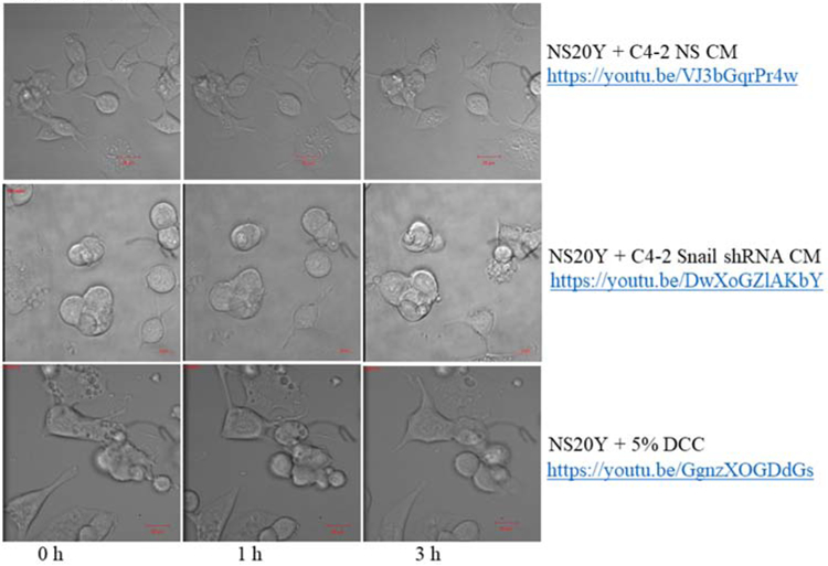
Figure 6. Visualization of neurite outgrowth in response to Snail-expressing cancer cells.

NS20Y neuronal cells were treated with conditioned media (CM) from C4-2 NS or C4-2 Snail shRNA cells for 4-6 hours. Treatment with 5% DCC was included as a negative control. The experimental and negative control nerve cells were photographed using time-lapse microscopy on a Zeiss LSM. The incubated nerve cells behavior was recorded using ZEN Black software. Images of nerve cells were taken every 60 seconds. Representative images are also shown at 0 h, 1 h, and 3 h. We observed that NS20Y nerve cells treated with CM from C4-2 NS prostate cancer cells (https://youtu.be/VJ3bGqrPr4w), displayed the highest cell to cell interaction, axonal elongation, and neurite formation compared to C4-2 Snail shRNA CM (https://youtu.be/DwXoGZlAKbY) or 5% DCC (https://youtu.be/GgnzXOGDdGs). Scale bars are 20 μm. All real time movies were recorded using 40X magnification.
