

Abstract
Background
Genetic studies have primarily been conducted in European ancestry populations, identifying dozens of loci associated with late-onset Alzheimer’s disease (AD). However, much of AD’s heritability remains unexplained; as the prevalence of AD varies across populations, the genetic architecture of the disease may also vary by population with the presence of novel variants or loci.
Methods
We conducted genome-wide analyses of AD in a sample of 2565 Caribbean Hispanics to better understand the genetic contribution to AD in this population. Statistical analysis included both admixture mapping and association testing. Evidence for differential gene expression within regions of interest was collected from independent transcriptomic studies comparing AD cases and controls in samples with primarily European ancestry.
Results
Our genome-wide association study of AD identified no loci reaching genome-wide significance. However, a genome-wide admixture mapping analysis that tests for association between a haplotype’s ancestral origin and AD status detected a genome-wide significant association with chromosome 3q13.11 (103.7–107.7Mb, P = 8.76E−07), driven by a protective effect conferred by the Native American ancestry (OR = 0.58, 95%CI = 0.47−0.73). Within this region, two variants were significantly associated with AD after accounting for the number of independent tests (rs12494162, P = 2.33E−06; rs1731642, P = 6.36E−05). The significant admixture mapping signal is composed of 15 haplotype blocks spanning 5 protein-coding genes (ALCAM, BBX, CBLB, CCDC54, CD47) and four brain-derived topologically associated domains, and includes markers significantly associated with the expression of ALCAM, BBX, CBLB, and CD47 in the brain. ALCAM and BBX were also significantly differentially expressed in the brain between AD cases and controls with European ancestry.
Conclusion
These results provide multiethnic evidence for a relationship between AD and multiple genes at 3q13.11 and illustrate the utility of leveraging genetic ancestry diversity via admixture mapping for new insights into AD.
Supplementary Information
The online version contains supplementary material available at 10.1186/s13195-021-00866-9.
Keywords: Admixture mapping, Alzheimer’s disease, Local ancestry, Genetic architecture, Genome scan, Multi-omics
Background
Late-onset Alzheimer’s disease (AD) is a leading cause of death in the USA, affecting approximately 1 in 10 Americans over the age of 65 years, with prevalence expected to double by 2050 [1]. Heritability estimates for AD range from 58 to 79% [2]; despite this strong genetic component, much of the underlying genetic variance remains to be explained [3]. Although the APOE ε4 allele is the strongest common genetic risk factor for AD [4, 5], dozens of loci have been associated with AD via genome-wide association studies (GWAS) [6, 7]. However, similar to other complex diseases, the vast majority of genetic discoveries for AD have been from GWAS performed in samples with predominantly European ancestry [8]. Much less is known about AD genetics across diverse populations, and in particular African Americans and Hispanic/Latino Americans who have increased risk of AD compared to European Americans [9–11].
Admixture mapping is a powerful alternative approach to GWAS for gene-mapping in recently admixed populations. Unlike widely used GWAS approaches that treat genetic ancestry differences as potential confounders in the analysis, admixture mapping leverages genetic ancestry differences [12–15]. With admixture mapping, regions of the genome with unusual local ancestry patterns relative to genome-wide averages are tested for association with a phenotype [16]. Admixture mapping is most powerful when both disease risk and trait-locus allele frequencies differ across groups, and it can be viewed as a complement to GWAS [17, 18].
Here, we performed genome scans of AD using both GWAS and admixture mapping approaches to identify regions associated with AD in Caribbean Hispanics, an admixed population with European, Native American, and African ancestry [19, 20]. Admixture mapping identified a genome-wide significant association between AD and Native American ancestry on 3q13.11, while GWAS identified no loci reaching genome-wide significance. Transcriptomic studies in samples with European ancestry nominate ALCAM and BBX as candidate protein-coding genes within the significant admixture mapping signal on 3q13.11, supported by the association between genetic variation and gene expression levels as well as differential expression between AD cases and controls. These results underline the power and challenges of leveraging genetic ancestry differences for new insight into the genetic architecture of late-onset AD in multiethnic populations.
Methods
Data
Genotype and phenotype data for 3067 participants in the Columbia University Study of Caribbean Hispanics and Late Onset Alzheimer’s Disease (CU Hispanics) were downloaded through dbGaP (Study Accession number phs000496.v1.p1), described in detail elsewhere [21]. The CU Hispanics study recruited subjects using both familial AD (22%) and sporadic case-control (78%) ascertainment. Subjects were excluded if they had any missing data for sex, AD status, APOE ε2/ ε3/ε4 genotypes, and either age-at-onset of dementia or age-at-last-evaluation.
European (from Utah) and African (Yorubans) samples from HapMap 3 [22] and Native American samples (Colombians, Pima, and Maya) from the Human Genome Diversity Project [23, 24] were used as reference populations. The reference datasets were merged using PLINK (v1.07) [25], resulting in 598,470 common autosomal single-nucleotide polymorphisms (SNPs). Genome coordinates were updated to build NCBI37/hg19 using LiftOver [26] to match the CU Hispanic data. The reference and CU Hispanics datasets were merged, randomly removing reference samples to balance ancestral population representation. Variants with a genotype missing rate > 5%, samples missing > 5% of genotypes, and 502 duplicated samples were excluded. Heterozygosity analysis identified 43 CU Hispanic outliers for both the F coefficient ( > 0.12, mean 0.02 ± 0.03) and heterozygosity rate (< 0.28, mean 0.32 ± 0.01), consistent with previous reports and pedigree documentation of consanguinity [27]. As both our admixture mapping and association tests adjust for genetic relatedness, keeping these samples had minimal impact on results (Additional file 1). The final combined dataset included 294,252 SNPs and 2754 samples: 2565 CU Hispanics plus 63 samples from each reference population. The overall genotyping rate was 0.993.
Genetic relatedness matrix
A genetic relatedness matrix (GRM) was estimated in a recursive manner using the PC-AiR and PC-Relate functions within the GENESIS R package [28–30]. The final combined data set was included in these analyses to improve inference of population structure. PC-AiR partitions subjects into unrelated and related sets based on kinship estimates from KING-robust [31], performs principal components (PC) analysis on the set of unrelated subjects, then projects PC values for the related set. PC-Relate adjusted the GRM for the first four PCs derived by PC-AiR, and the PC-AiR and PC-Relate steps were repeated using this adjusted GRM. The final GRM contains kinship coefficients that are robust to the population structure within our sample.
Ancestry proportions
As suggested by an established pipeline [32], the CU Hispanic and reference samples were phased jointly using ShapeIt2 [33] and 1000 Genomes phase 3 haplotypes [34] as reference. Local ancestry estimation was performed using RFMix (v1.5.4) [35]. Local ancestry values were averaged to estimate global European, African, and Native American ancestry proportions.
Admixture mapping
Admixture mapping was performed using a logistic mixed model for the AD phenotype, in which all European, African, and Native American ancestries were tested simultaneously. Admixture mapping was conducted using the GENESIS R package [30] available in Bioconductor [36]. We fit mixed models under the null hypothesis of no genetic association, adjusting for global ancestry proportions and APOE ε2 and ε4 allele dosages as fixed effects and the GRM as a random effect. The association between each admixture linkage disequilibrium (LD) block and the null model was evaluated by a score test. Recent admixture, as such observed in Hispanic/Latinos, creates long-range LD which dramatically reduces the number of independent tests in an admixture mapping genome scan, leading to a less-severe multiple testing correction. Genome-wide significance was defined as P < 5E−05 and suggestive evidence for significance was defined as P < 0.001, as suggested by previous studies of Hispanic populations [37, 38]. We evaluated the suitability of these significance thresholds by extending the method proposed by Shriner et al. 2011 [39] for three ancestral populations. We estimated the effective number of tests for each ancestral population by fitting autoregressive models to the vectors of African, European, and Native American local ancestry dosages in our sample (African: 251.1, European: 210.3, Native American: 281.2) and defined the final effective number of tests as the sum of the two largest values. This Bonferroni-corrected significance threshold of P < 9.39E−05 is slightly less conservative than our original threshold, suggesting it is well-suited for this sample.
Secondary admixture mapping analyses considered the effect of each reference group separately to identify which ancestral population was driving the significant signals. The coefficients of each lead SNP in the most significant LD-block were estimated, taking the allelic dosage of the ancestry driving the signal into account. Manhattan plots were prepared using the qqman R package [40], while regional association plots were generated using LocusZoom [41]. Additional sensitivity analyses assessed the robustness of our findings to age and sex covariate adjustment.
Association testing
SNPs and samples were submitted to the data cleaning procedures described above without the inclusion of reference samples, leaving 931,670 SNPs and 2565 CU Hispanic samples. We conducted the association testing for AD using a logistic mixed model implemented in the GENESIS R package [30]. Using the fitted null model described above, we tested the association between each SNP and the phenotype with a score test. Genome-wide significance was defined as P < 5E−08. Region-specific thresholds within the 3q13.11 locus for significant (P < 6.74E−05) and suggestive (P < 1.35E−05) evidence for association were adjusted for the effective number of tests, estimated by Genetic Type I error calculator [42].
Locus interpretation and gene prioritization
Conditional admixture mapping analyses were performed, applying the original model with further adjustment for allele dosage at SNPs of interest, individually and jointly. LD was estimated by both r2 and D’ using PLINK [25] in a set of 1349 unrelated CU Hispanics. LD plots based on the correlation statistic D’ by reference population were prepared using Haploview [43]. The Ensembl Variant Effect Predictor (v99 [44]) toolset generated SNP-level annotations within regions of interest.
The Accelerating Medical Partnerships for Alzheimer’s Disease (AMP-AD) project has provided a publicly available repository of multi-omic data aimed at finding genetic targets for AD therapeutics. We extracted significant cis expression quantitative trait loci (cis-eQTLs) from a recent AMP-AD study [45] (https://www.synapse.org/#!Synapse:syn17015233), representing RNA-sequencing data generated on brain samples from the Mayo study, Religious Orders Study, Rush Memory and Aging studies, and Mount Sinai Brain Bank study generated across 7 tissues types: cerebellum (N = 261), temporal cortex (N = 262), dorsolateral prefrontal cortex (N = 573), inferior gyrus (N = 230), superior temporal gyrus (N = 225), frontal pole (N =260), and parahippocampal gyrus (N = 225). We extracted evidence for differential gene expression in post-mortem brain tissues between those affected by AD and controls from another AMP-AD study [46] (https://www.synapse.org/#!Synapse:syn11914606), restricted to the meta-analysis results from the random effects model. A false discovery rate (FDR) cutoff of < 0.05 provided by the AMP-AD studies was applied to both the differential gene expression and eQTL results.
The genome is organized into topologically associated domains (TADs) in three-dimensional space, where genes within the same TAD are more likely to be regulated by common cis-regulatory elements and transcription factors. Genes within the same TAD as the haplotypes associated with AD were extracted from the 3D Genome Browser [47] and the human dorsolateral prefrontal cortex data (DLPFC) [48], again using the study-specific FDR < 0.05 as the significance threshold.
Genetic variation and patterns of LD vary across populations, and ideally colocalization analyses should use association and eQTL results representing the same population; unfortunately, large eQTL studies of Caribbean Hispanic populations are unavailable. Colocalization analyses comparing our admixture mapping or association studies are restricted to comparisons with the AMP-AD eQTLs representing samples with primarily European ancestry, which may identify relationships between eQTLs and AD risk shared between these populations [49, 50]. Approximate Bayes factor colocalization was performed using the Coloc package in R (v3.2-1) [50], which computes five posterior probabilities: PP0 = no association with either trait; PP1 = association with trait 1 but not trait 2; PP2 = association with trait 2 but not trait 1; PP3 = association with both traits, two independent causal SNPs; and PP4 = association with trait 1 and trait 2, one causal SNP shared for both traits. The LocusCompareR package in R (v1.0.0) [51] illustrated the correlation between admixture mapping or association results and eQTL data.
Results
Sample description
The CU Hispanics data represented 2565 subjects, where the 1174 cases were affected either by familial AD (22%) or sporadic AD (78%). Age-at-onset ranged from 44 to 100 years while the censoring age among the unaffected controls ranged from 35 to 100 years. The mean age and sex are similar across cases and controls (Table 1). The frequency of the protective APOE ε2 allele [52] is approximately 35% lower among cases, while the well-established risk allele ε4 [4, 5] is almost twice as common among cases than controls (Table 1). Global average ancestry proportion estimates vary widely across samples, from nearly zero to 0.99 per reference population (Fig. 1). Average ancestry proportions are 0.58 ± 0.17 European, 0.33 ± 0.19 African, and 0.09 ± 0.08 Native American ancestry.
Table 1.
Sample description
| Sample | N | Females (%) | Age (years) | ε2 (%) | ε4 (%) |
|---|---|---|---|---|---|
| Affected | 1174 | 64.1 | 74.9 (9.4) | 4.4 | 28.0 |
| Unaffected | 1391 | 67.7 | 72.5 (8.5) | 6.7 | 14.5 |
| Total | 2565 | 66.1 | 73.6 (9.0) | 5.6 | 20.7 |
Definitions: N = sample size, Age = mean ± standard deviation of age-at-onset of dementia (affected) or age-at-last-evaluation (unaffected), ε2 = frequency of the APOE ε2 allele, ε4 = frequency of the APOE ε4 allele
Fig. 1.

Estimated global ancestry proportions within the Caribbean Hispanics. X-axis: samples sorted by proportion of European ancestry, Y-axis: estimated global ancestry proportion. Colors correspond to reference populations: Blue for African, Purple for European, and Cyan for Native American
Admixture mapping and GWAS
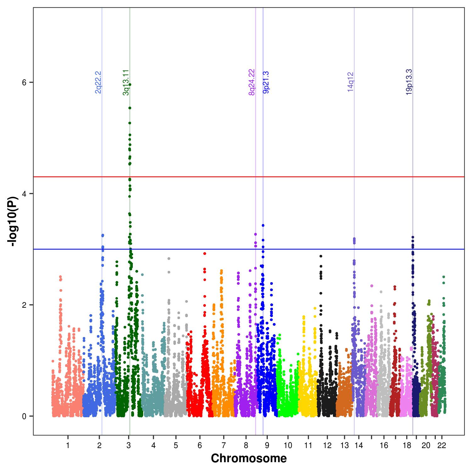
We identified a genome-wide significant association between AD and local ancestry at 3q13.11 (P < 5E−05; Table 2, Fig. 2). The 3q13.11 signal is supported by significant evidence of association across multiple LD-blocks (103.7 to 107.7Mb, min. P = 8.76E-07; Table 2), where the lead SNP rs10933849 is a common variant across the ancestral populations (alternate frequency: 0.56, 0.84, and 0.61 for 1000 Genomes phase 3 Africans, Europeans, and Native Americans, respectively). This region spans five protein-coding genes: ALCAM, CBLB, BBX, CCDC54, and CD47. Secondary analyses indicated that Native American ancestry at the lead SNP of each LD-block was associated with a protective effect against AD risk (OR 0.58–0.66; P < 3.24E-04; Additional file 2). Greater correlation is observed between 15 SNPs tagging the LD blocks within the 3q13.11 locus in the Native American reference data than in the European or African data (Additional file 3), providing further evidence that the admixture mapping signal between AD and 3q13.11 is driven by a Native American haplotype.
Table 2.
Evidence of association between local ancestry and Alzheimer’s disease in the Caribbean Hispanics
| Chr | Position | rsID | Gene | Consequence | Ancestry | OR (95% CI) | P |
|---|---|---|---|---|---|---|---|
| 2q22.2 | 142,486,253–143,387,612 | rs13024316 | LRP1B | Intron | EUR | 1.28 (1.13−1.45) | 6.82E−04 |
| 3q13.11 | 103,747,624–107,725,831 | rs10933849 | Intergenic | Intergenic | NAM | 0.58 (0.47−0.73) | 8.76E−07 |
| 6q22.31 | 123,548,997–123,838,033 | rs6940177 | TRDN | Intron | NAM | 1.44 (1.19−1.75) | 9.54E−04 |
| 8q24.22 | 135,308,849–135,856,404 | rs4308771 | RP11-513H8.1 | Intron | NAM | 1.36 (1.12−1.65) | 5.41E−04 |
| 9p21.3 | 22,207,037–22,870,294 | rs4977586 | Intergenic | Intergenic | NAM | 0.70 (0.56−0.87) | 4.56E−04 |
| 14q12 | 32,485,703–33,033,695 | rs1952961 | RP11-187E13.2 | Intron | AFR | 0.81 (0.71−0.93) | 7.24E−04 |
| 19p13.3 | 266,034–1,505,874 | rs3787017 | PALM | Intron | AFR | 1.29 (1.14−1.47) | 4.26E−04 |
Values are given for the SNP with the smallest P value per locus. Definitions: Chr = chromosome, Position = base pair position in NCBI37/hg19 genome build, Lead SNP = single-nucleotide polymorphism with the smallest P value within each LD-block, Ancestry = African (AFR), European (EUR), and Native American (NAM), OR = odds ratio, genome-wide significant evidence for association = P < 5E−05, suggestive evidence for association = P < 0.001. The block with significant evidence for association is highlighted in bold font. All intronic variants are canonical transcripts. Results for each block associated with Alzheimer’s disease at each locus are provided in Additional file 2
Fig. 2.

Association between Alzheimer’s disease and local ancestry among Caribbean Hispanics. Panel a: the joint European, African, and Native American ancestries admixture mapping analysis. Panels b, c, and d: results from single ancestry admixture mapping analyses for Native American, African, and European ancestries, respectively. Significant and suggestive thresholds represented by red and blue lines, respectively. Loci with significant or suggestive evidence of association with Alzheimer’s disease are highlighted with vertical bars labeled with their chromosomal position
Suggestive evidence of association between local ancestry and AD was observed at six additional loci: 2q22.2, 6q22.31, 8q24.22, 9p21.3, 14q12, and 19p13.3 (P < 0.001; Fig. 2, Table 2). LD-block-specific results for significant and suggestive associations with AD are provided in Additional file 2. Two LD-blocks with European background were responsible for the suggestive signal at 2q22.2, intersecting the gene LRP1B. The suggestive signal on 6q22.31 is driven by the Native American ancestry and was captured by a single LD-block within the TRDN gene. On 8q24.22, we observed three LD-blocks with Native American background driving the signal which spans the ZFAT gene. Two LD-blocks spanning the DMRTA1 gene were responsible for the signal on 9p21.3, driven by the Native American ancestry. The signal on 14q12, driven by the African ancestry, was captured by five LD-blocks implicating ARHGAP5 and AKAP6. Nine LD-blocks within a 1.3Mb region were responsible for the signal on 19p13.3 driven by African ancestry, implicating ABCA7 and dozens of other genes (Table 2). Sensitivity analyses revealed the admixture mapping results are robust to the inclusion of age and sex as covariates (Additional file 4). In contrast, traditional GWAS for AD did not identify any loci reaching genome-wide significance (P < 5E-08; Additional file 5).
Locus interpretation and gene prioritization
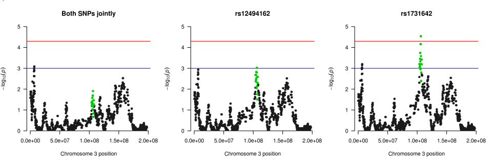
Targeted association testing within the 3q13.11 locus found two SNPs significantly associated with AD (rs12494162, P = 2.33E-06; rs1731642, P = 6.36E-05), and 22 SNPs with suggestive evidence of association with AD (P < 1.35E-03; Table 3). The first SNP, rs12494162, falls within an intron of lncRNA DUBR, while rs1731642 is an intergenic variant. These two SNPs, rs12494162 and rs1731642, are not in LD within our data (r2 = 0.003; D’= 0.17) and may represent independent association signals. This is consistent with LocusZoom plots of the admixture mapping and association signals at 3q13.11 using 1000 Genomes Native American estimates of LD (Fig. 3). The lead SNP rs12494162 is in LD with several other SNPs with evidence of association with AD, as expected. In contrast, the lead SNP from the admixture mapping analysis has modest evidence of LD with other SNPs on haplotypes associated with AD, as the admixture mapping signal is driven by differences in ancestry proportions rather than specific genotypes at the locus.
Table 3.
Variants with significant or suggestive evidence for association with Alzheimer’s disease within 3q13.11
| Chr | SNP | Position | Allele | Gene | Consequence | OR (95% CI) | P |
|---|---|---|---|---|---|---|---|
| 3 | rs1731642 | 103,811,750 | G | 38kb 5' of AC016970.1 | intergenic | 1.33 (1.17—1.53) | 6.36E−05 |
| 3 | rs9848147 | 104,111,719 | C | 338kb 5' of AC016970.1 | intergenic | 1.65 (1.30—2.08) | 7.02E−05 |
| 3 | rs2673478 | 104,158,244 | G | 385kb 5' of AC016970.1 | intergenic | 1.22 (1.09—1.38) | 1.33E−03 |
| 3 | rs16850638 | 104,594,467 | A | 491kb 5' of ALCAM | intergenic | 0.69 (0.57—0.84) | 4.24E−04 |
| 3 | rs12492893 | 104,667,061 | A | 419kb 5' of ALCAM | intergenic | 1.23 (1.10—1.38) | 5.45E−04 |
| 3 | rs9288795 | 104,683,753 | G | 402kb 5' of ALCAM | intergenic | 0.83 (0.74—0.92) | 1.21E−03 |
| 3 | rs16850772 | 104,736,837 | A | 349kb 5' of ALCAM | intergenic | 1.26 (1.12—1.41) | 1.91E−04 |
| 3 | rs9883825 | 104,745,091 | G | 341kb 5' of ALCAM | intergenic | 0.80 (0.71—0.90) | 3.55E−04 |
| 3 | rs13325696 | 104,755,481 | A | 330kb 5' of ALCAM | intergenic | 1.28 (1.13—1.45) | 2.29E−04 |
| 3 | rs1503079 | 104,759,032 | A | 327kb 5' of ALCAM | intergenic | 0.81 (0.72—0.91) | 4.92E−04 |
| 3 | rs1566720 | 104,761,408 | A | 324kb 5' of ALCAM | intergenic | 0.82 (0.73—0.92) | 1.13E−03 |
| 3 | rs1587707 | 104,786,133 | C | 300kb 5' of ALCAM | intergenic | 0.80 (0.71—0.89) | 2.06E−04 |
| 3 | rs2895295 | 104,790,415 | C | 295kb 5' of ALCAM | intergenic | 0.80 (0.71—0.90) | 2.32E−04 |
| 3 | rs10933809 | 104,800,350 | A | 285kb 5' of ALCAM | intergenic | 0.79 (0.70—0.89) | 1.19E−04 |
| 3 | rs1503089 | 104,805,417 | G | 280kb 5' of ALCAM | intergenic | 1.23 (1.09—1.39) | 9.28E−04 |
| 3 | rs1503075 | 104,806,853 | G | 279kb 5' of ALCAM | intergenic | 0.81 (0.72—0.92) | 1.23E−03 |
| 3 | rs1503158 | 104,815,105 | G | 271kb 5' of ALCAM | intergenic | 0.80 (0.71—0.90) | 5.52E−04 |
| 3 | rs13322578 | 105,002,637 | A | 83kb 5' of ALCAM | intergenic | 0.77 (0.66—0.89) | 5.90E−04 |
| 3 | rs9816851 | 105,012,805 | G | 73kb 5' of ALCAM | intergenic | 0.77 (0.67—0.89) | 6.64E−04 |
| 3 | rs9860520 | 106,416,700 | A | 9.1kb 3' of Y_RNA | intergenic | 0.79 (0.70—0.90) | 4.28E−4 |
| 3 | rs12489299 | 106,985,680 | A | DUBR | intron | 0.69 (0.57—0.83) | 1.70E−04 |
| 3 | rs12494162 | 107,036,379 | A | DUBR | Intron | 0.69 (0.60—0.80) | 2.33E−06 |
| 3 | rs7615167 | 107,514,134 | A | BBX | Intron | 1.56 (1.20—2.02) | 1.23E−03 |
| 3 | rs4855772 | 107,540,375 | G | 10kb 3' of BBX | intergenic | 1.80 (1.31—2.49) | 5.81E−04 |
Significance thresholds are based on the effective number of independent tests. Abbreviations: Chr = chromosome, SNP = single-nucleotide polymorphism identifier, Position = chromosome 3 position on the GRCh37/hg19 map, Allele = effect allele, OR = odds ratio, significant evidence of association = P < 6.7E-05, suggestive evidence of association = P < 1.4E-03. Significant results are highlighted in bold font
Fig. 3.

Patterns of linkage disequilibrium within the admixture mapping (top) and association (bottom) signals at 3q13.11. LocusZoom plots were drawn using the 1000 Genomes Native American estimates of linkage disequilibrium (r2; Nov. 2014). Chromosomal position on the hg19 map is shown on the X-axis, while the Y-axis provides evidence of association with Alzheimer’s disease as the –log10(P) value
Conditional admixture mapping analyses including both rs12494162 and rs1731642 as covariates eliminated the signal at 3q13.11 (P = 0.01), while analyses conditioned on either SNP alone only weakened the signals (Additional file 6), suggesting that admixture mapping and GWAS approaches may be tagging the same underlying variant. We assessed evidence of colocalization between eQTLs identified in DLPFC samples from subjects with primarily European ancestry and the admixture mapping and association signals at 3q13.11, as comparable studies representing Native Americans are unavailable. These analyses can only identify shared genetic architecture between eQTLs shared across populations and our admixture mapping or association results, which may represent fewer than half of eQTLs [49]. The leading eQTL within the 3q13.11 locus falls within a haplotype significantly associated with AD in the admixture mapping analysis (Fig. 4a): rs12629430 is significantly associated with the expression of lncRNA DUBR (Z = −4.47, FDR = 4.9E−04), lincRNA RP11.446H18.1 (Z = −6.22, FDR = 1.0E−07), and lincRNA RP11-446H18.6 (Z = −4.77, FDR = 1.4E−04). Colocalization analyses of the eQTL and admixture mapping signals did not reject the null hypothesis (PP0 = 0.9550). In contrast, the lead SNP from our targeted association testing within 3q13.11, rs12494162, is also an eQTL significantly associated with the expression of lincRNA RP11.446H18.1 (Z =−6.01, FDR = 3.4E−07), and lincRNA RP11-446H18.6 (Z =−4.44, FDR = 5.4E−04) (Fig. 4b). Colocalization analyses are not significant, but suggest association with both AD and gene expression here and weakly favor the model of independent SNPs driving these association (PP3 = 0.5070) rather than one shared SNP (PP4 = 0.4130).
Fig. 4.

Colocalization between cis-eQTLs and admixture mapping (a) and association results (b) at 3q13.11. a Colocalization results between significant admixture mapping haplotypes and eQTLs from dorsolateral prefrontal cortex (DLPFC) data. The color scale depicts extent of linkage disequilibrium (LD) with the lead cis-eQTL (rs12629430, purple diamond) in the 1000 Genomes Native American sample, which falls within a significant haplotype block. b Colocalization results between association test of 3q13.11 and cis-eQTLs from DLPFC data. The color scale depicts the amount of LD with the lead SNP from the association tests (rs12494162, purple diamond), based on 1000 Genomes Native American samples. The lead SNP rs1249162 is a significant cis-eQTL in the DLPFC data. Note: the difference in eQTL plots between panels a and b are due to differences in SNP marker density between admixture mapping and association testing
Within the 3q13.11 locus (chr3:103,747,624-107,725,831), we prioritized candidate protein-coding genes which fell either within one of the 15 LD-blocks associated with AD or within an intersecting TAD using the following features in transcriptomic studies representing European ancestry: (1) genes in which expression in brain tissue is significantly associated with cis-eQTL within the region of interest and (2) genes which are differentially expressed in the brain between AD cases and controls. The 3q13.11 region of interest spans five protein-coding genes: ALCAM, BBX, CBLB, CCDC54, and CD47 while four additional genes fall within the same TAD as BBX: IFT57, HHLA2, MYH15, and KIAA1524 (Additional file 7). A recent transcriptomic study [45] of 1694 brain samples identified 369 significant cis-eQTLs for IFT57, 182 significant cis-eQTLs for ALCAM, 118 significant cis-eQTLs for CBLB, 47 significant cis-eQTLs for CD47, 22 significant cis-eQTLs for BBX, and 6 significant cis-eQTLs for MYH15 (FDR < 0.05). The strongest cis-eQTL per gene is reported in Table 4, with all cis-eQTLs reported in Additional file 8. Another transcriptomic study [46] including an overlapping sample set of 2114 brain samples representing 478 cases and 300 controls identified significant evidence for differential gene expression in AD for both ALCAM (Z = 2.75, FDR = 2.76E-02) and BBX (Z = 3.73, FDR = 1.84E-03) (Additional file 9). While variation in the 3q13.11 region is associated with expression levels of ALCAM, BBX, CBLB, CD47 IFT57, and MYH15 in the brain, only ALCAM and BBX were significantly differentially expressed between AD cases and controls.
Table 4.
Significant cis expression quantitative loci (eQTLs) for candidate genes within 3q13.11 region of interest
| Position | SNP | Gene | Z | β | Allele | P | FDR |
|---|---|---|---|---|---|---|---|
| 105,108,867 | rs6797043 | ALCAM | -4.89 | -0.3429 | C | 1.29E-06 | 8.24E-05 |
| 107,583,197 | rs9875001 | BBX | 3.72 | 0.2620 | T | 2.17E-04 | 7.43E-03 |
| 105,488,134 | rs139969708 | CBLB | 4.01 | 0.4411 | T | 7.03E-05 | 2.85E-03 |
| 107,550,819 | rs1908324 | CD47 | 3.83 | 0.2512 | T | 1.45E-04 | 5.29E-03 |
| 107,917,824 | rs9857584 | IFT57 | 6.91 | 0.6714 | C | 1.33E-11 | 1.89E-09 |
| 107,823,224 | rs80003826 | MYH15 | -3.92 | -0.4498 | A | 9.85E-05 | 3.80E-03 |
Results are filtered to only include those with false discovery rate (FDR) < 0.05, restricted to the SNP with the smallest FDR value. Results for all significant eQTLs are presented in Additional file 8 and the original data can be found at https://www.synapse.org/#!Synapse:syn17015233. Legend: Position = GRCh37 position of the variant on chromosome 3, Z = Z statistic, β = estimated effect size, allele = effect allele, FDR = false discovery rate
Discussion
Admixture mapping of AD within a Caribbean Hispanic sample identified one genome-wide significant signal on 3q13.11 (P = 8.76E-07) and six unique suggestive signals at 2q22.2, 6q22.31, 8q24.22, 9p21.3, 14q12, and 19p13.3. The admixture mapping signal on 3q13.11 spanned 15 haplotype blocks, where the Native American ancestry is associated with reduced risk of AD. Association between the Native American ancestry and reduced risk of AD has previously been reported [53, 54]. Suggestive evidence of association between the 3q13.11 locus and AD has recently been reported in an African American GWAS involving nearly three times the sample size as our study [55], demonstrating the effectiveness of the admixture mapping approach as a complement to GWAS.
While admixture mapping provides insights into the genetic basis of disease in multiethnic populations, integration of AD transcriptomics allowed us to nominate candidate genes within 3q13.11. ALCAM and BBX, the genes with significant evidence for both brain eQTLs and differential expression between AD cases and controls, both have robust support in the literature for a functional relationship to AD. Proteomic studies suggest ALCAM, which plays a role in neuron-neuron adhesion and neurite growth networks, is dysregulated during the progression of AD [56]. ALCAM is also involved in blood-brain barrier disruption and T cell-dependent neurodegeneration [57], biological pathways implicated in the progression of AD [58]. Furthermore, ALCAM is a target gene of miR-142 which is significantly upregulated in the AD brain [59, 60]. BBX is differentially expressed in the entorhinal cortex and hippocampus and appears to play a role in the crosstalk between the peripheral blood and the central nervous system [61]. Multiple studies have shown that BBX is differentially expressed in the AD brain [61, 62], while another implicated BBX as a candidate Master Regulator responsible for AD progression [63].
Each of the loci harboring suggestive admixture mapping signals have also been previously associated with AD risk and/or pathology. The signal on 19p13.3 is driven by African ancestry and spans ABCA7, a gene in which coding changes have been associated with risk of AD in both African American and European American samples [64–67]. LRP1B within the 2q22.2 signal has been implicated in the production and presentation of amyloid beta (Aβ) [68], while multiple LRP1B haplotypes are associated with risk of developing AD in studies representing European Americans [69] and Caribbean Hispanics [70]. Variants on 14q13.1 near NPAS3 have been associated with AD biomarkers [71] and general cognitive function [72]. Variants in ZFAT on 8q24.22 have been associated with extreme longevity [73] and cerebrospinal fluid tau/Aβ42 levels [74]. Within 6q22.31, TRDN variants have implicated in cerebral Aβ deposition in APOE ε4 non-carriers [75] and rate of cognitive decline in AD [76]. Finally, 9p21.3 has previously been linked to AD risk [77], and variants within the region have been associated with both vascular dementia and AD [78].
Limitations
Our study has several limitations. Admixture mapping identifies regions associated with a given trait which must then be fine-mapped to identify the underlying risk variants. Colocalization analysis is not well powered in our study due to the poor representation of non-European populations in large eQTL data sets, as the genetic architecture of eQTLs can be ancestry specific [49, 79]. Fine-mapping analyses of whole-genome sequence data collected in this sample may allow the detection of the variants responsible for the admixture mapping signals. Publicly available datasets comparable in size or Native American ancestry proportions suitable for replication analyses are not available. Ongoing efforts, including AMP-AD and the Alzheimer’s Disease Sequencing Project, will provide data which may assist these efforts in the future.
Conclusions
Most AD GWAS have represented samples with European ancestry, and alternative strategies may detect additional genetic variants influencing AD in multiethnic populations. Caribbean Hispanics, despite being more likely to be diagnosed with AD [80, 81], have been underrepresented in AD genetics studies [82]. We illustrated the power of admixture mapping for detecting loci associated with AD in a Caribbean Hispanic sample, provided robust evidence for this association, and nominated several candidate genes with orthogonal functional and statistical evidence for a role in AD. Further investigation of these loci and nominated genes could lead to a better understanding of the genetic heterogeneity of AD in populations with significant Native American ancestry.
Supplementary Information
Additional file 1: Joint Photographic Group format .jpg file. Admixture mapping of Alzheimer’s disease in the Caribbean Hispanics excluding the heterozygosity outlier samples. Description: Joint European, African and Native American ancestries admixture mapping analysis, with chromosomal position on hg19 on the X-axis and –log10(P) values on the Y-axis. Significant and suggestive thresholds represented by red and blue lines, respectively. Loci with significant or suggestive evidence of association with Alzheimer’s disease are highlighted with vertical bars labeled with the chromosomal position of the peak.
Additional file 2: File format: Microsoft Word .docx file. Title: Linkage disequilibrium blocks within regions with either genome-wide significant or suggestive evidence for association between local ancestry and Alzheimer’s disease. Description: Regions reaching at least suggestive evidence of association with Alzheimer’s disease are defined and the evidence for that association is summarized. Abbreviations: Chr = chromosome, Position = physical positions based on GRCh37/hg19 map, SNP = single nucleotide polymorphism, Effect size (95%CI): odds ratio for AD followed by the 95% confidence interval.
Additional file 3: File format: Portable graphics format .png file. Title: Linkage disequilibrium patterns at 3q13.11 by ancestry. Description: Each panel illustrates the amount of linkage disequilibrium (D’) between pairs of markers in the 3q13.11 locus using different reference populations drawn from the 1000 Genomes data (Nov 2014).
Additional file 4: File format: Joint Photographic Group format .jpg file. Title: Admixture mapping of Alzheimer’s disease in the Caribbean Hispanics adjusted for age and sex. Description: Joint European, African and Native American ancestries admixture mapping analysis, with chromosomal position on hg19 on the X-axis and –log10(P) values on the Y-axis. Significant and suggestive thresholds represented by red and blue lines, respectively. Loci with significant or suggestive evidence of association with Alzheimer’s disease are highlighted with vertical bars labeled with the chromosomal position of the peak.
Additional file 5: File format: Portable graphics format .png file. Title: Genome-wide association testing results for Alzheimer’s disease. Description: Alzheimer’s disease status was tested for association with genotypes using a logistic regression model, adjusting for global ancestry proportions and APOE ε2 and ε4 allele dosages as fixed effects and the genetic relatedness matrix as a random effect. Genomic position on the hg19 map are provided on the X-axis and –log10(P) values on the Y-axis. The dotted horizontal line corresponds to a genome-wide significance threshold of 5E-08.
Additional file 6: File format: Portable graphics format .png file. Title: Conditional admixture mapping results at 3q13.11. Description: Each panel represents an admixture mapping analysis in the 3q13.11 locus, conditioned on the two single nucleotide polymorphisms (SNPs) associated with Alzheimer’s disease. The first panel shows the admixture mapping results with both SNPs are included in the analysis model, while the latter two adjust for only the named SNP. The X-axis represents the genomic position on chromosome 3 and the Y-axis represents –log10(P) values. The horizontal red line represents region-wide significance, while the blue line represents suggestive evidence of association. Green dots represent the locus reaching genome-wide significance in the original admixture mapping analysis.
Additional file 7: File format: Portable graphics format .png file. Title: Local context of admixture mapping and association signals at 3q13.11. Description: The top panel illustrates the admixture mapping testing for association with Alzheimer's disease status model. The second panel provides the association testing results for AD. The third panel provides the position of genes within the region of interest. The fourth panel illustrates the topologically associated domains (TADs; blue and gold bars) indicated by Hi-C experiments in DLPFC, where the heat map in magenta indicates the number of sequencing reads aligning to a pair of physical positions. Red horizontal lines represent the genome-wide significance threshold (P < 5E-05) and a blue line at the suggestive threshold (P < 0.001) used for admixture mapping. Sequence positions are aligned to the GRCh37/hg19 genome reference and are represented by the X-axis.
Additional file 8: File format: Microsoft Word .docx file. Title: Significant cis expression quantitative loci (eQTLs) for candidate genes within 3q13.11 region of interest. Description: Evidence that genotypes at a marker are associated with gene expression values. Results are filtered to only include those with false discovery rate < 0.05. Data are stored in the Synapse repository, Synapse ID: syn17015233, https://www.synapse.org/#!Synapse:syn17015233. Legend: Chr = chromosome, Position = GRCh37 position of the variant on chromosome 3, Z = Z statistic, FDR = false discovery rate, β = estimated effect size, A1 = allele 1, A2 = allele 2, A2freq = observed frequency of the A2 allele, Aup = allele associated with increased expression of the gene.
Additional file 9: File format: Microsoft Word .docx file. Title: Evidence of differential gene expression between those with and without Alzheimer's disease for 3q13.11 candidate genes. Description: Data are stored in the Synapse repository, Synapse ID: syn11914606, https://www.synapse.org/#!Synapse:syn11914606, file meta.anlz.ad_cntrl.tsv, which included no values for CCDC54 or HHLA2. Legend: z.fixed = Z statistic for the fixed effects model, p.fixed = p-value for fixed effect model, z.random = Z statistic for the random effects model, p.random = p-value for the random effects model, fdr.fixed = false discovery rate from the fixed effects model, fdr.random = false discovery rate from random effects model. Results reaching the significance level of fdr.random < 0.05 are highlighted in bold.
Acknowledgements
We thank the study participants and their families for their contribution to this research. The authors declare no conflicts of interest. Research funding was provided by the National Institutes of Health, National Institute on Aging (NIA) [grant number R01AG059737]. The Genetic Studies of Alzheimer’s disease in Caribbean Hispanics (EFIGA, dbGaP Study Accession phs000496.v1.p1) were funded by the NIA [grant numbers 5R37AG015473, RF1AG015473, R56AG051876]. We acknowledge the EFIGA study participants and the EFIGA research and support staff for their contributions to this study.
Abbreviations
- Aβ
Amyloid beta
- AD
Alzheimer’s disease
- AMP-AD
Accelerating Medical Partnerships for Alzheimer’s Disease
- CU Hispanics
Participants in the Columbia University Study of Caribbean Hispanics and Late Onset Alzheimer’s Disease study
- DLPFC
Dorsolateral prefrontal cortex
- eQTL
Expression quantitative trait locus
- FDR
False discovery rate
- GRM
Genetic relatedness matrix
- GWAS
Genome-wide association study
- LD
Linkage disequilibrium
- PC
Principal components
- SNP
Single-nucleotide polymorphism
- TAD
Topologically associated domain
Authors’ contributions
All authors made substantial contributions to the design of the work, interpretation of data, and critically revised the manuscript. EEB acquired the data, and ARVH and DX analyzed the data and drafted the work. All authors agree to be accountable for all aspects of the work in ensuring that questions related to the accuracy or integrity of any part of the work are appropriately investigated and resolved.
Funding
Research funding was provided by the National Institutes of Health, National Institute on Aging (NIA) [grant number R01AG059737].
Availability of data and materials
The genome scan data representing the Caribbean Hispanics are available through an application to the database of Genotypes and phenotypes (dbGaP; study ID: phs000496.v1.p1). These data are consented for general research use and are available through an application to dbGaP (https://www.ncbi.nlm.nih.gov/projects/gap/cgi-bin/study.cgi?study_id=phs000496.v1.p1). Analysis pipelines were based upon the GENESIS package in the R programming language (https://bioconductor.org/packages/release/bioc/html/GENESIS.html) and a published ancestry inference pipeline (https://github.com/armartin/ancestry_pipeline).
Declarations
Consent to publication
Not applicable. See Availability of data and material.
Ethics approval and consent to participate
This study was reviewed and approved by the University of Washington Institutional Review Board (STUDY ID: STUDY00000240). Data analysis was restricted to publically available data consented for general research use.
Competing interests
On behalf of all authors, the corresponding author states that there is no conflict of interest.
Footnotes
Publisher’s Note
Springer Nature remains neutral with regard to jurisdictional claims in published maps and institutional affiliations.
References
- 1.2020 Alzheimer’s disease facts and figures. Alzheimers Dement. 2020. [DOI] [PubMed]
- 2.Gatz M, Reynolds CA, Fratiglioni L, Johansson B, Mortimer JA, Berg S, Fiske A, Pedersen NL. Role of genes and environments for explaining Alzheimer disease. Arch Gen Psychiatry. 2006;63(2):168–174. doi: 10.1001/archpsyc.63.2.168. [DOI] [PubMed] [Google Scholar]
- 3.Ridge PG, Hoyt KB, Boehme K, Mukherjee S, Crane PK, Haines JL, et al. Assessment of the genetic variance of late-onset Alzheimer’s disease. Neurobiol Aging. 2016;41:200 e13–200 e20. doi: 10.1016/j.neurobiolaging.2016.02.024. [DOI] [PMC free article] [PubMed] [Google Scholar]
- 4.Corder EH, Saunders AM, Strittmatter WJ, Schmechel DE, Gaskell PC, Small GW, Roses A, Haines J, Pericak-Vance M. Gene dose of apolipoprotein E type 4 allele and the risk of Alzheimer’s disease in late onset families. Science. 1993;261(5123):921–923. doi: 10.1126/science.8346443. [DOI] [PubMed] [Google Scholar]
- 5.Strittmatter WJ, Saunders AM, Schmechel D, Pericak-Vance M, Enghild J, Salvesen GS, Roses AD. Apolipoprotein E: high-avidity binding to beta-amyloid and increased frequency of type 4 allele in late-onset familial Alzheimer disease. Proc Natl Acad Sci U S A. 1993;90(5):1977–1981. doi: 10.1073/pnas.90.5.1977. [DOI] [PMC free article] [PubMed] [Google Scholar]
- 6.Andrews SJ, Fulton-Howard B, Goate A. Interpretation of risk loci from genome-wide association studies of Alzheimer’s disease. Lancet Neurol. 2020;19(4):326–335. doi: 10.1016/S1474-4422(19)30435-1. [DOI] [PMC free article] [PubMed] [Google Scholar]
- 7.Bellenguez C, Grenier-Boley B, Lambert JC. Genetics of Alzheimer’s disease: where we are, and where we are going. Curr Opin Neurobiol. 2020;61:40–48. doi: 10.1016/j.conb.2019.11.024. [DOI] [PubMed] [Google Scholar]
- 8.Popejoy AB, Fullerton SM. Genomics is failing on diversity. Nature. 2016;538(7624):161–164. doi: 10.1038/538161a. [DOI] [PMC free article] [PubMed] [Google Scholar]
- 9.Reitz C, Mayeux R. Genetics of Alzheimer’s disease in Caribbean Hispanic and African American populations. Biol Psychiatry. 2014;75(7):534–541. doi: 10.1016/j.biopsych.2013.06.003. [DOI] [PMC free article] [PubMed] [Google Scholar]
- 10.Hohman TJ, Cooke-Bailey JN, Reitz C, Jun G, Naj A, Beecham GW, Liu Z, Carney RM, Vance JM, Cuccaro ML, Rajbhandary R, Vardarajan BN, Wang LS, Valladares O, Lin CF, Larson EB, Graff-Radford NR, Evans D, de Jager PL, Crane PK, Buxbaum JD, Murrell JR, Raj T, Ertekin-Taner N, Logue MW, Baldwin CT, Green RC, Barnes LL, Cantwell LB, Fallin MD, Go RCP, Griffith P, Obisesan TO, Manly JJ, Lunetta KL, Kamboh MI, Lopez OL, Bennett DA, Hardy J, Hendrie HC, Hall KS, Goate AM, Lang R, Byrd GS, Kukull WA, Foroud TM, Farrer LA, Martin ER, Pericak-Vance MA, Schellenberg GD, Mayeux R, Haines JL, Thornton-Wells TA, Alzheimer Disease Genetics Consortium Global and local ancestry in African-Americans: implications for Alzheimer’s disease risk. Alzheimers Dement. 2016;12(3):233–243. doi: 10.1016/j.jalz.2015.02.012. [DOI] [PMC free article] [PubMed] [Google Scholar]
- 11.Jun GR, Chung J, Mez J, Barber R, Beecham GW, Bennett DA, Buxbaum JD, Byrd GS, Carrasquillo MM, Crane PK, Cruchaga C, de Jager P, Ertekin-Taner N, Evans D, Fallin MD, Foroud TM, Friedland RP, Goate AM, Graff-Radford NR, Hendrie H, Hall KS, Hamilton-Nelson KL, Inzelberg R, Kamboh MI, Kauwe JSK, Kukull WA, Kunkle BW, Kuwano R, Larson EB, Logue MW, Manly JJ, Martin ER, Montine TJ, Mukherjee S, Naj A, Reiman EM, Reitz C, Sherva R, St. George-Hyslop PH, Thornton T, Younkin SG, Vardarajan BN, Wang LS, Wendlund JR, Winslow AR, Haines J, Mayeux R, Pericak-Vance MA, Schellenberg G, Lunetta KL, Farrer LA, Adams PM, Albert MS, Albin RL, Apostolova LG, Arnold SE, Asthana S, Atwood CS, Barmada MM, Barnes LL, Beach TG, Becker JT, Bigio EH, Bird TD, Blacker D, Boeve BF, Bowen JD, Boxer A, Burke JR, Cairns NJ, Cao C, Carlson CS, Carlsson CM, Carney RM, Carrasquillo MM, Carroll SL, Chui HC, Clark DG, Corneveaux J, Cribbs DH, Crocco EA, Cruchaga C, de Jager PL, DeCarli C, DeKosky ST, Demirci FY, Dick M, Dickson DW, Doody RS, Duara R, Ertekin-Taner N, Faber KM, Fairchild TJ, Fallon KB, Farlow MR, Ferris S, Frosch MP, Galasko DR, Gearing M, Geschwind DH, Ghetti B, Gilbert JR, Glass JD, Graff-Radford NR, Green RC, Growdon JH, Hakonarson H, Hamilton RL, Hardy J, Harrell LE, Head E, Honig LS, Huebinger RM, Huentelman MJ, Hulette CM, Hyman BT, Jarvik GP, Jicha GA, Jin LW, Karydas A, Kauwe JSK, Kaye JA, Kim R, Koo EH, Kowall NW, Kramer JH, LaFerla FM, Lah JJ, Leverenz JB, Levey AI, Li G, Lieberman AP, Lin CF, Lopez OL, Lyketsos CG, Mack WJ, Marson DC, Martiniuk F, Mash DC, Masliah E, McCormick WC, McCurry SM, McDavid AN, McKee AC, Mesulam M, Miller BL, Miller CA, Miller JW, Morris JC, Mukherjee S, Murrell JR, Myers AJ, O'Bryant S, Olichney JM, Pankratz VS, Parisi JE, Partch A, Paulson HL, Perry W, Peskind E, Petersen RC, Pierce A, Poon WW, Potter H, Quinn JF, Raj A, Raskind M, Reisberg B, Reisch JS, Reitz C, Ringman JM, Roberson ED, Rogaeva E, Rosen HJ, Rosenberg RN, Royall DR, Sager MA, Sano M, Saykin AJ, Schneider JA, Schneider LS, Seeley WW, Smith AG, Sonnen JA, Spina S, Stern RA, Tanzi RE, Thornton-Wells TA, Trojanowski JQ, Troncoso JC, Tsuang DW, van Deerlin VM, van Eldik LJ, Vardarajan BN, Vinters HV, Vonsattel JP, Weintraub S, Welsh-Bohmer KA, Williamson J, Wishnek S, Woltjer RL, Wright CB, Wu CK, Yu CE, Yu L, Zhang X. Transethnic genome-wide scan identifies novel Alzheimer’s disease loci. Alzheimers Dement. 2017;13(7):727–738. doi: 10.1016/j.jalz.2016.12.012. [DOI] [PMC free article] [PubMed] [Google Scholar]
- 12.McKeigue PM. Prospects for admixture mapping of complex traits. Am J Hum Genet. 2005;76(1):1–7. doi: 10.1086/426949. [DOI] [PMC free article] [PubMed] [Google Scholar]
- 13.Gouveia MH, Cesar CC, Santolalla ML, Anna HPS, Scliar MO, Leal TP, Araújo NM, Soares-Souza GB, Magalhães WCS, Mata IF, Ferri CP, Castro-Costa E, Mbulaiteye SM, Tishkoff SA, Shriner D, Rotimi CN, Tarazona-Santos E, Lima-Costa MF. Genetics of cognitive trajectory in Brazilians: 15 years of follow-up from the Bambui-Epigen Cohort Study of Aging. Sci Rep. 2019;9(1):18085. doi: 10.1038/s41598-019-53988-4. [DOI] [PMC free article] [PubMed] [Google Scholar]
- 14.Wang H, Cade BE, Sofer T, Sands SA, Chen H, Browning SR, Stilp AM, Louie TL, Thornton TA, Johnson WC, Below JE, Conomos MP, Evans DS, Gharib SA, Guo X, Wood AC, Mei H, Yaffe K, Loredo JS, Ramos AR, Barrett-Connor E, Ancoli-Israel S, Zee PC, Arens R, Shah NA, Taylor KD, Tranah GJ, Stone KL, Hanis CL, Wilson JG, Gottlieb DJ, Patel SR, Rice K, Post WS, Rotter JI, Sunyaev SR, Cai J, Lin X, Purcell SM, Laurie CC, Saxena R, Redline S, Zhu X. Admixture mapping identifies novel loci for obstructive sleep apnea in Hispanic/Latino Americans. Hum Mol Genet. 2019;28(4):675–687. doi: 10.1093/hmg/ddy387. [DOI] [PMC free article] [PubMed] [Google Scholar]
- 15.Ziyatdinov A, Parker MM, Vaysse A, Beaty TH, Kraft P, Cho MH, Aschard H. Mixed-model admixture mapping identifies smoking-dependent loci of lung function in African Americans. Eur J Hum Genet. 2020;28(5):656–668. doi: 10.1038/s41431-019-0545-8. [DOI] [PMC free article] [PubMed] [Google Scholar]
- 16.Shriner D. Overview of admixture mapping. Curr Protoc Hum Genet. 2013;Chapter 1:Unit 1 23. [DOI] [PMC free article] [PubMed]
- 17.Chakraborty R, Weiss KM. Admixture as a tool for finding linked genes and detecting that difference from allelic association between loci. Proc Natl Acad Sci U S A. 1988;85(23):9119–23. doi: 10.1073/pnas.85.23.9119. [DOI] [PMC free article] [PubMed] [Google Scholar]
- 18.Zhu X, Cooper RS, Elston RC. Linkage analysis of a complex disease through use of admixed populations. Am J Hum Genet. 2004;74(6):1136–1153. doi: 10.1086/421329. [DOI] [PMC free article] [PubMed] [Google Scholar]
- 19.Benn-Torres J, Bonilla C, Robbins CM, Waterman L, Moses TY, Hernandez W, Santos ER, Bennett F, Aiken W, Tullock T, Coard K, Hennis A, Wu S, Nemesure B, Leske MC, Freeman V, Carpten J, Kittles RA. Admixture and population stratification in African Caribbean populations. Ann Hum Genet. 2008;72(Pt 1):90–98. doi: 10.1111/j.1469-1809.2007.00398.x. [DOI] [PubMed] [Google Scholar]
- 20.Blue EE, Horimoto A, Mukherjee S, Wijsman EM, Thornton TA. Local ancestry at APOE modifies Alzheimer’s disease risk in Caribbean Hispanics. Alzheimers Dement. 2019;15(12):1524–1532. doi: 10.1016/j.jalz.2019.07.016. [DOI] [PMC free article] [PubMed] [Google Scholar]
- 21.Tosto G, Fu H, Vardarajan BN, Lee JH, Cheng R, Reyes-Dumeyer D, Lantigua R, Medrano M, Jimenez-Velazquez IZ, Elkind MSV, Wright CB, Sacco RL, Pericak-Vance M, Farrer L, Rogaeva E, St George-Hyslop P, Reitz C, Mayeux R. F-box/LRR-repeat protein 7 is genetically associated with Alzheimer’s disease. Ann Clin Transl Neurol. 2015;2(8):810–820. doi: 10.1002/acn3.223. [DOI] [PMC free article] [PubMed] [Google Scholar]
- 22.International HapMap C. Altshuler DM, Gibbs RA, Peltonen L, Altshuler DM, Gibbs RA, et al. Integrating common and rare genetic variation in diverse human populations. Nature. 2010;467(7311):52–58. doi: 10.1038/nature09298. [DOI] [PMC free article] [PubMed] [Google Scholar]
- 23.Cann HM, de Toma C, Cazes L, Legrand MF, Morel V, Piouffre L, Bodmer J, Bodmer WF, Bonne-Tamir B, Cambon-Thomsen A, Chen Z, Chu J, Carcassi C, Contu L, du R, Excoffier L, Ferrara GB, Friedlaender JS, Groot H, Gurwitz D, Jenkins T, Herrera RJ, Huang X, Kidd J, Kidd KK, Langaney A, Lin AA, Mehdi SQ, Parham P, Piazza A, Pistillo MP, Qian Y, Shu Q, Xu J, Zhu S, Weber JL, Greely HT, Feldman MW, Thomas G, Dausset J, Cavalli-Sforza LL. A human genome diversity cell line panel. Science. 2002;296(5566):261–262. doi: 10.1126/science.296.5566.261b. [DOI] [PubMed] [Google Scholar]
- 24.Cavalli-Sforza LL. The Human Genome Diversity Project: past, present and future. Nat Rev Genet. 2005;6(4):333–340. doi: 10.1038/nrg1579. [DOI] [PubMed] [Google Scholar]
- 25.Purcell S, Neale B, Todd-Brown K, Thomas L, Ferreira MA, Bender D, et al. PLINK: a tool set for whole-genome association and population-based linkage analyses. Am J Hum Genet. 2007;81(3):559–575. doi: 10.1086/519795. [DOI] [PMC free article] [PubMed] [Google Scholar]
- 26.Kuhn RM, Haussler D, Kent WJ. The UCSC genome browser and associated tools. Brief Bioinform. 2013;14(2):144–161. doi: 10.1093/bib/bbs038. [DOI] [PMC free article] [PubMed] [Google Scholar]
- 27.Vardarajan BN, Schaid DJ, Reitz C, Lantigua R, Medrano M, Jimenez-Velazquez IZ, et al. Inbreeding among Caribbean Hispanics from the Dominican Republic and its effects on risk of Alzheimer disease. Genet Med. 2015;17(8):639–643. doi: 10.1038/gim.2014.161. [DOI] [PMC free article] [PubMed] [Google Scholar]
- 28.Conomos MP, Reiner AP, Weir BS, Thornton TA. Model-free estimation of recent genetic relatedness. Am J Hum Genet. 2016;98(1):127–148. doi: 10.1016/j.ajhg.2015.11.022. [DOI] [PMC free article] [PubMed] [Google Scholar]
- 29.Conomos MP, Miller MB, Thornton TA. Robust inference of population structure for ancestry prediction and correction of stratification in the presence of relatedness. Genet Epidemiol. 2015;39(4):276–293. doi: 10.1002/gepi.21896. [DOI] [PMC free article] [PubMed] [Google Scholar]
- 30.Gogarten SM, Sofer T, Chen H, Yu C, Brody JA, Thornton TA, Rice KM, Conomos MP. Genetic association testing using the GENESIS R/Bioconductor package. Bioinformatics. 2019;35(24):5346–5348. doi: 10.1093/bioinformatics/btz567. [DOI] [PMC free article] [PubMed] [Google Scholar]
- 31.Manichaikul A, Mychaleckyj JC, Rich SS, Daly K, Sale M, Chen WM. Robust relationship inference in genome-wide association studies. Bioinformatics. 2010;26(22):2867–2873. doi: 10.1093/bioinformatics/btq559. [DOI] [PMC free article] [PubMed] [Google Scholar]
- 32.Martin ER, Tunc I, Liu Z, Slifer SH, Beecham AH, Beecham GW. Properties of global- and local-ancestry adjustments in genetic association tests in admixed populations. Genet Epidemiol. 2018;42(2):214–229. doi: 10.1002/gepi.22103. [DOI] [PMC free article] [PubMed] [Google Scholar]
- 33.Delaneau O, Coulonges C, Zagury JF. Shape-IT: new rapid and accurate algorithm for haplotype inference. BMC Bioinformatics. 2008;9(1):540. doi: 10.1186/1471-2105-9-540. [DOI] [PMC free article] [PubMed] [Google Scholar]
- 34.1000 Genomes Project Consortium, Auton A, Brooks LD, Durbin RM, Garrison EP, Kang HM, et al. A global reference for human genetic variation. Nature. 2015;526(7571):68–74. [DOI] [PMC free article] [PubMed]
- 35.Maples BK, Gravel S, Kenny EE, Bustamante CD. RFMix: a discriminative modeling approach for rapid and robust local-ancestry inference. Am J Hum Genet. 2013;93(2):278–288. doi: 10.1016/j.ajhg.2013.06.020. [DOI] [PMC free article] [PubMed] [Google Scholar]
- 36.Huber W, Carey VJ, Gentleman R, Anders S, Carlson M, Carvalho BS, Bravo HC, Davis S, Gatto L, Girke T, Gottardo R, Hahne F, Hansen KD, Irizarry RA, Lawrence M, Love MI, MacDonald J, Obenchain V, Oleś AK, Pagès H, Reyes A, Shannon P, Smyth GK, Tenenbaum D, Waldron L, Morgan M. Orchestrating high-throughput genomic analysis with Bioconductor. Nat Methods. 2015;12(2):115–121. doi: 10.1038/nmeth.3252. [DOI] [PMC free article] [PubMed] [Google Scholar]
- 37.Brown LA, Sofer T, Stilp AM, Baier LJ, Kramer HJ, Masindova I, Levy D, Hanson RL, Moncrieft AE, Redline S, Rosas SE, Lash JP, Cai J, Laurie CC, Browning S, Thornton T, Franceschini N. Admixture mapping identifies an Amerindian ancestry locus associated with albuminuria in hispanics in the United States. J Am Soc Nephrol. 2017;28(7):2211–2220. doi: 10.1681/ASN.2016091010. [DOI] [PMC free article] [PubMed] [Google Scholar]
- 38.Grinde KE, Brown LA, Reiner AP, Thornton TA, Browning SR. Genome-wide significance thresholds for admixture mapping studies. Am J Hum Genet. 2019;104(3):454–465. doi: 10.1016/j.ajhg.2019.01.008. [DOI] [PMC free article] [PubMed] [Google Scholar]
- 39.Shriner D, Adeyemo A, Rotimi CN. Joint ancestry and association testing in admixed individuals. PLoS Comput Biol. 2011;7(12):e1002325. doi: 10.1371/journal.pcbi.1002325. [DOI] [PMC free article] [PubMed] [Google Scholar]
- 40.Turner SD. qqman: an R package for visualizing GWAS results using Q-Q and manhattan plots. J Open Source Software. 2018;3(25):731. doi: 10.21105/joss.00731. [DOI] [Google Scholar]
- 41.Pruim RJ, Welch RP, Sanna S, Teslovich TM, Chines PS, Gliedt TP, Boehnke M, Abecasis GR, Willer CJ. LocusZoom: regional visualization of genome-wide association scan results. Bioinformatics. 2010;26(18):2336–2337. doi: 10.1093/bioinformatics/btq419. [DOI] [PMC free article] [PubMed] [Google Scholar]
- 42.Li MX, Yeung JM, Cherny SS, Sham PC. Evaluating the effective numbers of independent tests and significant p-value thresholds in commercial genotyping arrays and public imputation reference datasets. Hum Genet. 2012;131(5):747–756. doi: 10.1007/s00439-011-1118-2. [DOI] [PMC free article] [PubMed] [Google Scholar]
- 43.Barrett JC, Fry B, Maller J, Daly MJ. Haploview: analysis and visualization of LD and haplotype maps. Bioinformatics. 2005;21(2):263–265. doi: 10.1093/bioinformatics/bth457. [DOI] [PubMed] [Google Scholar]
- 44.McLaren W, Gil L, Hunt SE, Riat HS, Ritchie GR, Thormann A, et al. The Ensembl variant effect predictor. Genome Biol. 2016;17(1):122. doi: 10.1186/s13059-016-0974-4. [DOI] [PMC free article] [PubMed] [Google Scholar]
- 45.Sieberts SK, Perumal TM, Carrasquillo MM, Allen M, Reddy JS, Hoffman GE, et al. Large eQTL meta-analysis reveals differing patterns between cerebral cortical and cerebellar brain regions. Sci Data. 2020;7(1):340. doi: 10.1038/s41597-020-00642-8. [DOI] [PMC free article] [PubMed] [Google Scholar]
- 46.Logsdon BA, Perumal TM, Swarup V, Wang M, Funk C, Gaiteri C, et al. Meta-analysis of the human brain transcriptome identifies heterogeneity across human AD coexpression modules robust to sample collection and methological approach. 2019. [Google Scholar]
- 47.Wang Y, Song F, Zhang B, Zhang L, Xu J, Kuang D, Li D, Choudhary MNK, Li Y, Hu M, Hardison R, Wang T, Yue F. The 3D Genome Browser: a web-based browser for visualizing 3D genome organization and long-range chromatin interactions. Genome Biol. 2018;19(1):151. doi: 10.1186/s13059-018-1519-9. [DOI] [PMC free article] [PubMed] [Google Scholar]
- 48.Dixon JR, Selvaraj S, Yue F, Kim A, Li Y, Shen Y, Hu M, Liu JS, Ren B. Topological domains in mammalian genomes identified by analysis of chromatin interactions. Nature. 2012;485(7398):376–380. doi: 10.1038/nature11082. [DOI] [PMC free article] [PubMed] [Google Scholar]
- 49.Shang L, Smith JA, Zhao W, Kho M, Turner ST, Mosley TH, Kardia SLR, Zhou X. Genetic architecture of gene expression in European and African Americans: an eQTL mapping study in GENOA. Am J Hum Genet. 2020;106(4):496–512. doi: 10.1016/j.ajhg.2020.03.002. [DOI] [PMC free article] [PubMed] [Google Scholar]
- 50.Giambartolomei C, Vukcevic D, Schadt EE, Franke L, Hingorani AD, Wallace C, Plagnol V. Bayesian test for colocalisation between pairs of genetic association studies using summary statistics. PLoS Genet. 2014;10(5):e1004383. doi: 10.1371/journal.pgen.1004383. [DOI] [PMC free article] [PubMed] [Google Scholar]
- 51.Liu B, Gloudemans MJ, Rao AS, Ingelsson E, Montgomery SB. Abundant associations with gene expression complicate GWAS follow-up. Nat Genet. 2019;51(5):768–769. doi: 10.1038/s41588-019-0404-0. [DOI] [PMC free article] [PubMed] [Google Scholar]
- 52.Benjamin R, Leake A, McArthur FK, Ince PG, Candy JM, Edwardson JA, et al. Protective effect of apoE epsilon 2 in Alzheimer’s disease. Lancet. 1994;344(8920):473–474. doi: 10.1016/S0140-6736(94)91804-X. [DOI] [PubMed] [Google Scholar]
- 53.Benedet AL, Moraes CF, Camargos EF, Oliveira LF, Souza VC, Lins TC, Henriques AD, Carmo DGS, Machado-Silva W, Araújo CN, Córdova C, Pereira RW, Nóbrega OT. Amerindian genetic ancestry protects against Alzheimer’s disease. Dement Geriatr Cogn Disord. 2012;33(5):311–317. doi: 10.1159/000339672. [DOI] [PubMed] [Google Scholar]
- 54.Moreno DJ, Ruiz S, Rios A, Lopera F, Ostos H, Via M, et al. Association of GWAS top genes with late-onset Alzheimer’s disease in Colombian population. Am J Alzheimers Dis Other Demen. 2017;32(1):27–35. doi: 10.1177/1533317516679303. [DOI] [PMC free article] [PubMed] [Google Scholar]
- 55.Kunkle BW, Schmidt M, Klein HU, Naj AC, Hamilton-Nelson KL, Larson EB, Evans DA, de Jager PL, Crane PK, Buxbaum JD, Ertekin-Taner N, Barnes LL, Fallin MD, Manly JJ, Go RCP, Obisesan TO, Kamboh MI, Bennett DA, Hall KS, Goate AM, Foroud TM, Martin ER, Wang LS, Byrd GS, Farrer LA, Haines JL, Schellenberg GD, Mayeux R, Pericak-Vance MA, Reitz C, Writing Group for the Alzheimer’s Disease Genetics Consortium (ADGC) Abner E, Adams PM, Albin RL, Apostolova LG, Arnold SE, Atwood CS, Baldwin CT, Barber RC, Barral S, Beach TG, Becker JT, Beecham GW, Bigio EH, Bird TD, Blacker D, Boeve BF, Bowen JD, Boxer A, Burke JR, Burns JM, Cairns NJ, Cao C, Carlsson CM, Carney RM, Carrasquillo MM, Cribbs DH, Cruchaga C, Dick M, Dickson DW, Doody RS, Duara R, Faber KM, Fairchild TJ, Fallon KB, Fardo DW, Farlow MR, Ferris S, Frosch MP, Galasko DR, Gearing M, Geschwind DH, Ghetti B, Gilbert JR, Green RC, Growdon JH, Hakonarson H, Hamilton RL, Hardy J, Harrell LE, Honig LS, Huebinger RM, Huentelman MJ, Hulette CM, Jarvik GP, Jin LW, Karydas A, Katz MJ, Kauwe JSK, Keene CD, Kim R, Kramer JH, Lah JJ, Leung YY, Li G, Lieberman AP, Lipton RB, Lyketsos CG, Malamon J, Marson DC, Martiniuk F, Masliah E, McCormick WC, McCurry SM, McDavid AN, McDonough S, McKee AC, Mesulam M, Miller BL, Miller CA, Montine TJ, Mukherjee S, Myers AJ, O’Bryant SE, Olichney JM, Parisi JE, Peskind E, Pierce A, Poon WW, Potter H, Qu L, Quinn JF, Raj A, Raskind M, Reisberg B, Reisch JS, Ringman JM, Roberson ED, Rogaeva E, Rosen HJ, Royall DR, Sager MA, Schneider JA, Schneider LS, Seeley WW, Small S, Sonnen JA, Spina S, St George-Hyslop P, Stern RA, Tanzi RE, Troncoso JC, Tsuang DW, Valladares O, van Deerlin VM, Vardarajan BN, Vinters HV, Vonsattel JP, Weintraub S, Welsh-Bohmer KA, Wilhelmsen KC, Williamson J, Wingo TS, Woltjer RL, Wu CK, Younkin SG, Yu L, Yu CE, Zhao Y, Graff-Radford NR, Martinez I, Ayodele T, Logue MW, Cantwell LB, Jean-Francois M, Kuzma AB, Adams LD, Vance JM, Cuccaro ML, Chung J, Mez J, Lunetta KL, Jun GR, Lopez OL, Hendrie HC, Reiman EM, Kowall NW, Leverenz JB, Small SA, Levey AI, Golde TE, Saykin AJ, Starks TD, Albert MS, Hyman BT, Petersen RC, Sano M, Wisniewski T, Vassar R, Kaye JA, Henderson VW, DeCarli C, LaFerla FM, Brewer JB, Miller BL, Swerdlow RH, van Eldik LJ, Paulson HL, Trojanowski JQ, Chui HC, Rosenberg RN, Craft S, Grabowski TJ, Asthana S, Morris JC, Strittmatter SM, Kukull WA. Novel Alzheimer disease risk loci and pathways in African American individuals using the african genome resources panel: a meta-analysis. JAMA Neurol. 2021;78(1):102–113. doi: 10.1001/jamaneurol.2020.3536. [DOI] [PMC free article] [PubMed] [Google Scholar]
- 56.Zelaya MV, Perez-Valderrama E, de Morentin XM, Tunon T, Ferrer I, Luquin MR, et al. Olfactory bulb proteome dynamics during the progression of sporadic Alzheimer’s disease: identification of common and distinct olfactory targets across Alzheimer-related co-pathologies. Oncotarget. 2015;6(37):39437–39456. doi: 10.18632/oncotarget.6254. [DOI] [PMC free article] [PubMed] [Google Scholar]
- 57.Gonzalez H, Elgueta D, Montoya A, Pacheco R. Neuroimmune regulation of microglial activity involved in neuroinflammation and neurodegenerative diseases. J Neuroimmunol. 2014;274(1-2):1–13. doi: 10.1016/j.jneuroim.2014.07.012. [DOI] [PubMed] [Google Scholar]
- 58.Zenaro E, Piacentino G, Constantin G. The blood-brain barrier in Alzheimer’s disease. Neurobiol Dis. 2017;107:41–56. doi: 10.1016/j.nbd.2016.07.007. [DOI] [PMC free article] [PubMed] [Google Scholar]
- 59.Lau P, Bossers K, Janky R, Salta E, Frigerio CS, Barbash S, et al. Alteration of the microRNA network during the progression of Alzheimer’s disease. EMBO Mol Med. 2013;5(10):1613–1634. doi: 10.1002/emmm.201201974. [DOI] [PMC free article] [PubMed] [Google Scholar]
- 60.Ghanbari M, Munshi ST, Ma B, Lendemeijer B, Bansal S, Adams HH, Wang W, Goth K, Slump DE, den Hout MCGN, IJcken WFJ, Bellusci S, Pan Q, Erkeland SJ, Vrij FMS, Kushner SA, Ikram MA. A functional variant in the miR-142 promoter modulating its expression and conferring risk of Alzheimer disease. Hum Mutat. 2019;40(11):2131–2145. doi: 10.1002/humu.23872. [DOI] [PubMed] [Google Scholar]
- 61.Pang X, Zhao Y, Wang J, Zhou Q, Xu L, Kang, et al. The bioinformatic analysis of the dysregulated genes and microRNAs in entorhinal cortex, hippocampus, and blood for Alzheimer’s disease. Biomed Res Int. 2017;2017:9084507. doi: 10.1155/2017/9084507. [DOI] [PMC free article] [PubMed] [Google Scholar]
- 62.Wang Y, Wang Z. Identification of dysregulated genes and pathways of different brain regions in Alzheimer’s disease. Int J Neurosci. 2020;130(11):1082–1094. doi: 10.1080/00207454.2020.1720677. [DOI] [PubMed] [Google Scholar]
- 63.Aubry S, Shin W, Crary JF, Lefort R, Qureshi YH, Lefebvre C, Califano A, Shelanski ML. Assembly and interrogation of Alzheimer’s disease genetic networks reveal novel regulators of progression. Plos One. 2015;10(3):e0120352. doi: 10.1371/journal.pone.0120352. [DOI] [PMC free article] [PubMed] [Google Scholar]
- 64.Hollingworth P, Harold D, Sims R, Gerrish A, Lambert JC, Carrasquillo MM, et al. Common variants at ABCA7, MS4A6A/MS4A4E, EPHA1, CD33 and CD2AP are associated with Alzheimer’s disease. Nat Genet. 2011;43(5):429–435. doi: 10.1038/ng.803. [DOI] [PMC free article] [PubMed] [Google Scholar]
- 65.Lambert JC, Ibrahim-Verbaas CA, Harold D, Naj AC, Sims R, Bellenguez C, et al. Meta-analysis of 74,046 individuals identifies 11 new susceptibility loci for Alzheimer’s disease. Nat Genet. 2013;45(12):1452–1458. doi: 10.1038/ng.2802. [DOI] [PMC free article] [PubMed] [Google Scholar]
- 66.Reitz C, Jun G, Naj A, Rajbhandary R, Vardarajan BN, Wang LS, Valladares O, Lin CF, Larson EB, Graff-Radford NR, Evans D, de Jager PL, Crane PK, Buxbaum JD, Murrell JR, Raj T, Ertekin-Taner N, Logue M, Baldwin CT, Green RC, Barnes LL, Cantwell LB, Fallin MD, Go RCP, Griffith P, Obisesan TO, Manly JJ, Lunetta KL, Kamboh MI, Lopez OL, Bennett DA, Hendrie H, Hall KS, Goate AM, Byrd GS, Kukull WA, Foroud TM, Haines JL, Farrer LA, Pericak-Vance MA, Schellenberg GD, Mayeux R, Alzheimer Disease Genetics Consortium Variants in the ATP-binding cassette transporter (ABCA7), apolipoprotein E 4,and the risk of late-onset Alzheimer disease in African Americans. JAMA. 2013;309(14):1483–1492. doi: 10.1001/jama.2013.2973. [DOI] [PMC free article] [PubMed] [Google Scholar]
- 67.De Roeck A, Van Broeckhoven C, Sleegers K. The role of ABCA7 in Alzheimer’s disease: evidence from genomics, transcriptomics and methylomics. Acta Neuropathol. 2019;138(2):201–220. doi: 10.1007/s00401-019-01994-1. [DOI] [PMC free article] [PubMed] [Google Scholar]
- 68.Cam JA, Zerbinatti CV, Knisely JM, Hecimovic S, Li Y, Bu G. The low density lipoprotein receptor-related protein 1B retains beta-amyloid precursor protein at the cell surface and reduces amyloid-beta peptide production. J Biol Chem. 2004;279(28):29639–29646. doi: 10.1074/jbc.M313893200. [DOI] [PubMed] [Google Scholar]
- 69.Poduslo SE, Huang R, Spiro A., 3rd A genome screen of successful aging without cognitive decline identifies LRP1B by haplotype analysis. Am J Med Genet B Neuropsychiatr Genet. 2010;153B(1):114–119. doi: 10.1002/ajmg.b.30963. [DOI] [PubMed] [Google Scholar]
- 70.Shang Z, Lv H, Zhang M, Duan L, Wang S, Li J, Liu G, Ruijie Z, Jiang Y. Genome-wide haplotype association study identify TNFRSF1A, CASP7, LRP1B, CDH1 and TG genes associated with Alzheimer’s disease in Caribbean Hispanic individuals. Oncotarget. 2015;6(40):42504–42514. doi: 10.18632/oncotarget.6391. [DOI] [PMC free article] [PubMed] [Google Scholar]
- 71.Chouraki V, De Bruijn RF, Chapuis J, Bis JC, Reitz C, Schraen S, et al. A genome-wide association meta-analysis of plasma Abeta peptides concentrations in the elderly. Mol Psychiatry. 2014;19(12):1326–1335. doi: 10.1038/mp.2013.185. [DOI] [PMC free article] [PubMed] [Google Scholar]
- 72.Trampush JW, Yang ML, Yu J, Knowles E, Davies G, Liewald DC, et al. GWAS meta-analysis reveals novel loci and genetic correlates for general cognitive function: a report from the COGENT consortium. Mol Psychiatry. 2017;22(3):336–345. doi: 10.1038/mp.2016.244. [DOI] [PMC free article] [PubMed] [Google Scholar]
- 73.Miller JB, Ward E, Staley LA, Stevens J, Teerlink CC, Tavana JP, Cloward M, Page M, Dayton L, Alzheimer's Disease Genetics Consortium. Cannon-Albright LA, Kauwe JSK. Identification and genomic analysis of pedigrees with exceptional longevity identifies candidate rare variants. Neurobiol Dis. 2020;143:104972. doi: 10.1016/j.nbd.2020.104972. [DOI] [PMC free article] [PubMed] [Google Scholar]
- 74.Maxwell TJ, Corcoran C, Del-Aguila JL, Budde JP, Deming Y, Cruchaga C, et al. Genome-wide association study for variants that modulate relationships between cerebrospinal fluid amyloid-beta 42, tau, and p-tau levels. Alzheimers Res Ther. 2018;10(1):86. doi: 10.1186/s13195-018-0410-y. [DOI] [PMC free article] [PubMed] [Google Scholar]
- 75.Li QS, Parrado AR, Samtani MN, Narayan VA. Alzheimer’s disease neuroimaging I. Variations in the FRA10AC1 fragile site and 15q21 are associated with cerebrospinal fluid Abeta1-42 level. Plos One. 2015;10(8):e0134000. doi: 10.1371/journal.pone.0134000. [DOI] [PMC free article] [PubMed] [Google Scholar]
- 76.Sherva R, Gross A, Mukherjee S, Koesterer R, Amouyel P, Bellenguez C, Dufouil C, Bennett DA, Chibnik L, Cruchaga C, del-Aguila J, Farrer LA, Mayeux R, Munsie L, Winslow A, Newhouse S, Saykin AJ, Kauwe JSK, the Alzheimer's Disease Genetics Consortium. Crane PK, Green RC. Genome-wide association study of rate of cognitive decline in Alzheimer’s disease patients identifies novel genes and pathways. Alzheimers Dement. 2020;16(8):1134–1145. doi: 10.1002/alz.12106. [DOI] [PMC free article] [PubMed] [Google Scholar]
- 77.Zuchner S, Gilbert JR, Martin ER, Leon-Guerrero CR, Xu PT, Browning C, et al. Linkage and association study of late-onset Alzheimer disease families linked to 9p21.3. Ann Hum Genet. 2008;72(Pt 6):725–731. doi: 10.1111/j.1469-1809.2008.00474.x. [DOI] [PMC free article] [PubMed] [Google Scholar]
- 78.Emanuele E, Lista S, Ghidoni R, Binetti G, Cereda C, Benussi L, Maletta R, Bruni AC, Politi P. Chromosome 9p21.3 genotype is associated with vascular dementia and Alzheimer’s disease. Neurobiol Aging. 2011;32(7):1231–1235. doi: 10.1016/j.neurobiolaging.2009.07.003. [DOI] [PubMed] [Google Scholar]
- 79.Gay NR, Gloudemans M, Antonio ML, Abell NS, Balliu B, Park Y, et al. Impact of admixture and ancestry on eQTL analysis and GWAS colocalization in GTEx. Genome Biol. 2020;21(1):233. doi: 10.1186/s13059-020-02113-0. [DOI] [PMC free article] [PubMed] [Google Scholar]
- 80.Tang MX, Cross P, Andrews H, Jacobs DM, Small S, Bell K, Merchant C, Lantigua R, Costa R, Stern Y, Mayeux R. Incidence of AD in African-Americans, Caribbean Hispanics, and Caucasians in northern Manhattan. Neurology. 2001;56(1):49–56. doi: 10.1212/WNL.56.1.49. [DOI] [PubMed] [Google Scholar]
- 81.Reitz C, Mayeux R. Alzheimer disease: epidemiology, diagnostic criteria, risk factors and biomarkers. Biochem Pharmacol. 2014;88(4):640–651. doi: 10.1016/j.bcp.2013.12.024. [DOI] [PMC free article] [PubMed] [Google Scholar]
- 82.Vega IE, Cabrera LY, Wygant CM, Velez-Ortiz D, Counts SE. Alzheimer’s disease in the Latino community: intersection of genetics and social determinants of health. J Alzheimers Dis. 2017;58(4):979–992. doi: 10.3233/JAD-161261. [DOI] [PMC free article] [PubMed] [Google Scholar]
Associated Data
This section collects any data citations, data availability statements, or supplementary materials included in this article.
Supplementary Materials
Additional file 1: Joint Photographic Group format .jpg file. Admixture mapping of Alzheimer’s disease in the Caribbean Hispanics excluding the heterozygosity outlier samples. Description: Joint European, African and Native American ancestries admixture mapping analysis, with chromosomal position on hg19 on the X-axis and –log10(P) values on the Y-axis. Significant and suggestive thresholds represented by red and blue lines, respectively. Loci with significant or suggestive evidence of association with Alzheimer’s disease are highlighted with vertical bars labeled with the chromosomal position of the peak.
Additional file 2: File format: Microsoft Word .docx file. Title: Linkage disequilibrium blocks within regions with either genome-wide significant or suggestive evidence for association between local ancestry and Alzheimer’s disease. Description: Regions reaching at least suggestive evidence of association with Alzheimer’s disease are defined and the evidence for that association is summarized. Abbreviations: Chr = chromosome, Position = physical positions based on GRCh37/hg19 map, SNP = single nucleotide polymorphism, Effect size (95%CI): odds ratio for AD followed by the 95% confidence interval.
Additional file 3: File format: Portable graphics format .png file. Title: Linkage disequilibrium patterns at 3q13.11 by ancestry. Description: Each panel illustrates the amount of linkage disequilibrium (D’) between pairs of markers in the 3q13.11 locus using different reference populations drawn from the 1000 Genomes data (Nov 2014).
Additional file 4: File format: Joint Photographic Group format .jpg file. Title: Admixture mapping of Alzheimer’s disease in the Caribbean Hispanics adjusted for age and sex. Description: Joint European, African and Native American ancestries admixture mapping analysis, with chromosomal position on hg19 on the X-axis and –log10(P) values on the Y-axis. Significant and suggestive thresholds represented by red and blue lines, respectively. Loci with significant or suggestive evidence of association with Alzheimer’s disease are highlighted with vertical bars labeled with the chromosomal position of the peak.
Additional file 5: File format: Portable graphics format .png file. Title: Genome-wide association testing results for Alzheimer’s disease. Description: Alzheimer’s disease status was tested for association with genotypes using a logistic regression model, adjusting for global ancestry proportions and APOE ε2 and ε4 allele dosages as fixed effects and the genetic relatedness matrix as a random effect. Genomic position on the hg19 map are provided on the X-axis and –log10(P) values on the Y-axis. The dotted horizontal line corresponds to a genome-wide significance threshold of 5E-08.
Additional file 6: File format: Portable graphics format .png file. Title: Conditional admixture mapping results at 3q13.11. Description: Each panel represents an admixture mapping analysis in the 3q13.11 locus, conditioned on the two single nucleotide polymorphisms (SNPs) associated with Alzheimer’s disease. The first panel shows the admixture mapping results with both SNPs are included in the analysis model, while the latter two adjust for only the named SNP. The X-axis represents the genomic position on chromosome 3 and the Y-axis represents –log10(P) values. The horizontal red line represents region-wide significance, while the blue line represents suggestive evidence of association. Green dots represent the locus reaching genome-wide significance in the original admixture mapping analysis.
Additional file 7: File format: Portable graphics format .png file. Title: Local context of admixture mapping and association signals at 3q13.11. Description: The top panel illustrates the admixture mapping testing for association with Alzheimer's disease status model. The second panel provides the association testing results for AD. The third panel provides the position of genes within the region of interest. The fourth panel illustrates the topologically associated domains (TADs; blue and gold bars) indicated by Hi-C experiments in DLPFC, where the heat map in magenta indicates the number of sequencing reads aligning to a pair of physical positions. Red horizontal lines represent the genome-wide significance threshold (P < 5E-05) and a blue line at the suggestive threshold (P < 0.001) used for admixture mapping. Sequence positions are aligned to the GRCh37/hg19 genome reference and are represented by the X-axis.
Additional file 8: File format: Microsoft Word .docx file. Title: Significant cis expression quantitative loci (eQTLs) for candidate genes within 3q13.11 region of interest. Description: Evidence that genotypes at a marker are associated with gene expression values. Results are filtered to only include those with false discovery rate < 0.05. Data are stored in the Synapse repository, Synapse ID: syn17015233, https://www.synapse.org/#!Synapse:syn17015233. Legend: Chr = chromosome, Position = GRCh37 position of the variant on chromosome 3, Z = Z statistic, FDR = false discovery rate, β = estimated effect size, A1 = allele 1, A2 = allele 2, A2freq = observed frequency of the A2 allele, Aup = allele associated with increased expression of the gene.
Additional file 9: File format: Microsoft Word .docx file. Title: Evidence of differential gene expression between those with and without Alzheimer's disease for 3q13.11 candidate genes. Description: Data are stored in the Synapse repository, Synapse ID: syn11914606, https://www.synapse.org/#!Synapse:syn11914606, file meta.anlz.ad_cntrl.tsv, which included no values for CCDC54 or HHLA2. Legend: z.fixed = Z statistic for the fixed effects model, p.fixed = p-value for fixed effect model, z.random = Z statistic for the random effects model, p.random = p-value for the random effects model, fdr.fixed = false discovery rate from the fixed effects model, fdr.random = false discovery rate from random effects model. Results reaching the significance level of fdr.random < 0.05 are highlighted in bold.
Data Availability Statement
The genome scan data representing the Caribbean Hispanics are available through an application to the database of Genotypes and phenotypes (dbGaP; study ID: phs000496.v1.p1). These data are consented for general research use and are available through an application to dbGaP (https://www.ncbi.nlm.nih.gov/projects/gap/cgi-bin/study.cgi?study_id=phs000496.v1.p1). Analysis pipelines were based upon the GENESIS package in the R programming language (https://bioconductor.org/packages/release/bioc/html/GENESIS.html) and a published ancestry inference pipeline (https://github.com/armartin/ancestry_pipeline).