Abstract
The main causes of diffuse cystic lung diseases include lymphangioleiomyomatosis, pulmonary Langerhans cell histiocytosis, Birt-Hogg-Dubé syndrome, lymphoid interstitial pneumonia, light chain deposition disease, Pneumocystis jirovecii pneumonia, hypersensitivity pneumonitis, and desquamative interstitial pneumonia. Diffuse cystic lung diseases are rarely caused by a malignant process, which are secondary to metastases from sarcomas and gastrointestinal and gynecologic adenocarcinomas. Here, we present a rare case of invasive pulmonary adenocarcinoma associated with progressive diffusion of cystic lesions, revealed by chronic cough and progressive shortness of breath. It is important for clinicians to be aware of this unusual imaging manifestation of lung cancer, to avoid misdiagnoses.
Keywords: Cystic lung diseases, Pulmonary adenocarcinoma
Introduction
Cystic lung diseases represent a heterogeneous group of disorders that share in common the radiographic feature of multiple air-filled lucencies surrounded by a thin perceptible wall (<2 mm) and a well-defined interface with normal lung [1]. Tumoral causes of diffuse cystic lung disease are rare and mainly represented by metastasis from sarcoma or colorectal, pancreatic, and gynecologic adenocarcinomas [2]. Here, we present a rare case of invasive pulmonary adenocarcinoma associated with progressive diffusion of cystic lesions.
Case Report
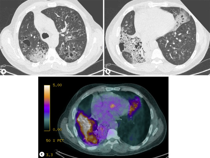
A 58-year-old man was referred for a 12-month history of chronic cough productive of clear sputum, progressive shortness of breath (Medical Research Council Dyspnea scale grade 1), and about 10 kg of weight loss. The patient had history of smoking discontinued 24 years ago and gastroesophageal reflux disease treated by ranitidine 300 mg per day. There were no severe comorbidities or environmental exposures and no familial history of chronic lung disease. On admission, resting oxygen saturation was 98%, and chest auscultation revealed decreased breath sounds in the right side. Thoracic CT scan showed extensive bilateral pulmonary cystic lesions surrounded by areas of ground-glass opacities with a marked pulmonary consolidation in the right lower lobe (Fig. 1a, b). Biological workup showed normal inflammatory markers, blood cell count, and serum proteins electrophoresis, negative human immunodeficiency virus serological testing, and negative autoimmune tests. Aspergillus serology was highly positive with both enzyme-linked immunosorbent assay (316 IU/mL) and Western blot. Bronchoalveolar lavage showed 680 cells/mm3, with 46% of macrophages, 46% of polynuclear neutrophils, 7% of eosinophils, 2% of lymphocytes, and no microorganism on direct examination and cultures. Immunohistochemical staining for CD1a was negative. Positron emission tomography-CT scan showed peak standard uptake values for 18-fluoro-deoxyglucose of 8.3 in the right lower lobe consolidation and 4.4 within diffuse cystic opacities (Fig. 1c).
Fig. 1.
CT scan with extensive bilateral pulmonary cystic opacities, surrounded by areas of ground-glass opacity (a); CT scan with extensive bilateral pulmonary cystic opacities, surrounded by areas of ground-glass opacity (b); PET-CT scan with intensely hypermetabolic right lower lobar fibrotic range (c).
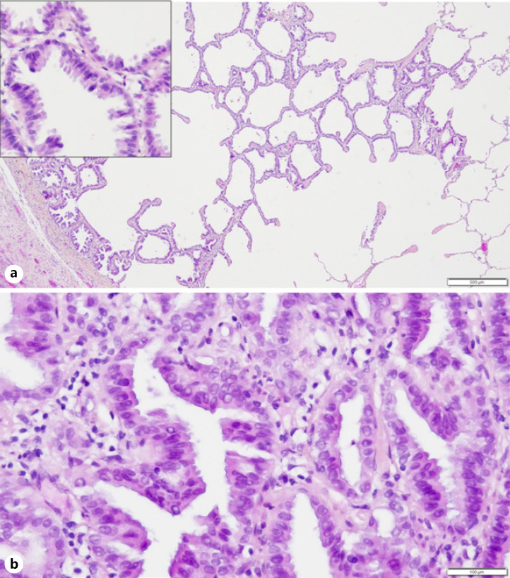
A few weeks later, the patient was readmitted for spontaneous right-sided pneumothorax, requiring chest tube placement. Video-assisted thoracoscopic lung biopsies demonstrated in situ adenocarcinoma in 3 foci of 1–4 mm diameter into an otherwise subnormal lung parenchyma (Fig. 2a). Finally, invasive pulmonary adenocarcinoma with lepidic, acinar, and papillary components was confirmed by a CT-guided biopsy in the right lower lobe (Fig. 2b). Immunohistochemical analysis showed positive TTF1 staining without ALK or ROS1 expression and no significant PDL1 expression in tumor cells (<1%). Biomolecular analysis in New Genome Sequencing on the biopsy specimen and plasma found a KRAS-G12V mutation (COSM520) and a TP53-G245S mutation (COSM6932) without any other mutations of therapeutic interest (EGFR, B-RAF, MET, ALK, or ERBB2 mutation).
Fig. 2.
a In situ adenocarcinoma in video-assisted thoracoscopic lung biopsy (hematoxylin-phloxine-saffron stain, Xobj 4). Inset, higher magnification highlights malignant nuclei (Xobj 40). b Invasive lung adenocarcinoma in a CT-guided biopsy (Hematoxylin-phloxine-saffron stain, Xobj 40).
Two hundred milligrams of itraconazole twice a day was initiated for invasive aspergillosis. According to recommendations, a chemotherapy associating carboplatin and pemetrexed was initiated but not well tolerated (importantly nauseas, headache, and asthenia). Observing a progression of ground-glass opacities and an increasing size of solid nodules and alveolar condensations on CT scan after 2 cures of chemotherapy, treatment was switched for second-line anti-PDL1 immunotherapy with atezolizumab. The CT scan after 3 cycles of atezolizumab concluded to a global stability of different lesions. Anti-PDL1 immunotherapy was well tolerated. However, cystic and ground-glass nodules progressed after 6 cycles of immunotherapy, and a third-line treatment by docetaxel was thus initiated without clinical and radiological improvement.
Discussion
The main conditions that are commonly referred to as diffuse cystic lung diseases include lymphangioleiomyomatosis, pulmonary Langerhans cell histiocytosis, Birt-Hogg-Dubé syndrome, lymphoid interstitial pneumonia, light chain deposition disease, Pneumocystis jirovecii pneumonia, hypersensitivity pneumonitis, and desquamative interstitial pneumonia [1]. Only 5 cases of primitive lung adenocarcinoma revealed by diffuse lung cysts, summarized in Table 1, have been previously reported in the literature [3, 4, 5, 6, 7]. The mechanism behind the formation of lung cysts in a tumor context is not clearly defined, and several hypotheses have been proposed. Accumulation of tumor cells in the terminal bronchioles could form a unidirectional valve, resulting in an expansion of the distal air spaces and the formation of cystic cavities. Another mechanism described is the infiltration of the vascular system by tumor cells, which can lead to ischemic necrosis of the bronchioles and alveoli and alveolar dilatation leading to the formation of cysts [3].
Table 1.
Summary of previously reported case reports of lung carcinoma presenting with diffuse cystic lesions
| Author | Case presentation | Evolution | ||
|---|---|---|---|---|
| Gui et al. [3] | 52-year-old Chinese woman CT scan: multiple cysts and nodules Anatomopathology (TBCB): Adenocarcinoma with EGFR mutation |
Significant improvement with afatinib therapy | ||
| Rogers et al. [4] | 65-year-old American woman CT scan: cysts in all lobes, areas of ground-glass opacity Anatomopathology (autopsy): invasive muncinous adenocarcinoma |
The patient developed progressive respiratory failure and died before treatment | ||
| Shannon et al. [5] | 56-year-old American woman CT scan: bilateral pulmonary cystic opacities Anatomopathology (VAT lung biopsies): multifocal, invasive, mucinous adenocarcinoma |
Chemotherapy with carboplatin and pemetrexed, but patient died 2 months later of respiratory failure |
||
| Kushima et al. [6] | 49-year-old Filipino man CT scan: multiloculated cystic lesions Anatomopathology (BAL): adenocarcinoma |
The patient died 4 weeks after diagnosis |
||
| Zhang et al. [7] | 39-year-old man CT scan: nodule in the left upper lobe and diffused cystic lesions Anatomopathology (bronchial biopsy): adenocarcinoma |
Significant improvement with cisplatin/gemcitabine chemotherapy |
||
TBCB, transbronchial lung cryobiopsy; VAT, video-assisted thoracoscopic; BAL, bronchoalveolar lavage.
Conclusion
This report highlights that lung adenocarcinoma might present as multiple cystic lesions on rare occasions. It is important for clinicians to be aware of this unusual imaging manifestation of lung cancer, to avoid misdiagnoses.
Statement of Ethics
Written informed consent was obtained from the patient for publication of this case report and any accompanying images.
Conflict of Interest Statement
The authors have no conflicts of interest to declare.
Funding Sources
No financial support was used for this case report.
Author Contributions
B.A., P.P., J.C., T.U., and F.G. searched the literature and wrote the manuscript. F.G. conceived and edited the manuscript. G.D.C. supervised the patient treatment, critically revised, and edited the manuscript. M.C.R. gave us input about pathology. All authors have made significant contributions to the manuscript and have reviewed it before submission. All authors have confirmed that the manuscript is not under consideration for review at any other journal. All authors have read and approved the final manuscript.
References
- 1.Gupta N, Vassallo R, Wikenheiser-Brokamp KA, McCormack FX. Diffuse cystic lung disease. Part I and II. Am J Respir Crit Care Med. 2015;191:1354–66. doi: 10.1164/rccm.201411-2094CI. [DOI] [PMC free article] [PubMed] [Google Scholar]
- 2.Seo JB, Im JG, Goo JM, Chung MJ, Kim MY. Atypical pulmonary metastases: spectrum of radiologic findings. Radiographics. 2001;21((2)):403–17. doi: 10.1148/radiographics.21.2.g01mr17403. [DOI] [PubMed] [Google Scholar]
- 3.Gui X, Ding J, Li Y, Yu M, Chen T, Huang M, et al. Lung carcinoma with diffuse cystic lesions misdiagnosed as pulmonary langerhans cell histocytosis: a case report. BMC Pulm Med. 2020;20:30. doi: 10.1186/s12890-020-1066-5. [DOI] [PMC free article] [PubMed] [Google Scholar]
- 4.Rogers C, Kent-Bramer J, Devaraj A, Nicholson AG, Molyneaux PL, Wells AU, et al. Rapidly progressive cystic lung disease. Am J Respir Crit Care Med. 2018;198:264. doi: 10.1164/rccm.201801-0161IM. [DOI] [PubMed] [Google Scholar]
- 5.Shannon VR, Nanda AS, Middleton LP, Faiz SA. Pulmonary mucinous cystadenocarcinoma presenting as extensive multifocal cystic lesions. Am J Respir Crit Care Med. 2017;195:1267–8. doi: 10.1164/rccm.201610-2106IM. [DOI] [PubMed] [Google Scholar]
- 6.Kushima H, Ishii H, Yokoyama A, Kadota J. Lung adenocarcinoma presenting with diffuse multiloculated cystic lesions. Intern Med. 2013;52:2375. doi: 10.2169/internalmedicine.52.1061. [DOI] [PubMed] [Google Scholar]
- 7.Zhang J, Zhao YL, Ye MX, Sun G, Wu H, Wu CG, et al. Rapidly progressive diffuse cystic lesions as a radiological hallmark of lung adenocarcinoma. J Thorac Oncol. 2012;7:457–8. doi: 10.1097/JTO.0b013e31823c5a39. [DOI] [PubMed] [Google Scholar]


