Abstract
Wilms' tumor is the most common malignant kidney tumor found in children. The Horseshoe kidney is the most common renal fusion malformation. However, Wilms' tumor is rarely identified in horseshoe kidney patients. Multimodal treatments in Wilms' tumor can play important roles in increasing the survival rate. In this study, we report the case of a 6-year-old boy in whom a Wilms' tumor was identified in a horseshoe kidney. The tumor was successfully treated with preoperative chemotherapy, followed by surgical resection.
Keywords: Wilms' tumor, Horseshoe kidney, Renal tumor, Children
Introduction
Nephroblastoma, also known as Wilms' tumor, is the most commonly identified pediatric renal mass, accounting for 87% of all renal masses and representing 7% of all malignant tumors identified in children [1]. The median age at which this tumor is identified is 3 years [2]. Wilms' tumor has been associated with a number of syndromes, including WAGR syndrome (Wilms' tumor, aniridia, genitourinary anomalies, and range of developmental delays), Beckwith-Wiedemann syndrome, Denys-Drash syndrome, and Edwards or Perlman syndrome [3]. Horseshoe kidney occurs in approximately 1 in 500 cases, with a male preponderance [4]. However, the occurrence of Wilms' tumor in horseshoe kidney is outstandingly rare, with an estimated incidence of approximately 0.4–0.9% of all Wilms' tumors [5]. In this report, we describe a rare case of Wilms' tumor identified in a horseshoe kidney to highlight the effectiveness of using preoperative chemotherapy during the treatment of this tumor.
Case Report
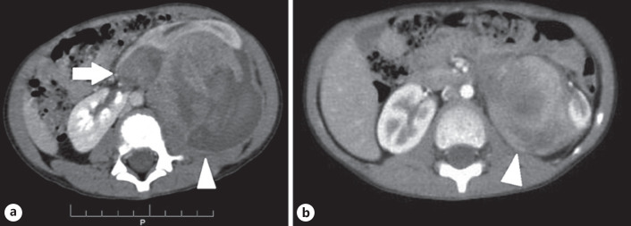
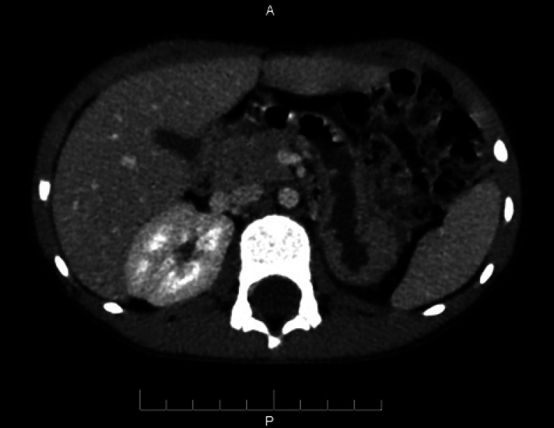
A 6-year-old boy presented to the hospital due to left upper abdominal pain after a falling accident. The patient's blood pressure and pulse were within the normal ranges. A physical examination revealed a large mass on the left upper abdomen. Laboratory studies demonstrated that complete blood counts, liver function tests, renal function tests, and urinalysis results were normal. An abdominal computed tomography (CT) scan was performed, which showed the existence of an isthmus connecting the right and left kidneys, anterior to the aorta and inferior vena cava. A large mass was observed, measuring 7 × 8 cm, located in the isthmus of the horseshoe kidney, which primarily developed toward the left side of the abdomen (Fig. 1a). This mass showed heterogeneous enhancement with less enhancement relative to the normal kidney parenchyma. The chest CT scan was normal. Due to the presence of a large kidney tumor in a child, an initial diagnosis of Wilms' tumor was established. Because this patient had both a large kidney tumor and horseshoe kidney, preoperative chemotherapy was used. The patient was treated with vincristine and actinomycin D for 12 weeks. An abdominal CT scan performed after 12 weeks of chemotherapy treatment showed that the tumor was 5 × 5 cm, suggesting a significant reduction in tumor size (Fig. 1b). The patient underwent surgical resection of the tumor, the isthmus, and the left kidney. A postsurgical pathological examination confirmed a Wilms' tumor of a favorable type, without abdominal lymph node metastasis. The patient completed 27 weeks of adjuvant chemotherapy with vincristine and actinomycin D. Two years later, no tumor recurrence or metastasis was observed on abdominal CT scans (Fig. 2). Chest CT scans performed 2 years after treatment also showed no lesions associated with suspected metastasis in either lung (Fig. 1, 2).
Fig. 1.
Abdominal CT images revealed the presence of a large mass located in the isthmus of the horseshoe kidney, which primarily developed toward the left side of the abdomen (a, arrow). This mass showed heterogeneous enhancement, with less enhancement relative to the normal kidney parenchyma (a, arrowhead). Abdominal CT image after 12 weeks of chemotherapy treatment revealed a significant reduction in tumor size (b, arrowhead). CT, computed tomography.
Fig. 2.
Abdominal CT image showing no metastasis or local recurrent lesion. CT, computed tomography.
Discussion
During the fetal period, the kidneys develop and ascend, developing first in the pelvis and then gradually ascending into position below the thorax, on either side of the lumbar spine [6]. During this ascending process, the kidneys also rotate, which typically occurs by the gestational ninth week [6]. Renal fusion anomalies may occur during this process [7]. The isthmus of the horseshoe kidney may contain functioning renal parenchyma or a fibrous band [8]. In up to 80% of cases of horseshoe kidney, the isthmus contains functional renal parenchyma tissue, and in >90% of cases, fusion occurs at the lower pole [6]. Patients with horseshoe kidney are often asymptomatic and are typically discovered incidentally, often due to symptoms secondary to pelvic ureteric junction obstruction and infection [9]. These patients are thought to be at increased risk of developing malignancies, such as renal cell carcinoma, Wilms' tumor, and carcinoids, among which renal cell carcinoma is the most common [7]. However, Wilms' tumor is the most common malignant kidney tumor identified in children [10]. The risk of Wilms' tumor in children with horseshoe kidney is 2–6 times that of children in the general population [11]. Approximately 50% of Wilms' tumors in horseshoe kidney develop from the isthmus, likely due to the abnormal proliferation of metanephric blastema in the isthmus [7]. The same anomaly that causes the development of horseshoe kidney may also lead to the development of Wilms' tumor [12]. Patients with Wilms' tumors are often asymptomatic; approximately 10% are discovered incidentally after trauma, whereas 25% present with microscopic hematuria or hypertension secondary to renin production [13]. Ultrasound is used to diagnose horseshoe kidney, whereas CT and magnetic resonance imaging are often used for staging purposes [14]. On ultrasound, the mass presents as a large renal mass, which can be either solid or cystic, with large hypoechoic areas due to central necrosis and cyst formation [13]. Areas characterized by fat deposits, calcification, or hemorrhage may appear [13]. On CT, the tumors are lower density and enhance less than the normal renal parenchyma [15]. Tumors are often characterized by heterogeneous contrast enhancement and may feature punctuated calcifications [15]. On magnetic resonance imaging, the tumors have low signal intensity on T1-weighted images, with either low or high signal intensity on T2-weighted images and restricted diffusivity on diffusion-weighted images [13]. CT is also used for the detection of lung metastasis or local recurrence [13]. Wilms' tumors contain variable quantities of embryonic renal elements, such as blastema, epithelium, and stroma [16]. Wilms' tumor can be divided into 2 types, based on prognosis: favorable (over 90%) and unfavorable (6–10%) [13]. Histopathological analysis is the current gold standard for diagnosing Wilms' tumor. Surgery, chemotherapy, and radiotherapy are also used to treat Wilms' tumor [10]. The National Wilms Tumor Study Group (NWTSG)/Children's Oncology Group (COG) and the International Society of Paediatric Oncology (SIOP) have established the major guidelines regarding the management of Wilms' tumor [17]. SIOP recommends using preoperative chemotherapy to reduce the tumor size and prevent intraoperative spillage due to tumor rupture [10]. In contrast, the NWTSG/COG recommends the application of primary surgery before any adjuvant treatments [17]. The overall survival of children with Wilms' tumor in horseshoe kidney appears to be similar to that among children with Wilms' tumor in normal kidneys [5].
Although multiple guidelines may be used for the management of Wilms' tumor, this patient was treated with chemotherapy to reduce the tumor size before surgery. Because the tumor arose from the isthmus and primarily developed to the left, the left kidney, isthmus, and tumor were all removed completely. After 2 years of follow-up, no evidence of tumor recurrence or metastasis was observed.
Conclusion
Horseshoe kidney is a common congenital kidney anomaly. Patients with horseshoe kidney have an increased risk of Wilms' tumor than patients with normal kidneys. Imaging plays a pivotal role in the diagnosis and follow-up posttreatment. Neoadjuvant chemotherapy can reduce the size of the tumor prior to surgery while maintaining normal kidney parenchyma.
Statement of Ethics
All treatments and examinations followed the guidance of the Declaration of Helsinki. Informed consent for treatment was obtained from the legal guardian of the patient. The legal guardian of the patient has given written informed consent to publish the case, including the publication of images.
Conflict of Interest Statement
The authors have no conflicts of interest to declare.
Funding Sources
This project was not supported by any grant or funding agencies.
Author Contributions
Doan Tien Luu and Nguyen Minh Duc contributed equally to this article as co-first authors. Doan Tien Luu, Nguyen Minh Duc, and Thieu-Thi Tra My contributed to the acquisition of data and writing of the manuscript. Nguyen Minh Duc and Thieu-Thi Tra My provided supervision, assessment, and interpretation of data, along with mentorship. All authors approved the final manuscript.
References
- 1.Lee JS, Sanchez TR, Wootton-Gorges S. Malignant renal tumors in children. J Kidney Cancer VHL. 2015 May 10;2((3)):84–9. doi: 10.15586/jkcvhl.2015.29. [DOI] [PMC free article] [PubMed] [Google Scholar]
- 2.Bozlu G, Çıtak EÇ. Evaluation of renal tumors in children. Turk J Urol. 2018 May;44((3)):268–73. doi: 10.5152/tud.2018.70120. [DOI] [PMC free article] [PubMed] [Google Scholar]
- 3.T Treger TD, Chowdhury T, Pritchard-Jones K, Behjati S. The genetic changes of Wilms tumour. Nat Rev Nephrol. 2019 Apr;15((4)):240–51. doi: 10.1038/s41581-019-0112-0. [DOI] [PubMed] [Google Scholar]
- 4.Glodny B, Petersen J, Hofmann KJ, Schenk C, Herwig R, Trieb T, et al. Kidney fusion anomalies revisited: clinical and radiological analysis of 209 cases of crossed fused ectopia and horseshoe kidney. BJU Int. 2009 Jan;103((2)):224–35. doi: 10.1111/j.1464-410X.2008.07912.x. [DOI] [PubMed] [Google Scholar]
- 5.Lee SH, Bae MH, Choi SH, Lee JS, Cho YS, Joo KJ, et al. Wilms' tumor in a horseshoe kidney. Korean J Urol. 2012 Aug;53((8)):577–80. doi: 10.4111/kju.2012.53.8.577. [DOI] [PMC free article] [PubMed] [Google Scholar]
- 6.Taghavi K, Kirkpatrick J, Mirjalili SA. The horseshoe kidney: surgical anatomy and embryology. J Pediatr Urol. 2016 Oct;12((5)):275–80. doi: 10.1016/j.jpurol.2016.04.033. [DOI] [PubMed] [Google Scholar]
- 7.Shah HU, Ojili V. Multimodality imaging spectrum of complications of horseshoe kidney. Indian J Radiol Imaging. 2017 Apr−Jun;27((2)):133–40. doi: 10.4103/ijri.IJRI_298_16. [DOI] [PMC free article] [PubMed] [Google Scholar]
- 8.Natsis K, Piagkou M, Skotsimara A, Protogerou V, Tsitouridis I, Skandalakis P. Horseshoe kidney: a review of anatomy and pathology. Surg Radiol Anat. 2014 Aug;36((6)):517–26. doi: 10.1007/s00276-013-1229-7. [DOI] [PubMed] [Google Scholar]
- 9.Cascio S, Sweeney B, Granata C, Piaggio G, Jasonni V, Puri P. Vesicoureteral reflux and ureteropelvic junction obstruction in children with horseshoe kidney: treatment and outcome. J Urol. 2002 Jun;167((6)):2566–8. [PubMed] [Google Scholar]
- 10.Bhatnagar S. Management of Wilms' tumor: NWTS vs SIOP. J Indian Assoc Pediatr Surg. 2009 Jan;14((1)):6–14. doi: 10.4103/0971-9261.54811. [DOI] [PMC free article] [PubMed] [Google Scholar]
- 11.Tkocz M, Kupajski M. Tumour in horseshoe kidney − different surgical treatment shown in five example cases. Contemp Oncol. 2012;16((3)):254–7. doi: 10.5114/wo.2012.29295. [DOI] [PMC free article] [PubMed] [Google Scholar]
- 12.Neville H, Ritchey ML, Shamberger RC, Haase G, Perlman S, Yoshioka T. The occurrence of Wilms tumor in horseshoe kidneys: a report from the national wilms tumor study group (NWTSG) J Pediatr Surg. 2002 Aug;37((8)):1134–7. doi: 10.1053/jpsu.2002.34458. [DOI] [PubMed] [Google Scholar]
- 13.Dumba M, Jawad N, McHugh K. Neuroblastoma and nephroblastoma: a radiological review. Cancer Imaging. 2015 Apr 8;15((1)):5. doi: 10.1186/s40644-015-0040-6. [DOI] [PMC free article] [PubMed] [Google Scholar]
- 14.Servaes SE, Hoffer FA, Smith EA, Khanna G. Imaging of Wilms tumor: an update. Pediatr Radiol. 2019 Oct;49((11)):1441–52. doi: 10.1007/s00247-019-04423-3. [DOI] [PubMed] [Google Scholar]
- 15.Olukayode A, Richard I, Rachael A, Babajide O, Gbolahan O. Pattern of computed tomography scan findings in children with Wilms' tumor in a tertiary hospital in Lagos, Nigeria. Indian J Med Paediatr Oncol. 2014;35((1)):31. doi: 10.4103/0971-5851.133713. [DOI] [PMC free article] [PubMed] [Google Scholar]
- 16.Lonergan GJ, Martínez-León MI, Agrons GA, Montemarano H, Suarez ES. Nephrogenic rests, nephroblastomatosis, and associated lesions of the kidney. Radiographics. 1998 Jul−Aug;18((4)):947–68. doi: 10.1148/radiographics.18.4.9672980. [DOI] [PubMed] [Google Scholar]
- 17.Wang J, Li M, Tang D, Gu W, Mao J, Shu Q. Current treatment for Wilms tumor: COG and SIOP standards. World J Ped Surg. 2019;2((3)):e000038. [Google Scholar]


